Abstract
Cytosolic phospholipase A2 (GIVA cPLA2) is the only PLA2 that exhibits a marked preference for hydrolysis of arachidonic acid containing phospholipid substrates releasing free arachidonic acid and lysophospholipids and giving rise to the generation of diverse lipid mediators involved in inflammatory conditions. Thus, the development of potent and selective GIVA cPLA2 inhibitors is of great importance. We have developed a novel class of such inhibitors based on the 2-oxoester functionality. This functionality in combination with a long aliphatic chain or a chain carrying an appropriate aromatic system, such as the biphenyl system, and a free carboxyl group leads to highly potent and selective GIVA cPLA2 inhibitors (X I(50) values 0.00007–0.00008) and docking studies aid in understanding this selectivity. A methyl 2-oxoester, with a short chain carrying a naphthalene ring, was found to preferentially inhibit the other major intracellular PLA2, the calcium-independent PLA2. In RAW264.7 macrophages, treatment with the most potent 2-oxoester GIVA cPLA2 inhibitor resulted in over 50% decrease in KLA-elicited prostaglandin D2 production. The novel, highly potent and selective GIVA cPLA2 inhibitors provide excellent tools for the study of the role of the enzyme and could contribute to the development of novel therapeutic agents for the treatment of inflammatory diseases.
Introduction
In mammals, the phospholipase A2 (PLA2) superfamily consists of six types of diverse enzymes: GIV PLA2 [cytosolic PLA2 (cPLA2)], GVI PLA2 [calcium-independent PLA2 (iPLA2)], several groups of secreted PLA2 (sPLA2), two groups of platelet-activating factor-acetylhydrolases PLA2 (PAF-AHs), GXV PLA2 (lysosomal PLA2), and GXVI PLA2 (adipose PLA2)1. Among all these enzymes, cPLA2 is the only PLA2 that exhibits a marked preference for hydrolysis of arachidonic acid at the sn-2 position of phospholipid substrates2. The activation of cPLA2 results in the production of arachidonic acid and lysophospholipids giving rise to the generation of diverse lipid mediators, such as leukotrienes, prostaglandins, lysophosphatidic acid etc3. Since many of them are involved in the response to inflammation, the regulation of cPLA2 is of great importance in chronic inflammatory conditions1, 4. In a recent review article, Leslie has summarized the physiological function and the role of cPLA2 in diseases5. The most recent studies on inherited GIVA cPLA2 deficiency demonstrate the fundamental role of this enzyme in eicosanoid formation and cellular responses in human circulation6.
It was thirty years ago, when the first cytosolic PLA2 activity (now attributed to GIVA cPLA2 or cPLA2 α) was reported in human neutrophils and platelets7, 8. The purification, sequence, and cloning of the first human cPLA2 was reported in 19919, 10. GIVA cPLA2 contains 749 amino acids, is an 85 kDa protein, and consists of an N-terminal C2 domain and a C-terminal catalytic domain. The crystal structure of GIVA cPLA2 was solved by Dessen et al. in 199911. The catalytic domain of GIVA cPLA2 utilizes an unusual catalytic dyad, Ser-228/Asp-549, located in the α/β hydrolase domain, to catalyze the hydrolysis of the substrate phospholipid12, 13.
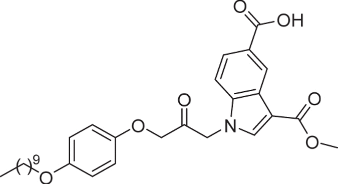
The diverse bioactive lipids produced by the cPLA2 activity regulate normal physiological processes and disease pathogenesis, and as a consequence, great attention has been given to the development of selective GIVA cPLA2 inhibitors. The structural diversity of the synthetic inhibitors is summarized in a number of review articles1, 14–16. The first synthetic inhibitor of GIVA cPLA2 was an arachidonic acid derivative, arachidonoyl trifluoromethyl ketone, containing an activated carbonyl functionality17. Shionogi developed a series of pyrrolidine-based inhibitors, including pyrrophenone (1, Fig. 1), following a high throughput screening approach18, 19. Wyeth has expended major efforts to develop novel indole-based inhibitors, for example, ecopladib (2a, Fig. 1), efipladib (2b, Fig. 1) and giripladib (2c, Fig. 1) as novel therapeutics for inflammatory diseases20–23. Giripladib was the most promising among them as it was advanced into a Phase II clinical trial for osteoarthritis, however in 2007 the trial was terminated due to gastrointestinal side effects24. A structurally related GIVA cPLA2 inhibitor is currently on phase I/II clinical study in healthy volunteers and patients with moderate to severe dermatitis and the estimated date of completion is June 201725. Our groups have designed and developed long chain 2-oxoamides based on unnatural amino acids, for example compound 3, as GIVA cPLA2 inhibitors26–31. Lehr and coworkers studied a variety of activated carbonyl-based indol-1-yl-propan-2-ones, for example compound 4 (Fig. 1) containing a variety of substituents on the heterocyclic ring to optimize the enzyme-inhibitor binding32–36. Recently, we have reported the new thiazolyl ketone GK47037 (5, Fig. 1) as a GIVA cPLA2 inhibitor, while Tomoo and colleagues demonstrated a new series of indole-based inhibitors, such as inhibitor 6 38.
Figure 1.
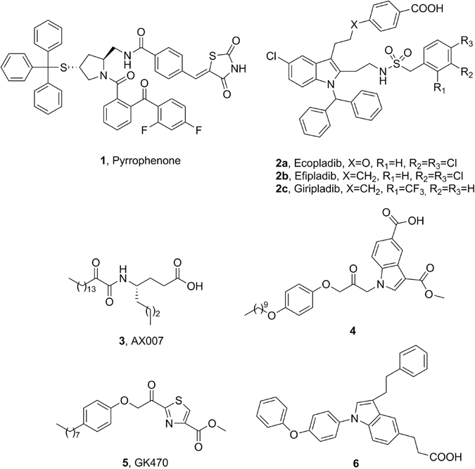
Common inhibitors of phospholipases A2.
To fully understand the role that each particular PLA2 type plays in physiological and pathological conditions, and to develop new candidates for the treatment of various inflammatory diseases, potent and selective GIVA cPLA2 inhibitors are needed. In this work, we present a novel class of potent and selective GIVA cPLA2 inhibitors and our studies on their synthesis and study of their in vitro inhibitory potency and selectivity.
Results
Design and synthesis of inhibitors
Upon activation by intracellular calcium binding to the C2 domain of GIVA cPLA2, the enzyme is translocated to the surface of the phospholipid membrane where it extracts a single phospholipid substrate into the active site39, 40. Then, the catalytic active site serine attacks the ester bond of the phospholipid substrate initiating the hydrolysis step. A number of the existing potent GIVA cPLA2 inhibitors, for example arachidonoyl trifluoromethyl ketone17, 2-oxoamides26–31, indolyl-propanones32–36, thiazolyl ketones37 contain an activated carbonyl group able to interact with the active site serine. In our quest for novel potent and selective GIVA cPLA2 inhibitors, we envisaged that the 2-oxoester (or α-keto ester) functionality could serve as such an activated carbonyl group. In 1990, it was demonstrated that α-keto ester derivatives of N-protected amino acids and peptides inhibit serine and cysteine proteinases41, while peptidyl α-keto esters inhibit the serine proteases porcine pancreatic elastase and human neutrophil elastase42. Later on, various peptide α-keto-esters and α-keto acids were reported as inhibitors of calpains and other cysteine proteases43 and of hepatitis C virus NS3 protease44. It is quite clear that a potential GIVA cPLA2 inhibitor, in addition to a functionality targeting the active site serine, should contain a lipophilic chain able to mimic the interactions of the substrate arachidonoyl chain with the lipophilic binding site of the enzyme. In addition, a free carboxyl group may contribute significantly to the overall binding of the inhibitor to the enzyme. As we have proposed in the past26, and according to the results of our mechanistic studies using a combination of hydrogen-deuterium exchange mass spectrometry with molecular dynamics simulations31, such a carboxyl group may interact with the side chain of the enzyme residue Arg-200. Taken together, we designed compounds containing a 2-oxoester functionality, a lipophilic chain and a free carboxyl group (Fig. 2).
Figure 2.

Design of 2-oxoesters.
A variety of 2-hydroxy acids, required for the synthesis of 2-oxoesters, were synthesized as described in Fig. 3. Aldehydes 7a-d were converted into cyanohydrins 8a-d and consequently to 2-hydroxy methyl esters 9a-d by treatment with HCl in methanol. 2-Hydroxy acids 11a-d were obtained by alkaline hydrolysis of 9a-d. In addition, 2-hydroxy methyl esters 9a,b,e were oxidized to the corresponding 2-oxoesters 10a,b,e (Fig. 3). Free 2-oxohexadecanoic acid 12e was synthesized by mild alkaline hydrolysis of 10e using aqueous Cs2CO3 in methanol, as depicted in Fig. 3.
Figure 3.
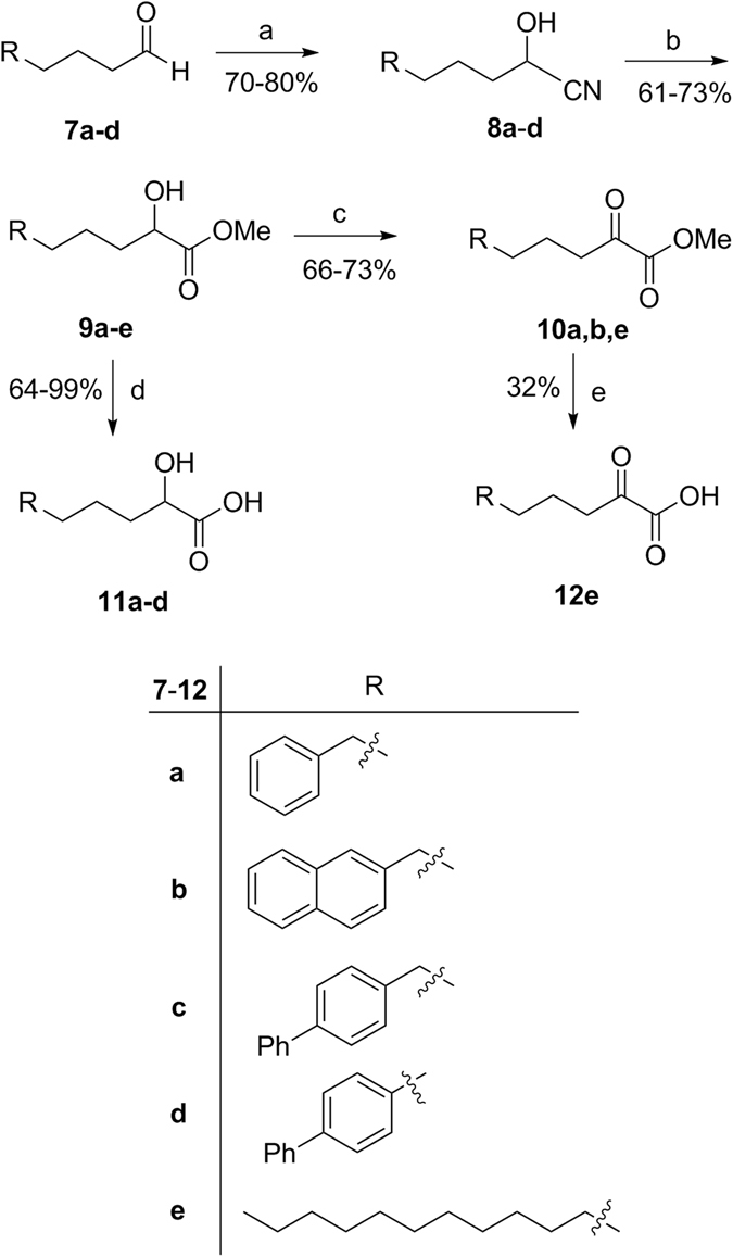
Synthesis of 2-hydroxy acids and 2-oxoacids. (a) (i) aq. sol. NaHSO3, CH2Cl2, (ii) KCN, H2O; (b) 4 N HCl/CH3OH; (c) Dess-Martin periodinane reagent, dry CH2Cl2; (d) NaOH 1 N, CH3OH; (e) 20% aq. sol. Cs2CO3, CH3OH.
The general route for the synthesis of the designed 2-oxoesters carrying a free carboxyl group is quite straightforward and is depicted in Fig. 4. The key-step was the reaction between the cesium salt of the appropriate 2-hydroxy acids 11a, 11c, 11d and 13a,b with omega-bromo esters 14a,b. The resulting 2-hydroxy esters 15a-h ware then oxidized to the corresponding 2-oxoesters 16a-h using preferably the Dess-Martin periodinane reagent45. Removal of the tert-butyl ester protecting group under acidic conditions led to the target compounds 17a-h.
Figure 4.
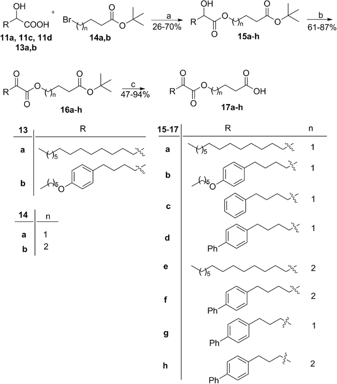
Synthesis of 2-oxoesters. (a) i. 20% aq. sol. Cs2CO3, THF, H2O, ii. Br(CH2)nCH2CH2COOBut, DMF, reflux overnight; (b) Dess-Martin periodinane reagent, dry CH2Cl2; (c) 50% CF3COOH in CH2Cl2.
2-Oxoester 19 carrying an ethyl ester group and 2-hydroxyester 20 carrying a free carboxyl group were synthesized as depicted in Fig. 5.
Figure 5.
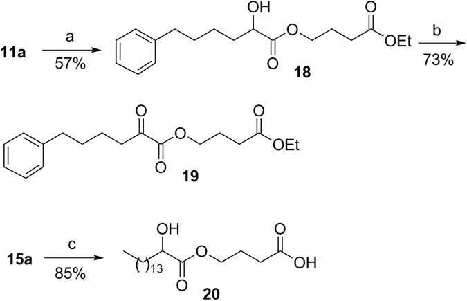
Synthesis of compounds 19 and 20. (a) i. 20% aq. sol. Cs2CO3, THF, H2O, ii. BrCH2CH2CH2COOEt, DMF, reflux overnight; (b) Dess-Martin periodinane reagent, dry CH2Cl2; (c) 50% CF3COOH in CH2Cl2.
In vitro inhibition of GIVA cPLA2, GVIA iPLA2 and GV sPLA2
All synthesized 2-oxoesters were tested for their in vitro activity on recombinant human GIVA cPLA2 using mixed micelle assays. In addition, their selectivity over human GVIA iPLA2 and GV sPLA2 was also studied using group specific mixed micelle assays. The activity of these PLA2s was tested on mixed-micelles containing 100 µM PAPC and 400 µM Triton-X.
The in vitro inhibition of human GIVA cPLA2, GVIA iPLA2 and GV sPLA2 was carried out using previously described mixed micelle-based assays27, 28, 30. The inhibition results are presented in Table 1, either as percent inhibition or as X I(50) values. At first, the percent of inhibition for each PLA2 enzyme at 0.091 mole fraction of each inhibitor was determined. Then, the X I(50) values were measured for compounds that displayed greater than 95% inhibition of GIVA cPLA2. The X I(50) is the mole fraction of the inhibitor in the total substrate interface required to inhibit the enzyme activity by 50%.
Table 1.
In vitro inhibitory potency and selectivity of 2-oxoesters.
| Entry | No | Structure | GIVA cPLA2 | GVIA iPLA2 | GV sPLA2 | ClogP | ||
|---|---|---|---|---|---|---|---|---|
| % Inhibitiona | X I(50) | % Inhibitiona | X I(50) | % Inhibitiona | ||||
| 1 | 10e |

|
68.2 ± 2.7 | 69.4 ± 12.2 | 27.5 ± 0.9 | 6.51 | ||
| 2 | 12e |

|
78.4 ± 3.5 | <25 | <25 | 5.63 | ||
| 3 | 17a |

|
>95 | 0.00008 ± 0.00001 | <25 | <25 | 6.76 | |
| 4 | 20 |

|
<25 | <25 | 41.0 ± 0.2 | 6.82 | ||
| 5 | 17b |

|
>95 | 0.00289 ± 0.00043 | <25 | 52.6 ± 5.3 | 5.46 | |
| 6 | 16c |

|
<25 | <25 | <25 | 4.58 | ||
| 7 | 19 |

|
27.3 ± 4.8 | <25 | <25 | 3.87 | ||
| 8 | 17d |

|
>95 | 0.00068 ± 0.00007 | <25 | <25 | 4.78 | |
| 9 | 17e |

|
> 95 | 0.00007 ± 0.00001 | 25 | <25 | 6.68 | |
| 10 | 17f |

|
> 95 | 0.000078 ± 0.00001 | 65 ± 3.4 | <25 | 4.70 | |
| 11 | 17g |

|
> 95 | 0.0065 ± 0.002 | 84 ± 1.5 | <25 | 4.25 | |
| 12 | 17h |

|
> 95 | 0.0010 ± 0.0003 | 94 ± 1.4 | <25 | 4.17 | |
| 13 | 10a |

|
<25 | 72 ± 4 | <25 | |||
| 14 | 10b |

|
55 ± 4.0 | > 95 | 0.0052 ± 0.0007 | <25 | 3.81 | |
| 15 | 4 |
|
>95 | 0.00008 ± 0.000005c | 8.50 | |||
a% Inhibition at 0.091 mole fraction of each inhibitor. bIC50 4.3 nM in a vesicle assay32.
Representative curves for the concentration dependence of the inhibition of GIVA cPLA2 by 2-oxoesters 17a, 17b and 17d were fit to sigmoidal curves and are presented in Fig. 6.
Figure 6.
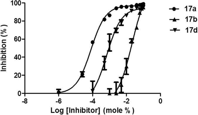
Inhibition curves for 17a, 17b and 17d. The curves were generated using GraphPad Prism with a nonlinear regression targeted at symmetrical sigmoidal curves based on plots of % inhibition versus log(inhibitor concentration). The reported X I(50) values were calculated from the resultant plots.
Discussion
Methyl 2-oxopalmitate 10e (entry 1, Table 1) weakly inhibited, at a high concentration, both the intracellular enzymes GIVA cPLA2 and GVIA iPLA2. However, 2-oxopalmitic acid 12e (entry 2, Table 1) inhibited weakly, but selectively, GIVA cPLA2. Interestingly, when the 2-oxoester functionality was combined with a long aliphatic chain together with a free carboxyl group at a distance of three carbon atoms, potent inhibition of GIVA cPLA2 was observed and the inhibitor 17a (GK161) showed a X I(50) value of 0.00008 (entry 3, Table 1). In addition, this inhibitor was selective and did not inhibit the activities of GVIA iPLA2 and the secreted GV sPLA2. This selectivity is in agreement with our previous observations that 2-oxoamides containing a free carboxyl group selectively inhibit GIVA cPLA2 28, 30. Given that for the most potent 2-oxoamides present X I(50) values are not lower than 0.00330, the present 2-oxoester was proven to be a much more potent inhibitor of GIVA cPLA2. The corresponding 2-hydroxy ester derivative 20 did not present any inhibition of either GIVA cPLA2 or GVIA iPLA2 (entry 4, Table 1), demonstrating the importance of the oxoester functionality for the inhibition.
When the long aliphatic chain was replaced by a chain of a similar size containing an aromatic ring, the inhibitory activity over GIVA cPLA2 was considerably reduced (entry 5, Table 1). Compounds 16c and 19 containing a medium chain carrying an aromatic ring and a protected carboxyl group (either ethyl ester or tert-butyl ester) totally abolished any inhibitory activity (entries 6 and 7, Table 1). In accord with our expectation, the replacement of the long aliphatic chain by a more drug-like chain of four carbon atoms carrying a biphenyl system led again to a potent and selective inhibition of GIVA cPLA2 (entry 8, Table 1). Inhibitor 17d (GK200) was found to be eight times less potent than 17a showing a X I(50) value of 0.00068. To extend the structure-activity relationship studies, we either increased the distance between the free carboxyl group and the oxoester functionality or decreased the distance between the aromatic rings and the oxoester functionality. Inhibitor 17e (GK433) proved to be highly potent, slightly better than 17a, presenting a X I(50) value of 0.00007 (entry 9, Table 1). The importance of the four-carbon atoms distance between the free carboxyl group and the oxoester functionality was clearly demonstrated by the inhibitor 17f (GK452), which presented highly potent inhibition of GIVA cPLA2 with a X I(50) value of 0.000078 (entry 10, Table 1). Decrease of the distance between the biphenyl aromatic system and the oxoester functionality (compounds 17g and 17h) resulted in considerable reduction of the potency (entries 11 and 12, Table 1). All the highly potent GIVA cPLA2 inhibitors 17a, 17d, 17e and 17f presented selectivity, because none of them exhibited any appreciable inhibition of GVIA iPLA2. In addition, none of the synthesized and tested 2-oxoesters inhibited GV sPLA2.
Since both the intracellular enzymes GIVA cPLA2 and GVIA iPLA2 are serine hydrolases and both utilize a catalytic dyad in their catalytic mechanism, it is likely that cross reactivity may be observed for inhibitors designed to carry a functionality targeting the active site serine. Indeed, such cross reactivity has been observed for several inhibitors containing an activated carbonyl group initially developed to target GIVA cPLA2. For example, arachidonoyl trifluoromethyl ketone was found to inhibit not only GIVA cPLA2, but also GVIA iPLA2. It is apparent that the presence of other groups able to develop appropriate hydrophobic and/or hydrophilic interactions contributes to the overall binding of the inhibitor to the enzyme, determining the inhibitory selectivity over GIVA cPLA2 or GVIA iPLA2. We have previously shown that pentafluoroethyl or trifluoromethyl ketones of a four-carbon atom chain carrying an aromatic ring are selective inhibitors of GVIA iPLA2 46–48. Inspired by the structures of FKGK1146 and FKGK1847, we designed simple methyl 2-oxoesters with a linker of four methylene groups between the activated carbonyl group and the aromatic ring. Unfortunately, compound 10a (entry 13, Table 1) carrying a phenyl ring only weakly inhibited GVIA iPLA2 at a high concentration. On the contrary, compound 10b (GK451) (entry 14, Table 1) carrying a naphthalene ring presented interesting inhibition of GVIA iPLA2 with a X I(50) value of 0.0052. At the same time, it presents selectivity, because it only weakly inhibits GIVA cPLA2 at a high concentration (55% at 0.091 mole fraction), while it does not inhibit at all GV sPLA2.
To better understand the interaction of 2-oxoesters with GIVA cPLA2 and GVIA iPLA2, the most potent GIVA cPLA2 inhibitor 17f was docked in the active site of either GIVA cPLA2 or GVIA iPLA2. For the docking calculations, the structures of GIVA cPLA2 and GVIA iPLA2 with two different fluoroketone compounds in the active site were used (GK174: orange color in Fig. 7a and FKGK18: magenta color in Fig. 7b). The binding mode of these two fluoroketones was validated using H/D exchange and MD simulations in a previously published study49. A theoretical score of 10.2 kcal/mol indicated that 17f is a tight binder for GIVA cPLA2. The oxoester moiety forms hydrogen-bonding with the oxyanion hole (Gly197/Gly198), while the carboxylic moiety interacts with Arg200, which was found to stabilize the phosphate group of a phospholipid substrate molecule39. Compared to GK174 (orange color in Fig. 7a) the addition of the carboxylic moiety is responsible for increasing the potency of 17f by 10-fold. This compound exhibits no activity towards GVIA iPLA2 and it received a low theoretical binding score of 6.3 kcal/mol indicating that is a weak binder. Compared to fluoroketone FKGK18 (magenta color in Fig. 7b) the addition of the carboxylic moiety increases the size of the compound and it cannot be accommodated in the active site of GVIA iPLA2.
Figure 7.
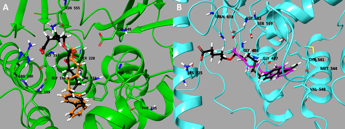
Binding mode of inhibitor 17f in the active site of (a) GIVA cPLA2 and (b) GVIA iPLA2.
All the above data, clearly demonstrate that 2-oxoesters consisting of a quite long chain (aliphatic or incorporating aromatic systems like the biphenyl system) in combination with a free carboxyl group at a distance of four or three carbon-atoms from the oxoester functionality are highly potent and selective inhibitors of GIVA cPLA2. Decreasing the size of the synthetic compound and eliminating the free carboxyl group may change the selectivity. Indeed, a methyl 2-oxoester based on a short chain carrying a naphthalene ring was found to inhibit preferentially GVIA iPLA2. In other words, it seems that the selectivity of compounds based on the 2-oxoester functionality may be tuned choosing the structural features that ensure the appropriate interactions with each enzyme (either GIVA cPLA2 or GVIA iPLA2).
To compare our novel highly potent 2-oxoester inhibitors of GIVA cPLA2 with the existing inhibitors, we studied the benchmark GIVA cPLA2 inhibitor 4 in our mixed-micelle assay. This inhibitor, developed by Lehr32, is the most potent inhibitor in the literature presenting an IC50 value of 4.3 nM in a vesicle assay32. In the mixed micelle assay, it was proved equipotent with oxoester 17a with a X I(50) value of 0.00008 (entry 15, Table 1). In addition, several 2-oxoesters were found to be more potent than the other benchmark GIVA cPLA2 inhibitor 1 (pyrrophenone), which presents an X I(50) value of 0.00226, 27. Another important property of a GIVA cPLA2 inhibitor, is the ClogP value, which is a measure of the hydrophobicity. ClogP represents the calculated partition coefficient in octanol/water on a logarithmic scale. Usually, GIVA cPLA2 inhibitors suffer from high lipophilicity. For example, the ClogP value of inhibitor 4 is 8.50, while pyrrophenone 1 and giripladib 2c present high lipophilicities too (ClogP 8.29 and 10.75, respectively). Inhibitors with such high values are not expected to present favorable ADME properties according to Lipinski’s rule of five50. Although 2-oxoesters 17a and 17e contain a long aliphatic chain, they present lower lipophilicity (ClogP 6.76 and 6.68, respectively), while the 2-oxoesters 17d and 17f carrying the biphenyl system have considerably lower ClogP values (4.78 and 4.70, respectively). The logP value of 17f, measured by HPLC, was found 3.5. Thus, the lipophilicity of 17f is encouraging and this inhibitor is the first example of a highly potent GIVA cPLA2 inhibitor, which presents a ClogP value lower than 5.
The cellular effect of the most potent GIVA cPLA2 inhibitor 17f on eicosanoid biosynthesis was studied in macrophages. RAW264.7 macrophages were used as a model system to determine if 17f displays inhibitory activity toward GIVA cPLA2 in vivo. It is well established that the toll-like receptor 4 (TLR4)-specific agonist Kdo2-lipid A (KLA) leads to GIVA cPLA2 activation51, 52 and release of arachidonic acid in macrophages that is then converted into eicosanoids by cyclooxygenase-253–55. Previous work has demonstrated that the major eicosanoid produced by KLA stimulated RAW264.7 macrophages is prostaglandin D2 (PGD2)56. The high levels of PGD2 compared to background in culture supernatants following KLA stimulation makes it an ideal marker for GIVA cPLA2 activity in macrophages. Inhibitor 17f did not show cellular toxicity at any concentrations tested as measured by trypan blue exclusion (data not shown). RAW264.7 macrophages were pre-treated with vehicle control, DMSO or 17f (5 μM) for one hour prior to stimulation with KLA (100 ng/mL). Culture supernatants were collected after 24 hours for eicosanoid quantification by LC-MS/MS. Treatment with inhibitor 17f resulted in over 50% decrease in KLA-elicited PGD2 production by macrophages (Fig. 8). A similar reduction in other minor products including PGE2, PGF2α, 11-HETE and 15-HETE was observed (data not shown), suggesting that the inhibition was not specific to PGD2. This data is consistent with 17f inhibition of GIVA cPLA2 in living cells.
Figure 8.
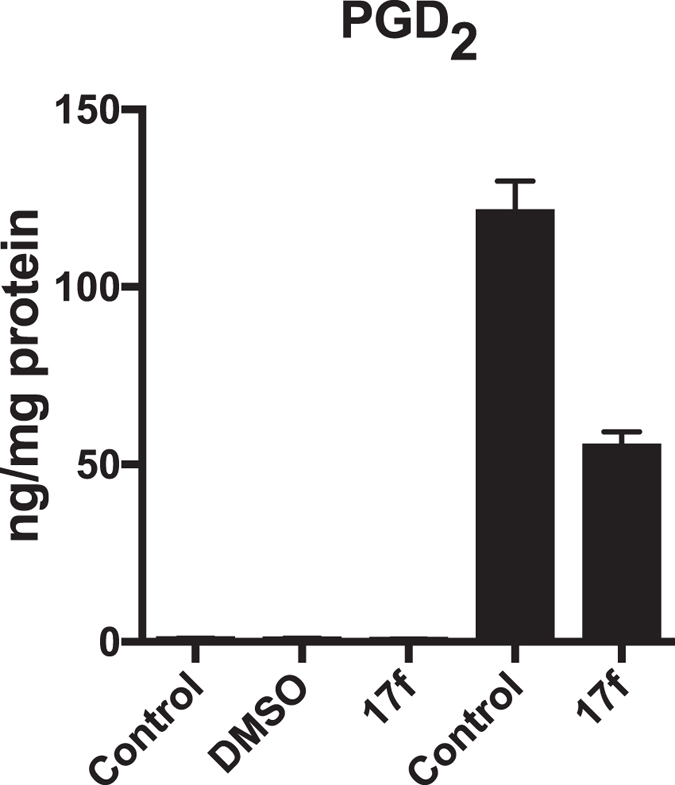
Inhibitor 17f inhibits KLA-elicited prostaglandin D2 biosynthesis by macrophages. Macrophages were pre-treated with media (control), vehicle control (DMSO, 0.1%) or inhibitor 17f (5 μM) 1 hr before KLA (100 ng/mL, ■) or mock (□) treatment. Supernatants were collected 24 hr following stimulation for eicosanoid quantification. Graph displays the mean ± SEM of a single experiment containing technical duplicates that is representative of 2 independent experiments. * indicates statistical difference compared to KLA treatment (P ≤ 0.05).
In conclusion, we describe a novel class of GIVA cPLA2 inhibitors based on the 2-oxoester functionality. This reactive functionality in combination with a long aliphatic chain or a chain carrying an appropriate aromatic system, such as the biphenyl system, and a free carboxyl group leads to highly potent and selective GIVA cPLA2 inhibitors. Inhibitors 17a, 17e and 17f present X I(50) values of 0.00007–0.00008 and are equipotent to the most potent known GIVA cPLA2 inhibitor. In particular, inhibitors incorporating the biphenyl system, like 17f, present interesting favorable lipophilicity (ClogP values lower than 5). The novel highly potent and selective GIVA cPLA2 inhibitors may be excellent tools for the study of the role of the enzyme in cells and in animals and may contribute to the development of novel medicinal agents for the treatment of inflammatory diseases.
Methods
General
Chromatographic purification of products was accomplished using Merck Silica Gel 60 (70–230 or 230–400 mesh). Thin-layer chromatography (TLC) was performed on Silica Gel 60 F254 aluminum plates. TLC pots were visualized with UV light and/or phosphomolybdic acid in EtOH. Melting points were determined using a Büchi 530 apparatus and were uncorrected. 1H and 13C NMR spectra were recorded on a Varian Mercury (200 MHz and 50 MHz respectively) in CDCl3. Chemical shifts are given in ppm, and coupling constants (J) in Hz. Peak multiplicities are described as follows: s, singlet, d, doublet, t, triplet and m, multiplet. Electron spray ionization (ESI) mass spectra were recorded on a Finnigan, Surveyor MSQ Plus spectrometer. Dichloromethane was dried by standard procedures and stored over molecular sieves. All other solvents and chemicals were reagent grade and used without further purification. The purity of all compounds subjected to biological tests was determined by analytical HPLC, and was found to be ≥95%. HPLC analyses were carried out on a Shimadzu LC-2010AHT system and a Merck Chromolith Performance (100 × 4.6 mm) analytical column, using H2O/MeOH 10/90 v/v, at a flow rate of 1.0 mL/min. HRMS spectra were recorded on a Bruker Maxis Impact QTOF Spectrometer.
Compounds 8a 57, 8b 57, 9e 58, 11a 57, 11b 57, 13a 59, 13b 60 have been described elsewhere and their analytical data are in accordance with literature.
Synthesis of cyanohydrins 8c,d
To a stirred solution of aldehyde 7c,d (1.0 mmol) in CH2Cl2 (1.4 mL), an aqueous solution of NaHSO3 (0.25 mL, 1.5 mmol) was added and the mixture was stirred for 30 min at room temperature. The organic solvent was evaporated under reduced pressure and H2O (1 mL) was added. The mixture was cooled to 0 °C and an aqueous solution of KCN (0.25 mL, 1.5 mmol) was added within 2 h under vigorous stirring. The reaction was stirred for 18 h at room temperature and then, water (10 mL) was added and extracted with CH2Cl2 (3 × 10 mL). The combined organic phases was washed with brine (30 mL), dried over Na2SO4 and evaporated under reduced pressure. The residue was purified by flash column chromatography [ethyl acetate (EtOAc)/petroleum ether (bp 40–60 °C), 2:8].
6-([1,1′-Biphenyl]-4-yl)-2-hydroxyhexanenitrile (8c)
Yield 80%; White solid; mp: 85–87 °C; 1H NMR (200 MHz, CDCl3): δ 7.67–7.14 (m, 9 H), 4.45 (t, J = 6.9 Hz, 1 H), 3.90 (br s, 1 H), 2.69 (t, J = 7.1 Hz, 2 H), 1.89 (q, J = 7.2 Hz, 2 H), 1.79–1.39 (m, 4 H); 13C NMR (50 MHz, CDCl3): δ 141.0, 140.9, 138.8, 129.0, 128.7, 127.0, 126.9, 126.5, 119.9, 61.1, 35.2, 34.9, 30.6, 24.2; MS (m/z, ESI): [M + NH4]+ calcd. for C18H19NO, 283.2; found, 283.3; analysis (calcd., found for C18H19NO): C (81.47, 81.18), H (7.22, 7.41), N (5.28, 5.33).
5-([1,1′-Biphenyl]-4-yl)-2-hydroxypentanenitrile (8d)
Yield 76%; White solid; mp: 80–82 °C; 1H NMR (200 MHz, CDCl3): δ 7.64–7.23 (m, 9 H), 4.48 (t, J = 6.9 Hz, 1 H), 3.80 (br s, 1 H), 2.64 (t, J = 7.0 Hz, 2 H), 1.84 (q, J = 7.0 Hz, 2 H), 1.69–1.57 (m, 2 H); 13C NMR (50 MHz, CDCl3): δ 141.3, 140.8, 138.4, 129.1, 128.7, 127.6, 127.2, 126.8, 118.5, 61.9, 35.1, 34.5, 23.8; MS (m/z, ESI): [M + NH4]+ calcd. for C17H17NO 269.2; found, 269.2; analysis (calcd., found for C17H17NO): C (81.24, 81.02), H (6.82, 6.99), N (5.57, 5.69).
Synthesis of 2-hydroxy esters 9a-d
Cyanohydrin 8a-d (1 mmol) was dissolved in methanolic solution of HCl (10 mL, 4 N) and the reaction mixture was stirred for 24 h at room temperature. The organic solvent was evaporated in vacuo and the remaining solid was dissolved in diethyl ether (10 mL) and re-evaporated. Dilution and evaporation was repeated twice. Then, the product was purified by flash column chromatography [EtOAc-petroleum ether (bp 40–60 °C), 2:8].
Methyl 2-hydroxy-6-phenylhexanoate (9a)
Yield 61%; Yellow oil; 1H NMR (200 MHz, CDCl3): δ 7.37–7.04 (m, 5 H), 4.23–4.10 (m, 1 H), 3.77 (s, 3 H), 2.74 (br s, 1 H), 2.62 (t, J = 7.1 Hz, 2 H), 1.92–1.25 (m, 6 H); 13C NMR (50 MHz, CDCl3): δ 175.7, 142.3, 128.3, 128.2, 125.6, 70.3, 52.4, 35.7, 34.1, 31.1, 24.4; MS (m/,z ESI): [M + NH4]+ calcd. for C13H18O3 240.2 found, 240.2; analysis (calcd., found for C13H18O3): C (70.24, 70.01), H (8.16, 8.29).
Methyl 2-hydroxy-6-(naphthalen-2-yl)hexanoate (9b)
Yield 73%; Colorless oil; 1H NMR (200 MHz, CDCl3): δ 7.90–7.20 (m, 7 H), 4.30–4.02 (m, 1 H), 3.76 (s, 3 H), 3.35 (br s, 1 H), 2.97–2.75 (m, 2 H), 1.97–1.34 (m, 6 H); 13C NMR (50 MHz, CDCl3): δ 175.6, 139.8, 133.5, 127.7, 127.5, 127.3, 127.2, 126.2, 125.8, 125.0, 70.3, 52.4, 35.8, 34.1, 30.9, 24.4; MS (m/z, ESI): [M + Na]+ calcd. for C17H20O3 295.1, found, 295.2; analysis (calcd., found for C17H20O3): C (74.97, 74.72), H (7.40, 7.62).
Methyl 6-([1,1′-biphenyl]-4-yl)-2-hydroxyhexanoate (9c)
Yield 69%; Colorless oil; 1H NMR (200 MHz, CDCl3): δ 7.70–7.06 (m, 9 H), 4.45 (t, J = 7.0 Hz, 1 H), 3.79 (s, 3 H), 3.00 (br s, 1 H), 2.69 (t, J = 7.1 Hz, 2 H), 1.89 (q, J = 7.5 Hz, 2 H), 1.79–1.36 (m, 4 H); 13C NMR (50 MHz, CDCl3): δ 175.7, 141.0, 140.9, 138.7, 129.0, 128.7, 127.0, 126.9, 70.4, 52.5, 35.2, 34.9, 30.7, 24.2; MS (m/z, ESI): [M + Na]+ calcd. for C19H22O3 321.1, found, 321.2; analysis (calcd., found for C19H22O3): C (80.82, 80.61), H (7.85, 7.98).
Methyl 5-([1,1′-biphenyl]-4-yl)-2-hydroxypentanoate (9d)
Yield 71%; Colorless oil; 1H NMR (200 MHz, CDCl3): δ 7.69–7.18 (m, 9 H), 4.40 (t, J = 6.9 Hz, 1 H), 3.76 (s, 3 H), 3.54 (brs, 1 H), 2.65 (t, J = 7.1 Hz, 2 H), 1.84 (q, J = 7.1 Hz, 2 H), 1.64–1.36 (m, 2 H); 13C NMR (50 MHz, CDCl3): δ 176.1, 141.3, 140.8, 138.2, 129.0, 128.9, 127.5, 127.0, 126.8, 70.3, 52.3, 35.5, 34.9, 24.4; MS (m/z, ESI): [M + Na]+ calcd. for C18H20O3 307.1, found, 307.2; analysis (calcd., found for C18H20O3): C (76.03, 75.83), H (7.09, 7.27).
Synthesis of 2-oxoesters 10α, 10b, 10e, 16a-h, 19
To a stirred solution of 2-hydroxy esters 9a, 9b, 9e, 15a-h, 18 (1 mmol) in dry CH2Cl2 (10 mL) was added Dess-Martin periodinane (1.1 mmol, 0.47 g) and the reaction mixture was stirred for 1.5 h at room temperature. Then, CH2Cl2 (5 mL) was added and the organic phase was washed with a mixture of Na2S2O3 10% and NaHCO3 10% (15 mL, 1:1, v/v). Τhe aqueous phase was washed with CH2Cl2 (15 mL) and all the organic phases were collected, dried (Na2SO4) and evaporated under reduced pressure. The residue was purified by flash column chromatography [EtOAc-petroleum ether (bp 40–60 °C), 2:8].
Methyl 2-oxo-6-phenylhexanoate (10a, GK437)
Yield 66%; Colorless oil; 1H NMR (200 MHz, CDCl3): δ 7.40–7.08 (m, 5 H), 3.84 (s, 3 H), 2.85 (t, J = 6.4 Hz, 2 H), 2.62 (t, J = 6.5 Hz, 2 H), 1.78–1.58 (m, 4 H); 13C NMR (50 MHz, CDCl3): δ 194.0, 161.4, 141.8, 128.3, 128.1, 125.8, 52.9, 39.1, 35.5, 30.6, 22.5; MS (m/z, ESI): [M + NH4]+ calcd. for C13H16O3 238.1, found, 238.2; HRMS (m/z, ESI): [M + Na]+ calcd. for C13H16O3, 243.0992; found, 243.0994; analysis (calcd., found for C13H16O3): C (70.89, 70.58), H (7.32, 7.46).
Methyl 6-(naphthalen-2-yl)-2-oxohexanoate (10b, GK451)
Yield 73%; Colorless oil; 1H NMR (200 MHz, CDCl3): δ 7.90–7.10 (m, 7 H), 3.85 (s, 3 H), 2.92–2.71 (m, 4 H), 1.83–1.49 (m, 4 H); 13C NMR (50 MHz, CDCl3): δ 194.0, 161.4, 139.2, 133.5, 131.9, 127.9, 127.5, 127.4, 127.2, 126.3, 125.9, 125.1, 52.9, 39.1, 35.6, 30.4, 22.5; MS (m/z, ESI): [(M + NH4)+] calcd. for C17H18O3 288.2, found, 288.2; HRMS (m/z, ESI): [M + Na]+ calcd. for C17H18O3, 293.1148; found, m/z 293.1149; analysis (calcd., found for C17H18O3): C (75.53, 75.32), H (6.71, 6.95).
Methyl 2-oxohexadecanoate (10e)
Yield 73%; White solid; mp: 53–55 °C; 1H NMR (200 MHz, CDCl3): δ 3.85 (s, 3 H), 2.82 (t, J = 7.2 Hz, 2 H), 1.70–1.51 (m, 2 H), 1.37–1.16 (m, 22 H), 0.86 (t, J = 7.0 Hz, 3 H); 13C NMR (50 MHz, CDCl3): δ 194.3, 161.5, 52.8, 39.3, 31.9, 29.6, 29.5, 29.4, 29.3, 29.2, 28.9, 22.9, 22.6, 14.1; MS (m/z, ESI): [M + NH4]+ calcd. for C17H32O3 302.3; found, 302.361.
4-(tert-Butoxy)-4-oxobutyl 2-oxohexadecanoate (16a)
Yield 87%; Colorless oil, 1H NMR (200 MHz, CDCl3): δ 4.27 (t, J = 6.0 Hz, 2 H), 2.81 (t, J = 7.8 Hz, 2 H), 2.33 (t, J = 6.0 Hz, 2 H), 2.04 (quint, J = 6.0 Hz, 2 H), 1.70–1.50 (m, 2 H), 1.44 (s, 9 H), 1.40–1.15 (m, 22 H), 0.86 (t, J = 7.0 Hz, 3 H); 13C NMR (50 MHz, CDCl3): δ 194.5, 171.8, 161.2, 80.6, 65.2, 39.3, 31.9, 31.6, 29.6, 29.5, 29.4, 29.3, 29.2, 28.9, 28.0, 23.8, 22.9, 22.6, 14.1; MS (m/z, ESI): [M + NH4]+ calcd. for C24H44O5 430.4; found, 430.4; analysis (calcd., found for C24H44O5): C (69.86, 69.6), H (10.75, 10.92).
Compound 16b was not isolated and used directly in the next step.
4-(tert-Butoxy)-4-oxobutyl 2-oxo-6-phenylhexanoate (16c, GK192)
Yield 77%; Yellow oil; 1H NMR (200 MHz, CDCl3): δ 7.35–7.10 (m, 5 H), 4.26 (t, J = 8.0 Hz, 2 H), 2.84 (t, J = 6.0 Hz, 2 H), 2.70–2.55 (m, 2 H), 2.32 (t, J = 6.0 Hz, 2 H), 2.00 (quint, J = 6.0 Hz, 2 H), 1.70–1.60 (m, 4 H), 1.44 (s, 9 H), 13C NMR (50 MHz, CDCl3): δ 194.1, 171.7, 161.1, 141.8, 128.3, 128.2, 125.8, 80.6, 65.3, 39.1, 35.5, 31.6, 30.6, 28.0, 23.8, 22.5; HRMS (m/z, ESI): [M + Na]+calcd. for C20H28O5 371.1829; found, 371.1831; analysis (calcd., found for C20H28O5): C (68.94, 68.66), H (8.10, 8.29).
4-tert-Butoxy-4-oxobutyl 6-(biphenyl-4-yl)-2-oxohexanoate (16d)
Yield 86%; Colorless oil; 1H NMR (200 MHz, CDCl3): δ 7.96–7.16 (m, 9 H), 4.28 (t, J = 6.4 Hz, 2 H), 2.97–2.83 (m, 2 H), 2.77–2.62 (m, 2 H), 2.42–2.28 (m, 2 H), 2.11–1.55 (m, 6 H), 1.45 (s, 9 H); 13C NMR (50 MHz, CDCl3): δ 194.1, 171.8, 161.0, 140.9, 138.7, 129.0, 128.9, 128.7, 128.6, 127.2, 127.0, 126.9, 80.6, 65.3, 39.1, 35.1, 31.6, 30.5, 28.0, 23.7, 22.5; MS (m/z, ESI): [M + Na]+ calcd. for C26H32O5 447.2; found, 447.0; analysis (calcd., found for C26H32O5): C (73.56, 73.35), H (7.60, 7.78).
5-(tert-Butoxy)-5-oxopentyl 2-oxohexadecanoate (16e)
Yield 78%; White oil; 1H NMR (200 MHz, CDCl3): δ 4.23 (t, J = 6.9 Hz, 2 H), 2.79 (t, J = 7.3 Hz, 2 H), 2.24 (t, J = 6.9 Hz, 2 H), 1.80–1.45 (m, 4 H), 1.44 (s, 9 H), 1.30–1.15 (s, 24 H), 0.85 (t, J = 7.0 Hz, 3 H); 13C NMR (50 MHz, CDCl3): δ 194.8, 172.6, 161.5, 80.5, 66.0, 39.5, 35.0, 32.1, 29.9, 29.8, 29.6, 29.5, 29.1, 28.3, 27.9, 23.1, 22.9, 21.6, 14.3; MS (m/z, ESI): [M + NH4]+ calcd. for C25H46O5 444.4; found, 444.3; analysis (calcd., found for C25H46O5): C (70.38, 70.17), H (10.87, 11.05).
5-(tert-Butoxy)-5-oxopentyl 6-([1,1′-biphenyl]-4-yl)-2-oxohexanoate (16f)
Yield 63%; Colorless oil; 1H NMR (200 MHz, CDCl3): δ 7.66–7.19 (m, 9 H), 4.25 (t, J = 6.0 Hz, 2 H), 2.87 (t, J = 6.3 Hz, 2 H), 2.68 (t, J = 6.1 Hz, 2 H), 2.26 (t, J = 7.0 Hz, 2 H), 1.86–1.53 (m, 8 H), 1.44 (s, 9 H); 13C NMR (50 MHz, CDCl3): δ 194.3, 172.4, 161.1, 141.0, 138.8, 129.0, 128.8, 128.7, 127.2, 127.0, 126.9, 80.3, 65.9, 39.1, 35.2, 34.8, 30.6, 28.1, 27.7, 22.5, 21.3; MS (m/z, ESI): [M + NH4]+ calcd. for C27H34O5 456.3; found, 456.3; analysis (calcd., found for C27H34O5): C (73.95, 73.75), H (7.81, 7.99).
4-(tert-Butoxy)-4-oxobutyl 5-([1,1′-biphenyl]-4-yl)-2-oxopentanoate (16 g)
Yield 65%; Colorless oil; 1H NMR (200 MHz, CDCl3): δ 7.64–7.20 (m, 9 H), 4.27 (t, J = 6.4 Hz, 2 H), 2.88 (t, J = 7.2 Hz, 2 H), 2.70 (t, J = 7.5 Hz, 2 H), 2.33 (t, J = 7.2 Hz, 2 H), 2.10–1.90 (m, 4 H), 1.44 (s, 9 H); 13C NMR (50 MHz, CDCl3): δ 194.3, 172.1, 161.3, 141.2, 140.5, 139.3, 129.1, 128.9, 127.4, 127.3, 127.2, 80.9, 65.6, 38.8, 34.6, 31.9, 28.3, 24.7, 24.0; MS (m/z, ESI): [M + NH4]+ calcd. for C25H30O5 428.2; found, 428.3; analysis (calcd., found for C25H30O5): C (73.15, 72.97), H (7.37, 7.56).
5-(tert-Butoxy)-5-oxopentyl 5-([1,1′-biphenyl]-4-yl)-2-oxopentanoate (16 h)
Yield 61%; Colorless oil; 1H NMR (200 MHz, CDCl3): δ 7.64–7.20 (m, 9 H), 4.24 (t, J = 6.0 Hz, 2 H), 2.87 (t, J = 6.3 Hz, 2 H), 2.69 (t, J = 7.5 Hz, 2 H), 2.25 (t, J = 5.9 Hz, 2 H), 2.20–1.90 (m, 2 H), 1.90–1.54 (m, 4 H), 1.44 (s, 9 H); 13C NMR (50 MHz, CDCl3); δ 194.4, 172.7, 161.3, 141.2, 140.5, 139.3, 129.1, 128.9, 127.4, 127.3, 127.2, 80.6, 66.2, 38.8, 35.1, 34.6, 28.3, 27.9, 24.7, 21.6; MS (m/z, ESI): [M + NH4]+ calcd. for C26H32O5 442.3; found, 442.3; analysis (calcd., found for C26H32O5): C (73.56, 73.32), H (7.60, 7.82).
4-Ethoxy-4-oxobutyl 2-oxo-6-phenylhexanoate (19, GK194)
Yield 73%; Yellowish oil, 1H NMR (200 MHz, CDCl3): δ 7.30–7.10 (m, 5 H), 4.28 (t, J = 8.0 Hz, 2 H), 4.13 (q, J = 6.0 Hz, 2 H) 2.84 (t, J = 6.0 Hz, 2 H), 2.70–2.55 (m, 2 H), 2.41 (t, J = 8.0 Hz, 2 H), 2.15–1.95 (m, 2 H), 1.70–1.55 (m, 4 H), 1.24 (t, J = 6.0 Hz, 3 H); 13C NMR (50 MHz, CDCl3): δ 194.0, 172.4, 161.0, 141.8, 128.3, 128.2, 125.7, 65.2, 60.5, 39.0, 35.5, 30.6, 30.5, 23.6, 22.5, 14.1; MS (m/z, ESI): [M + NH4]+calcd. for C18H24O5 338.2; found, 338.2;HRMS (m/z, ESI): [M + Na]+ calcd for C18H24O5 343.1516; found, 343.1512; analysis (calcd., found for C18H24O5): C (67.48, 67.19), H (7.55, 7.61).
Synthesis of 2-hydroxy acids 11α-e
To a stirred solution of 2-hydroxy ester 9a-e (1 mmol) in methanol (10 mL), aqueous NaOH (1.1 mL, 1 N) was added and the reaction mixture was stirred overnight at room temperature. The organic solvent was evaporated in vacuo to dryness and then aqueous HCl 1 N was added until acidic pH. The aqueous phase was washed with EtOAc (3 × 10 mL). Finally, the organic phase was dried (Na2SO4) and evaporated under reduced pressure.
6-([1,1′-Biphenyl]-4-yl)-2-hydroxyhexanoic acid (11c)
Yield 99%; White solid; mp: 143–145 °C; 1H NMR (200 MHz, CDCl3): δ 10.43 (s, 1 H), 7.67–7.02 (m, 9 H), 5.00 (br s, 1 H) 4.22 (t, J = 6.0 Hz, 1 H), 2.59 (t, J = 7.2 Hz, 2 H), 2.03–1.37 (m, 6 H); 13C NMR (50 MHz, CDCl3): δ 176.6, 141.2, 140.6, 138.1, 129.1, 128.3, 128.2, 126.5, 126.4, 69.8, 34.9, 33.6, 30.8, 24.3; MS (m/z, ESI): [M-H]− calcd. for C18H20O3 283.1; found, 283.1; analysis (calcd., found for C18H20O3): C (76.03, 75.85), H (7.09, 7.25).
5-([1,1′-Biphenyl]-4-yl)-2-hydroxypentanoic acid (11d)
Yield 64% (over two steps); Light violet viscous oil; 1H NMR (200 MHz, CDCl3): δ 10.60 (s, 1 H), 7.66–7.06 (m, 9 H), 4.98 (s, 1 H), 4.29 (t, J = 6.0 Hz, 1 H), 2.68 (t, J = 6.4 Hz, 2 H), 1.99–1.68 (m, 4 H); 13C NMR (50 MHz, CDCl3): δ 178.4, 141.3, 141.2, 139.0, 129.1, 129.0, 128.9, 127.3, 127.2, 70.3, 35.3, 33.9, 26.8; MS (m/z, ESI): [M-H]− calcd. for C17H18O3 269.1; found, 269.1; analysis (calcd., found for C17H18O3): C (75.53, 75.31), H (6.71, 6.87).
2-Oxohexadecanoic acid (12e)
To a stirred solution of 9e (0.35 mmol, 100 mg) in MeOH (3.5 mL), aqueous Cs2CO3 20% (w/v) (1.7 mL, 1.0 mmol) was added, and the reaction mixture was stirred at room temperature. The reaction progress was monitored by TLC, until completion. The organic solvent was evaporated in vacuo to dryness, water was added (10 mL) and then aqueous HCl 1 N was added until acidic pH. The aqueous phase was washed with EtOAc (3 × 10 mL). Finally, the organic phase was dried over Na2SO4 and evaporated under reduced pressure. Yield 32%; White solid; mp: 66–68 °C; 1H NMR (200 MHz, CDCl3): δ 9.02 (br s, 1 H), 2.93 (t, J = 7.2 Hz, 2 H), 1.76–1.51 (m, 2 H), 1.43–1.05 (m, 22 H), 0.88 (t, J = 6.6 Hz, 3 H); 13C NMR (50 MHz, CDCl3): δ 194.8, 162.5, 39.3, 31.9, 29.6, 29.5, 29.4, 29.3, 29.2, 28.9, 22.9, 22.7, 14.1; MS (m/z, ESI): [M-H]− calcd. for C16H30O3 269.2; found, 269.262.
Synthesis of 2-hydroxy esters 15a-h and 18
To a stirred solution of 2-hydroxy acids 11a, 11c, 11d, 13a,b (1 mmol) in tetrahydrofuran (THF) (6 mL), water (0.6 mL) and few drops of aqueous CsCO3 20% (w/v) were added in order to adjust pH in neutral value. The organic solvent was evaporated in vacuo and the residue was dissolved in N,N-dimethylformamide (DMF) (15 mL). Subsequently, tert-butyl 5-bromoalkanooate 14a,b or ethyl 4-bromobutyrate (1.2 mmol) was added and the reaction mixture was refluxed for 72 h. Water (20 mL) was then added and the reaction mixture was washed with EtOAc (2 × 20 mL). The organic phase was dried (Na2SO4) and evaporated under reduced pressure. The residue was purified by flash column chromatography [EtOAc-petroleum ether (bp 40–60 °C), 1:9 or 2:8].
4-(tert-Butoxy)-4-oxobutyl 2-hydroxyhexadecanoate (15a)
Yield 44%; Yellow oil, 1H NMR (200 MHz, CDCl3): δ 4.25–4.10 (m, 3 H), 2.73 (br s, 1 H), 2.30 (t, J = 6.0 Hz, 2 H), 1.94 (qu, J = 6.0 Hz, 2 H), 1.60–1.45 (m, 2 H), 1.43 (s, 9 H), 1.40–1.20 (m, 24 H), 0.86 (t, J = 6.0 Hz, 3 H); 13C NMR (50 MHz, CDCl3): δ 175.3, 171.9, 80.6, 70.4, 64.5, 34.4, 31.9, 31.7, 29.6, 29.5, 29.4, 29.3, 28.0, 24.7, 24.0, 22.6, 14.1; MS (m/z, ESI): [M + NH4]+ cald. for C24H46O5 432.4; found, 432.3; analysis (calcd., found for C24H46O5): C (69.52, 69.36), H (11.18, 11.29).
4-(tert-Butoxy)-4-oxobutyl 6-(4-(hexyloxy)phenyl)-2-hydroxyhexanoate (15b)
Yield 35%; Yellowish oil, 1H NMR (200 MHz, CDCl3): δ 7.08 (d, J = 8.6 Hz, 2 H), 6.82 (d, J = 8.6 Hz, 2 H), 4.27–4.12 (m, 3 H), 3.93 (t, J = 6.0 Hz, 2 H), 2.78 (br s, 1 H), 2.58 (t, J = 7.0 Hz, 2 H), 2. 30 (t, J = 7.0 Hz, 2 H), 1.95 (q, J = 7.0 Hz, 2 H), 1.85–1.47 (m, 8 H), 1.46 (s, 9 H) 1.45–1.20 (m, 6 H), 0.91 (t, J = 7.0 Hz, 3 H); 13C NMR (50 MHz, CDCl3): δ 175.1, 172.8, 157.1, 134.1, 129.1, 114.2, 80.3, 70.3, 67.9, 64.4, 34.7, 34.2, 31.5, 31.3, 30.3, 29.2, 28.0, 25.7, 24.4, 23.6, 22.5, 14.0; MS (m/z, ESI): [M + NH4]+ calcd. for C26H42O6 468.3; found, 468.1; analysis (calcd., found for C26H42O6): C (69.30, 69.08), H (9.40, 9.61).
4-(tert-Butoxy)-4-oxobutyl 2-hydroxy-6-phenylhexanoate (15c)
Yield 50%; Yellow oil, 1H NMR (200 MHz, CDCl3): δ 7.33–7.08 (m, 5 H), 4.23–4.10 (m, 3 H), 2.86 (br s, 1 H), 2.61 (t, J = 7.0 Hz, 2 H), 2.28 (t, J = 7.0 Hz, 2 H), 2.00–1.75 (m, 2 H), 1.70–1.45 (m, 6 H), 1.44 (s, 9 H); 13C NMR (50 MHz, CDCl3): δ 175.1, 162.8, 142.3, 128.3, 128.1, 125.6, 80.3, 70.3, 64.5, 36.5, 35.7, 34.2, 31.5, 28.0, 24.4, 23.9; MS (m/z, ESI): [M + NH4]+ calcd. for C20H30O5 368.2; found, 368.3; analysis (calcd., found for C20H30O5): C (68.55, 68.34), H (8.63, 8.81).
4-tert-Butoxy-4-oxobutyl 6-(biphenyl-4-yl)-2-hydroxyhexanoate (15d)
Yield 61%; Oil, 1H NMR (200 MHz, CDCl3): δ 7.64–7.19 (m, 9 H), 4.41–4.00 (m, 3 H), 2.75 (br s, 1 H), 2.67 (t, J = 7.4 Hz, 2 H), 2.30 (t, J = 7.4 Hz, 2 H), 2.03–1.48 (m, 8 H), 1.45 (s, 9 H); 13C NMR (50 MHz, CDCl3): δ 175.1, 171.8, 141.4, 138.5, 129.5, 128.7, 128.6, 126.9, 126.8, 80.5, 70.2, 64.5, 35.3, 34.2, 31.6, 31.1, 28.0, 24.4, 23.9; MS (m/z, ESI): [M + Na]+ calcd. for C26H34O5 449.2; found, 449.2; analysis (calcd., found for C26H34O5): C (73.21, 73.00), H (8.03, 8.21).
5-(tert-Butoxy)-5-oxopentyl 2-hydroxyhexadecanoate (15e)
Yield 48%; Light yellow oil; 1H NMR (200 MHz, CDCl3): δ 4.19–4.05 (m, 3 H), 2.84 (br s, 1 H), 2.21 (t, J = 6.7 Hz, 2 H), 1.70–1.45 (m, 8 H), 1.40 (s, 9 H), 1.30–1.15 (m, 22 H), 0.83 (t, J = 6.3 Hz, 3 H); 13C NMR (50 MHz, CDCl3): δ 175.6, 172.7, 80.5, 70.7, 65.3, 35.0, 34.6, 32.1, 29.9, 29.8, 29.7, 29.6, 29.5, 28.2, 28.1, 25.0, 22.9, 21.6, 14.3; MS (m/z, ESI): [M + NH4]+ calcd. for C25H48O5 446.4; found, 446.3; analysis (calcd., found for C25H48O5): C (70.05, 69.89), H (11.29, 11.44).
5-(tert-Butoxy)-5-oxopentyl 6-([1,1′-biphenyl]-4-yl)-2-hydroxyhexanoate (15f)
Yield 70%; Colorless oil; 1H NMR (200 MHz, CDCl3): δ 7.64–7.14 (m, 9 H), 4.24–4.03 (m, 3 H), 2.70–2.54 (m, 3 H), 2.24 (t, J = 6.6 Hz, 2 H), 1.86–1.53 (m, 10 H), 1.44 (s, 9 H); 13C NMR (50 MHz, CDCl3): δ 175.3, 172.5, 141.5, 138.6, 129.5, 128.8, 128.7, 127.0, 126.9, 80.3, 70.3, 65.2, 35.4, 34.8, 34.3, 31.1, 28.1, 27.9, 24.5, 21.4; MS (m/z, ESI): [M + NH4]+ calcd. for C27H36O5 458.3; found, 458.2; analysis (calcd., found for C27H36O5): C (73.61, 73.37), H (8.24, 8.39).
4-(tert-Butoxy)-4-oxobutyl 5-([1,1′-biphenyl]-4-yl)-2-hydroxypentanoate (15g)
Yield 26%; Colorless oil; 1H NMR (200 MHz, CDCl3): δ 7.64–7.20 (m, 9 H), 4.21 (m, 3 H), 2.85 (br s, 1 H), 2.69 (t, J = 7.0 Hz, 2 H), 2.29 (t, J = 7.2 Hz, 2 H), 2.06–1.66 (m, 6 H), 1.45 (s, 9 H); 13C NMR (50 MHz, CDCl3): δ 175.2, 172.6, 141.1, 139.2, 129.5, 128.8, 127.3, 127.2, 127.0, 80.8, 69.9, 64.8, 35.9, 34.8, 34.4, 28.4, 26.7, 21.5; MS (m/z, ESI): [M + NH4]+ calcd. for C25H32O5 430.3; found, 430.3; analysis (calcd., found for C25H32O5): C (72.79, 72.60), H (7.82, 7.92).
5-(tert-Butoxy)-5-oxopentyl 5-([1,1′-biphenyl]-4-yl)-2-hydroxypentanoate (15h)
Yield 54%; Colorless oil; 1H NMR (200 MHz, CDCl3): δ 7.64–7.18 (m, 9 H), 4.30–4.08 (m, 3 H), 2.87 (br s, 1 H), 2.68 (t, J = 6.0 Hz, 2 H), 2.23 (t, J = 5.9 Hz, 2 H), 1.90–1.50 (m, 8 H), 1.44 (s, 9 H); 13C NMR (50 MHz, CDCl3): δ 175.5, 172.8, 141.3, 139.0, 129.0, 128.9, 127.3, 127.2, 127.1, 80.6, 70.5, 65.5, 35.3, 35.1, 34.2, 28.3, 28.1, 26.8, 21.7; MS (m/z, ESI): [M + NH4]+ calcd. for C26H34O5 444.3; found, 444.3; analysis (calcd., found for C26H34O5): C (73.21, 73.07), H (8.03, 8.19).
4-Ethoxy-4-oxobutyl 2-hydroxy-6-phenylhexanoate (18)
Yield 57%; Yellow oil, 1H NMR (200 MHz, CDCl3): δ 7.30–7.05 (m, 5 H), 4.24–4.00 (m, 5 H), 2.92 (br s, 1 H), 2.61 (t, J = 7.1 Hz, 2 H), 2.36 (t, J = 6.0 Hz, 2 H), 1.96 (t, J = 7.1 Hz, 2 H), 1.80–1.60 (m, 4 H), 1.60–1.40 (m, 2 H), 1.24 (t, J = 6.0 Hz, 3 H); 13C NMR (50 MHz, CDCl3): δ 175.1, 172.5, 142.2, 128.2, 128.1, 125.6, 70.2, 64.4, 60.5, 35.6, 34.1, 31.0, 30.5, 24.4, 23.8, 14.1; MS (m/z, ESI): [M + NH4]+ calcd. for C18H26O5 340.2; found, 340.3; analysis (calcd., found for C18H26O5): C (67.06, 66.93), H (8.13, 8.28).
Synthesis of compounds 17a-h and 20
A solution of tert-butyl ester 16a-h and 15a (1 mmol) in 50% trifluoroacetic acid (TFA) in CH2Cl2 (10 mL) was stirred for 1 h at room temperature. The organic solvent was evaporated under reduced pressure and then CH2Cl2 was added and re-evaporated twice. The product was purified by precipitation with a mixture of EtOAc and petroleum ether (5:95, v/v, 10 mL) or by column chromatography (CH2Cl2-MeOH, 95:5).
4-((2-Oxohexadecanoyl)oxy)butanoic acid (17a, GK161)
Yield 85%; White solid; mp: 76–78 °C; 1H NMR (200 MHz, CDCl3): δ 9.25 (br s, 1 H), 4.32 (t, J = 6.0 Hz, 2 H), 2.82 (t, J = 6.0 Hz, 2 H), 2.51 (t, J = 6.0 Hz, 2 H), 2.15 (q, J = 6.0 Hz, 2 H), 1.80–1.50 (m, 2 H), 1.50–1.20 (m, 22 H), 0.88 (t, J = 7.0 Hz, 3 H); 13C NMR (50 MHz, CDCl3): δ 194.3, 178.6, 161.1, 65.0, 39.3, 31.9, 30.3, 29.6, 29.6, 29.4, 29.3, 29.3, 28.9, 23.4, 22.9, 22.7, 14.1; HRMS (m/z, ESI): [M-H]− calcd. for C20H36O5 355.2490; found, 355.2487; analysis (calcd., found for C20H36O5): C (67.38, 67.12), H (10.18, 10.39).
4-((6-(4-(Hexyloxy)phenyl)-2-oxohexanoyl)oxy)butanoic acid (17b, GK186)
Yield 54%; Low melting point white solid; 1H NMR (200 MHz, CDCl3): δ 9.20 (br s, 1 H), 7.04 (d, J = 8.6 Hz, 2 H), 6.77 (d, J = 8.6 Hz, 2 H), 4.26 (t, J = 7.0 Hz, 2 H), 3.89 (t, J = 7.0 Hz, 2 H), 2.81 (t, J = 7.0 Hz, 2 H), 2.60–2.45 (m, 2 H), 2.39 (t, J = 7.0 Hz, 2 H), 2.00 (q, J = 7.0 Hz, 2 H), 1.72 (t, J = 7.0 Hz, 2 H), 1.65–1.50 (m, 4 H), 1.48–1.35 (m, 2 H), 1.35–1.20 (m, 4 H), 0.86 (t, J = 7.0 Hz, 3 H); 13C NMR (50 MHz, CDCl3): δ 194.4, 178.1, 161.8, 157.1, 134.1, 129.1, 114.2, 67.9, 65.4, 34.7, 34.2, 31.5, 30.3, 29.2, 28.0, 25.7, 24.4, 23.6, 22.5, 14.0; MS (m/z, ESI): [M-H]− calcd. for C22H32O6 391.2; found, 391.4; HRMS (m/z, ESI): [M-H]− calcd. for C22H32O6 391.2126; found, 391.2122; analysis (calcd., found for C22H32O6): C (67.32, 67.13), H (8.22, 8.39).
4-((2-Oxo-6-phenylhexanoyl)oxy)butanoic acid (17c)
Yield 60%; Colorless oil; 1H NMR (200 MHz, CDCl3): δ 9.23 (br s, 1 H), 7.32–7.05 (m, 5 H), 4.29 (t, J = 6.0 Hz, 2 H), 2.84 (t, J = 8.0 Hz, 2 H), 2.63 (t, J = 8.0 Hz, 2 H), 2.48 (t, J = 6.0 Hz, 2 H), 2.06 (q, J = 8.0 Hz, 2 H), 1.75–1.55 (m, 4 H); 13C NMR (50 MHz, CDCl3): δ 194.3, 178.9, 161.3, 142.1, 128.6, 128.1, 126.1, 65.3, 39.3, 35.8, 30.8, 30.5, 23.6, 22.7; MS (m/z, ESI): [M + NH4]+ calcd. for C16H20O5 310.2; found, 310.1; analysis (calcd., found for C16H20O5): C (65.74, 65.53), H (6.90, 7.08).
4-(6-(Biphenyl-4-yl)-2-oxohexanoyloxy)butanoic acid (17d, GK200)
Yield 94%; White solid; mp: 101–103 °C; 1H NMR (200 MHz, CDCl3): δ 9.25 (br s, 1 H),7.63–7.17 (m, 9 H), 4.37–4.21 (m, 2 H), 2.93–2.79 (m, 2 H), 2.75–2.58 (m, 2 H), 2.55–2.40 (m, 2 H), 2.14–1.95 (m, 2 H), 1.81–1.59 (m, 4 H); 13C NMR (50 MHz, CDCl3): δ 194.0, 178.7, 160.9, 141.0, 140.2, 138.7, 128.8, 128.7, 127.2, 127.0, 126.9, 65.0, 39.1, 35.1, 30.5, 30.2, 23.3, 22.5; MS (m/z, ESI): [M-H]− calcd. for C22H24O5 367.2; found, 367.3; HRMS (m/z, ESI): [M-H]− calcd. for C22H24O5 367.1551; found, 367.1544; analysis (calcd., found for C22H24O5): C (71.72, 71.49), H (6.57, 6.79).
5-((2-Oxohexadecanoyl)oxy)pentanoic acid (17e, GK433)
Yield 66%; White solid; mp: 77–79 °C; 1H NMR (200 MHz, CDCl3): δ 9.28 (br s, 1 H), 4.25 (t, J = 6.0 Hz, 2 H), 2.80 (t, J = 7.3 Hz, 2 H), 2.40 (t, J = 6.8 Hz, 2 H), 1.88–1.46 (m, 6 H), 1.34–1.15 (m, 22 H), 0.85 (t, J = 7.0 Hz, 3 H); 13C NMR (50 MHz, CDCl3): δ 194.8, 179.6, 161.4, 65.9, 39.6, 33.6, 32.1, 29.9, 29.8, 29.7, 29.6, 29.5, 29.2, 27.9, 23.2, 22.9, 21.2, 14.4; HRMS (m/z, ESI): [M-H]− calcd. for C21H38O5 369.2646; found, 369.2660; analysis (calcd., found for C21H38O5): C (68.07, 67.82), H (10.34, 10.52).
5-((6-([1,1′-Biphenyl]-4-yl)-2-oxohexanoyl)oxy)pentanoic acid (17f, GK452)
Yield 91%; White solid; mp: 83–85 °C; 1H NMR (200 MHz, CDCl3): δ 9.25 (bs, 1 H), 7.64–7.12 (m, 9 H), 4.25 (t, J = 7.0 Hz, 2 H), 2.87 (t, J = 6.0 Hz, 2 H), 2.67 (t, J = 5.9 Hz, 2 H), 2.40 (t, J = 6.3 Hz, 2 H), 1.89–1.54 (m, 8 H); 13C NMR (50 MHz, CDCl3): δ 194.2, 179.0, 161.1, 141.0, 138.7, 128.8, 128.7, 127.0, 126.9, 65.7, 39.1, 35.2, 33.2, 30.6, 27.6, 22.5, 20.9; MS (m/z, ESI): [M + NH4]+ calcd. for C23H26O5 400.2; found, 400.2; HRMS (m/z, ESI): [M + Na]+ calcd. for C23H26O5 405.1672; found, 405.1677; analysis (calcd., found for C23H26O5): C (72.23, 72.04), H (6.85, 6.99).
4-((5-([1,1′-Biphenyl]-4-yl)-2-oxopentanoyl)oxy)butanoic acid (17g, GK457)
Yield 47%; Light yellow solid; mp: 58–60 °C; 1H NMR (200 MHz, CDCl3): δ 10.06 (br s, 1 H), 7.66–7.18 (m, 9 H), 4.29 (t, J = 6.3 Hz, 2 H), 2.88 (t, J = 7.2 Hz, 2 H), 2.70 (t, J = 8.0 Hz, 2 H), 2.49 (t, J = 7.2 Hz, 2 H), 2.15–1.90 (m, 4 H); 13C NMR (50 MHz, CDCl3): δ 194.1, 178.2, 161.1, 141.2, 140.2, 139.3, 129.1, 128.9, 127.4, 127.3, 127.2, 65.3, 38.7, 34.5, 30.4, 24.6, 23.6; HRMS (m/z, ESI): [M + Na]+ calcd. for C21H22O5 377.1359; found, 377.1357; analysis (calcd., found for C21H22O5): C (71.17, 71.02), H (6.26, 6.45).
5-((5-([1,1′-Biphenyl]-4-yl)-2-oxopentanoyl)oxy)pentanoic acid (17h, GK458)
Yield 65%; Light yellow solid; mp: 88–90 °C; 1H NMR (200 MHz, CDCl3): δ 9.81 (br s, 1 H), 7.65–7.18 (m, 9 H), 4.25 (t, J = 6.9 Hz, 2 H), 2.88 (t, J = 7.2 Hz, 2 H), 2.71 (t, J = 7.5 Hz, 2 H), 2.40 (t, J = 6.7 Hz, 2 H), 2.12–1.90 (m, 2 H), 1.81–1.65 (m, 4 H); 13C NMR (50 MHz, CDCl3): δ 194.4, 179.5, 161.3, 141.2, 140.5, 139.3, 129.1, 128.9, 127.4, 127.3, 127.2, 66.0, 38.8, 34.6, 33.6, 27.9, 24.7, 21.2. HRMS (m/z, ESI): [M + Na]+ calcd. for C22H24O5 391.1516; found, 391.1504; analysis (calcd., found for C22H24O5): C (71.72, 71.48), H (6.57, 6.71).
4-((2-Hydroxyhexadecanoyl)oxy)butanoic acid (20, GK515)
Yield 85%; Low melting point white solid; 1H NMR (200 MHz, CDCl3): δ 9.26 (br s, 1 H), 4.30–4.10 (m, 3 H), 2.78 (br s, 1 H), 2.46 (t, J = 6.0 Hz, 2 H), 2.10–1.90 (m, 2 H), 1.85–1.50 (m, 4 H), 1.50–1.10 (m, 22 H), 0.87 (t, J = 7.0 Hz, 3 H); 13C NMR (50 MHz, CDCl3): δ 178.2, 175.4, 70.5, 64.4, 34.4, 31.9, 30.3, 29.7, 29.6, 29.5, 29.3, 24.8, 23.7, 22.7, 14.1; HRMS (m/z, ESI): [M-H]− calcd. for C20H38O5 357.2646; found, 357.2639; analysis (calcd., found for C20H38O5): C (67.00, 66.81), H (10.68, 10.89).
In vitro PLA2 activity assay
The activities of human GVIA iPLA2, GIV cPLA2 and GV sPLA2 were determine using a group-specific mixed micelle modified Dole assay27, 28, 30. The substrate was prepared using slightly different conditions for each enzyme to achieve optimum activity: (i) GIVA cPLA2 mixed micelle substrate consisted of 400 μM Triton X-100, 95.3 μM PAPC, 1.7 μM arachidonyl-1-14C PAPC, and 3 μM phosphatidyl inositol (4,5)-bisphosphate (PIP2) in a buffer containing 100 mM 4-(2-hydroxyethyl)-1-piperazineethanesulfonic acid (HEPES) pH 7.5, 90 μM CaCl2, 2 mM dithiothreitol (DTT), and 0.1 mg/ml bovine serum albumin (BSA); (ii) GVIA iPLA2 mixed micelle substrate consisted of 400 μM Triton X-100, 98.3 μM 1-palmitoyl-2-arachidonylphosphatidylcholine (PAPC), and 1.7 μM arachidonyl-1-14C PAPC in a buffer containing 100 mM HEPES pH 7.5, 2 mM adenosine triphosphate (ATP), and 4 mM DTT; and (iii) GV sPLA2 mixed micelles substrate consisted of 400 μM Triton X-100, 98.3 μM PAPC, and 1.7 μM arachidonyl-1-14C PAPC in a buffer containing 50 mM tris(hydroxymethyl)aminomethane hydrochloride (Tris-HCl) pH 8.0, and 5 mM CaCl2. The compounds were initially screened at 0.091 mole fraction (5 μL of 5 mM inhibitor in dimethyl sulfoxide (DMSO)) in substrate (495 µL). X I(50) was determined for compounds exhibiting greater than 95% inhibition. Inhibition curves were generated using GraphPad Prism 5.0 and the non-linear regression by plotting percentage of inhibition vs log (mole fraction) to calculate the reported X I(50) and its associated error.
Docking Calculations
Enzyme structures were optimized using the PPW. The structures of the inhibitors were sketched using Maestro sketcher and they were optimized using LigPrep. Glide was used for the rigid-docking of the compounds into the enzyme active site. The grid required for the docking procedure was generated using a scaling factor of 1.0 and partial charge cutoff of 0.25, while X, Y, Z dimensions of the inner box were set to 12 Å. For the inhibitor docking a scaling factor of 0.8 and partial charge cutoff of 0.15 were used that allow complete flexibility of the structures. The poses were selected according to the binding mode and the XP GScore. The Glide Extra-Precision (XP) scoring function was used for the calculations63.
Macrophage Eicosanoid Production
RAW264.7 murine macrophage cells (ATCC #TIB-71) were maintained at 37 °C, 5% CO2 in DMEM (Life Technologies 11995–065) containing 10% FBS (Gemini), 100 U/mL penicillin/streptomycin, 1 mM sodium pyruvate and 4 mM L-glutamine. Macrophages were plated in 12-well tissue culture plates in 1 mL phenol red-free DMEM (Life Technologies) at a concentration of 5 × 10⁵ macrophages per well and were allowed to adhere for 24 hours. Wells receiving inhibitor treatment were spiked with 17f to a final concentration of 5 μM and incubated for 1 hour at 37 °C. Kdo2-Lipid A (KLA; Avanti Polar Lipids) was then added to a final concentration of 100 ng/mL. Supernatants were collected at 24 hours for eicosanoid quantification. Cells were washed 2 times with 1 mL PBS and then collected in 1 mL PBS for determination of total protein concentration using a Pierce BCA assay kit (ThermoFisher). Supernatants and cellular material were stored at −80 °C until analysis. Supernatants were thawed on ice, and then spiked with 100 μL of an internal standard mix in ethanol (100 pg/μL; Cayman). Samples were purified via solid-phase extraction (SPE) and prepared for eicosanoid analysis as described in detail previously64. Briefly, following SPE, 10 μL of each sample was separated by reversed-phase liquid chromatography over 5.3 minutes using a gradient of the mobile phase A [water:acetonitrile:acetic acid (60:40:0.02; v/v/v)] and mobile phase B [acetonitrile:isopropanol (50:50; v/v)] on a 2.1 × 100 mm Acuity UPLC ® BEH Shield RP18 1.7 μm column. Online UPLC-electrospray ionization MS/MS quantitation of eicosanoids was performed on a QTRAP 6500 hybrid quadrupole/linear ion-trap mass spectrometer (AB Sciex) via multiple reaction monitoring (MRM) in negative ion mode. Eicosanoids were quantified by comparing the MRM signal and retention time to a pure standard. GraphPad Prism 7.0 was used for statistical analysis. Statistical significance was determined by one-way ANOVA analysis of variance and a Dunnett’s post-test comparing all columns to KLA treatment, P ≤ 0.05.
Acknowledgements
This research has been co-financed by the European Union (European Regional Development Fund-ERDF) and Greek national funds through the Operational Program “Competitiveness and Entrepreneurship” of the National Strategic Reference Framework (NSRF) - Research Funding Program: “Phospholipases A2 inhibitors: Developing a drug pipeline for the treatment of inflammatory neurological disorders” (G.K.) and by NIH Grant GM20501 (E.A.D). M.G.K. would like to thank the National Scholarship Foundation (IKY) for a fellowship.
Author Contributions
G.K. and E.A.D. contributed to conception and design of the work, interpreted the data, and wrote the manuscript. M.G.K. contributed to in vitro assay experiments, data analyses, and manuscript preparation and editing. G.G., V.M., G.Koutoulogenis, E.B. contributed to synthesis. D.L., V.D.M. and B.S. contributed to in vitro assay experiments. A.N. contributed to macrophage eicosanoid production experiments.
Competing Interests
The authors declare that they have no competing interests.
Footnotes
Publisher's note: Springer Nature remains neutral with regard to jurisdictional claims in published maps and institutional affiliations.
Contributor Information
Edward A. Dennis, Email: edennis@ucsd.edu
George Kokotos, Email: gkokotos@chem.uoa.gr.
References
- 1.Dennis EA, Cao J, Hsu YH, Magrioti V, Kokotos G. Phospholipase A2 enzymes: physical structure, biological function, disease implication, chemical inhibition, and therapeutic intervention. Chem. Rev. 2011;111:6130–6185. doi: 10.1021/cr200085w. [DOI] [PMC free article] [PubMed] [Google Scholar]
- 2.Ghosh M, Tucker DE, Burchett SA, Leslie CC. Properties of the group IV phospholipase A2 family. Prog. Lipid Res. 2006;45:487–510. doi: 10.1016/j.plipres.2006.05.003. [DOI] [PubMed] [Google Scholar]
- 3.Dennis EA, Norris PC. Eicosanoid storm in infection and inflammation. Nat. Rev. Immunol. 2015;15:511–523. doi: 10.1038/nri3859. [DOI] [PMC free article] [PubMed] [Google Scholar]
- 4.Murakami M, et al. Recent progress in phospholipase A2 research: from cells to animals to humans. Prog. Lipid Res. 2011;50:152–192. doi: 10.1016/j.plipres.2010.12.001. [DOI] [PubMed] [Google Scholar]
- 5.Leslie CC. Cytosolic phospholipase A2: physiological function and role in disease. J. Lipid Res. 2015;56:1386–1402. doi: 10.1194/jlr.R057588. [DOI] [PMC free article] [PubMed] [Google Scholar]
- 6.Kirkby NS, et al. Inherited human group IVA cytosolic phospholipase A2 deficiency abolishes platelet, endothelial, and leucocyte eicosanoid generation. FASEB J. 2015;29:4568–4578. doi: 10.1096/fj.15-275065. [DOI] [PMC free article] [PubMed] [Google Scholar]
- 7.Alonso F, Henson PM, Leslie CC. A cytosolic phospholipase in human neutrophils that hydrolyzes arachidonoyl-containing phosphatidylcholine. Biochim. Biophys. Acta. 1986;878:273–280. doi: 10.1016/0005-2760(86)90156-6. [DOI] [PubMed] [Google Scholar]
- 8.Kramer RM, et al. Solubilization and properties of Ca2+-dependent human platelet phospholipase A2. Biochim. Biophys. Acta. 1986;878:394–403. doi: 10.1016/0005-2760(86)90248-1. [DOI] [PubMed] [Google Scholar]
- 9.Clark JD, et al. A novel arachidonic acid-selective cytosolic PLA2 contains a Ca2+-dependent translocation domain with homology to PKC and GAP. Cell. 1991;65:1043–1051. doi: 10.1016/0092-8674(91)90556-E. [DOI] [PubMed] [Google Scholar]
- 10.Kramer RM, Roberts EF, Manetta J, Putnam JE. The Ca2+-sensitive cytosolic phospholipase A2 is a 100-kda protein in human monoblast U937 cells. J. Biol. Chem. 1991;266:5268–5272. [PubMed] [Google Scholar]
- 11.Dessen A, et al. Crystal structure of human cytosolic phospholipase A2 reveals a novel topology and catalytic mechanism. Cell. 1999;97:349–360. doi: 10.1016/S0092-8674(00)80744-8. [DOI] [PubMed] [Google Scholar]
- 12.Sharp JD, et al. Serine 228 is essential for catalytic activities of 85-kDa cytosolic phospholipase A2. J. Biol. Chem. 1994;269:23250–23254. [PubMed] [Google Scholar]
- 13.Pickard RT, et al. Identification of essential residues for the catalytic function of 85-kDa cytosolic phospholipase A2. J. Biol. Chem. 1996;271:19225–19231. doi: 10.1074/jbc.271.32.19225. [DOI] [PubMed] [Google Scholar]
- 14.Ong W-Y, Farooqui T, Kokotos G, Farooqui AA. Synthetic and natural inhibitors of phospholipases A2: Their importance for understanding and treatment of neurological disorders. ACS Chem. Neurosci. 2015;6:814–831. doi: 10.1021/acschemneuro.5b00073. [DOI] [PubMed] [Google Scholar]
- 15.Magrioti V, Kokotos G. Phospholipase A2 inhibitors for the treatment of inflammatory diseases: a patent review (2010–present) Expert Opin. Ther. Pat. 2013;23:333–344. doi: 10.1517/13543776.2013.754425. [DOI] [PubMed] [Google Scholar]
- 16.Kokotou MG, Limnios D, Nikolaou A, Psarra A, Kokotos G. Inhibitors of phospholipase A2 and their therapeutic potential: an update on patents (2012–2016) Expert Opin. Ther. Pat. 2017;27:217–225. doi: 10.1080/13543776.2017.1246540. [DOI] [PubMed] [Google Scholar]
- 17.Street IP, et al. Slow- and tight-binding inhibitors of the 85-kda human phospholipase A2. Biochemistry. 1993;32:5935–5940. doi: 10.1021/bi00074a003. [DOI] [PubMed] [Google Scholar]
- 18.Seno K, et al. Pyrrolidine inhibitors of human cytosolic phospholipase A2. J. Med. Chem. 2000;43:1041–1044. doi: 10.1021/jm9905155. [DOI] [PubMed] [Google Scholar]
- 19.Eno K, et al. Pyrrolidine inhibitors of human cytosolic phospholipase A2. Part 2: synthesis of potent and crystallized 4-triphenylmethylthio derivative ‘pyrrophenone’. Bioorg. Med. Chem. Lett. 2001;11:587–590. doi: 10.1016/S0960-894X(01)00003-8. [DOI] [PubMed] [Google Scholar]
- 20.McKew JC, et al. Inhibition of cytosolic phospholipase A2α: hit to lead optimization. J. Med. Chem. 2006;49:135–158. doi: 10.1021/jm0507882. [DOI] [PubMed] [Google Scholar]
- 21.Lee KL, et al. Discovery of Ecopladib, an indole inhibitor of cytosolic phospholipase A2α. J. Med. Chem. 2007;50:1380–1400. doi: 10.1021/jm061131z. [DOI] [PubMed] [Google Scholar]
- 22.Lee KL, et al. Benzenesulfonamide indole inhibitors of cytosolic phospholipase A2α: optimization of in vitro potency and rat pharmacokinetics for oral efficacy. Bioorg. Med. Chem. 2008;16:1345–1358. doi: 10.1016/j.bmc.2007.10.060. [DOI] [PubMed] [Google Scholar]
- 23.McKew JC, et al. Indole cytosolic phospholipase A2α inhibitors: discovery and in vitro and in vivo characterization of 4-{3-[5-chloro-2-(2-{[(3,4-dichlorobenzyl)sulfonyl]amino}ethyl)-1-(diphenylmethyl)-1h-indol-3-yl]propyl} benzoic acid, Efipladib. J. Med. Chem. 2008;51:3388–3413. doi: 10.1021/jm701467e. [DOI] [PubMed] [Google Scholar]
- 24.http://ClinicalTrials.gov/ Identifier: NCT00396955.
- 25.http://ClinicalTrials.gov/ Identifier: NCT02795832.
- 26.Kokotos G, et al. Novel 2-oxoamide inhibitors of human group IVA phospholipase A2. J. Med. Chem. 2002;45:2891–2893. doi: 10.1021/jm025538p. [DOI] [PubMed] [Google Scholar]
- 27.Kokotos G, et al. Inhibition of group IVA cytosolic phospholipase A2 by novel 2-oxoamides in vitro, in cells and in vivo. J. Med. Chem. 2004;47:3615–3628. doi: 10.1021/jm030485c. [DOI] [PubMed] [Google Scholar]
- 28.Stephens D, et al. Differential inhibition of group IVA and group VIA phospholipases A2 by 2-oxoamides. J. Med. Chem. 2006;49:2821–2828. doi: 10.1021/jm050993h. [DOI] [PMC free article] [PubMed] [Google Scholar]
- 29.Yaksh TL, et al. Systemic and intrathecal effects of a novel series of phospholipase A2 inhibitors on hyperalgesia and spinal prostaglandin E2 release. J. Pharmacol. Exper. Ther. 2006;316:466–475. doi: 10.1124/jpet.105.091686. [DOI] [PubMed] [Google Scholar]
- 30.Six DA, et al. Structure-activity relationship of 2-oxoamide inhibition of group IVA cytosolic phospholipase A2 and group V secreted phopholipase A2. J. Med. Chem. 2007;50:4222–4235. doi: 10.1021/jm0613673. [DOI] [PubMed] [Google Scholar]
- 31.Burke JE, et al. Location of inhibitors bound to group IVA phospholipase A2 determined by molecular dynamics and deuterium exchange mass spectrometry. J. Am. Chem. Soc. 2009;131:8083–8091. doi: 10.1021/ja900098y. [DOI] [PMC free article] [PubMed] [Google Scholar]
- 32.Ludwig J, Bovens S, Brauch C, Elfringhoff AS, Lehr M. Design and synthesis of 1-indol-1-yl-propan-2-ones as inhibitors of human cytosolic phospholipase A2α. J. Med. Chem. 2006;49:2611–2620. doi: 10.1021/jm051243a. [DOI] [PubMed] [Google Scholar]
- 33.Hess M, Elfringhoff AS, Lehr M. 1-(5-Carboxy- and 5-carbamoylindol-1-yl)propan-2-ones as inhibitors of human cytosolic phospholipase A2α: bioisosteric replacement of the carboxylic acid and carboxamide moiety. Bioorg. Med. Chem. 2007;15:2883–2891. doi: 10.1016/j.bmc.2007.02.016. [DOI] [PubMed] [Google Scholar]
- 34.Fritsche A, Elfringhoff AS, Fabian J, Lehr M. 1-(2-Carboxyindol-5-yloxy)propan-2-ones as inhibitors of human cytosolic phospholipase A2α: synthesis, biological activity, metabolic stability, and solubility. Bioorg. Med. Chem. 2008;16:3489–3500. doi: 10.1016/j.bmc.2008.02.019. [DOI] [PubMed] [Google Scholar]
- 35.Bovens S, et al. 1-(5-Carboxyindol-1-yl) propan-2-one inhibitors of human cytosolic phospholipase A2α: Effect of substituents in position 3 of the indole scaffold on inhibitory potency, metabolic stability, solubility, and bioavailability. J. Med. Chem. 2010;53:8298–8308. doi: 10.1021/jm101094p. [DOI] [PubMed] [Google Scholar]
- 36.Drews A, et al. 1-(5-carboxyindol-1-yl)propan-2-one inhibitors of human cytosolic phospholipase A2α with reduced lipophilicity: Synthesis, biological activity, metabolic stability, solubility, bioavailability, and topical in vivo activity. J. Med. Chem. 2010;53:5165–5178. doi: 10.1021/jm1001088. [DOI] [PubMed] [Google Scholar]
- 37.Kokotos G, et al. Inhibition of group IVA cytosolic phospholipase A2 by thiazolyl ketones in vitro, ex vivo, and in vivo. J. Med. Chem. 2014;57:7523–7535. doi: 10.1021/jm500192s. [DOI] [PubMed] [Google Scholar]
- 38.Tomoo T, et al. Design, synthesis, and biological evaluation of 3‑(1-aryl‑1H‑indol-5-yl)propanoic acids as new indole-based cytosolic phospholipase A2α inhibitors. J. Med. Chem. 2014;57:7244–7262. doi: 10.1021/jm500494y. [DOI] [PubMed] [Google Scholar]
- 39.Mouchlis VD, Bucher D, McCammon JA, Dennis EA. Membranes serve as allosteric activators of phospholipase A2, enabling it to extract, bind, and hydrolyze phospholipid substrates. PNAS. 2015;112:E516eE525. doi: 10.1073/pnas.1424651112. [DOI] [PMC free article] [PubMed] [Google Scholar]
- 40.Mouchlis VD, Dennis EA. Membrane and inhibitor interactions of intracellular phospholipases A2. Adv. Biol. Regul. 2015;61:17–24. doi: 10.1016/j.jbior.2015.11.011. [DOI] [PMC free article] [PubMed] [Google Scholar]
- 41.Angelastro MR, Mehdi S, Burkhart JP, Peet NP, Bey P. α-Diketone and α-keto ester derivatives of N-protected amino acids and peptides as novel inhibitors of cysteine and serine proteinases. J. Med. Chem. 1990;33:11–13. doi: 10.1021/jm00163a002. [DOI] [PubMed] [Google Scholar]
- 42.Peet NP, et al. Synthesis of peptidyl fluoromethyl ketones and peptidyl α-keto esters as inhibitors of porcine pancreatic elastase, human neutrophil elastase, and rat and human neutrophil cathepsin G. J. Med. Chem. 1990;33:394–407. doi: 10.1021/jm00163a063. [DOI] [PubMed] [Google Scholar]
- 43.Li Z, et al. Peptide α-keto ester, α-keto amide, and α-keto acid inhibitors of calpains and other cysteine proteases. J. Med. Chem. 1993;36:3472–3480. doi: 10.1021/jm00074a031. [DOI] [PubMed] [Google Scholar]
- 44.Han W, Hu Z, Jiang X, Decicco CP. α-Ketoamides, α-ketoesters and α-diketones as HCV NS3 protease inhibitors. Bioorg. Med. Chem. Lett. 2000;10:711–713. doi: 10.1016/S0960-894X(00)00074-3. [DOI] [PubMed] [Google Scholar]
- 45.Burkhart JP, Peet NP, Bey P. Oxidation of α-hydroxy esters to α-keto esters using the Dess-Martin periodinane reagent. Tetrahedron Lett. 1988;29:3433–3436. doi: 10.1016/0040-4039(88)85182-7. [DOI] [Google Scholar]
- 46.Baskakis C, et al. Synthesis of polyfluoro ketones for selective inhibition of human phospholipase A2 enzymes. J. Med. Chem. 2008;51:8027–8037. doi: 10.1021/jm800649q. [DOI] [PMC free article] [PubMed] [Google Scholar]
- 47.Kokotos G, et al. Potent and selective fluoroketone inhibitors of group VIA calcium-independent phospholipase A2. J. Med. Chem. 2010;53:3602–3610. doi: 10.1021/jm901872v. [DOI] [PMC free article] [PubMed] [Google Scholar]
- 48.Magrioti V, et al. New potent and selective polyfluoroalkyl ketone inhibitors of GVIA calcium-independent phospholipase A2. Bioorg. Med. Chem. 2013;21:5823–5829. doi: 10.1016/j.bmc.2013.07.010. [DOI] [PMC free article] [PubMed] [Google Scholar]
- 49.Mouchlis VD, et al. Development of potent and selective inhibitors for group VIA calcium-independent phospholipase A2 guided by molecular dynamics and structure-activity relationships. J. Med. Chem. 2016;59:4403–4414. doi: 10.1021/acs.jmedchem.6b00377. [DOI] [PMC free article] [PubMed] [Google Scholar]
- 50.Lipinski C, Lombardo F, Dominy B, Feeney P. Experimental and computational approaches to estimate solubility and permeability in drug discovery and development settings. Adv. Drug Deliv. Rev. 1997;23:3–25. doi: 10.1016/S0169-409X(96)00423-1. [DOI] [PubMed] [Google Scholar]
- 51.Six DA, Dennis EA. The expanding superfamily of phospholipase A2 enzymes: classification and characterization. Biochim. Biophys. Acta. 2000;1488:1–19. doi: 10.1016/S1388-1981(00)00105-0. [DOI] [PubMed] [Google Scholar]
- 52.Humes JL, et al. The diminished production of arachidonic acid oxygenation products by elicited mouse peritoneal macrophages: possible mechanisms. J. Immunol. 1980;124:2110–2116. [PubMed] [Google Scholar]
- 53.Funk CD. Prostaglandins and leukotrienes: advances in eicosanoid biology. Science. 2001;294:1871–1875. doi: 10.1126/science.294.5548.1871. [DOI] [PubMed] [Google Scholar]
- 54.Simmons DL, Botting RM, Hla T. Cyclooxygenase isozymes: the biology of prostaglandin synthesis and inhibition. Pharmacol. Rev. 2004;56:387–437. doi: 10.1124/pr.56.3.3. [DOI] [PubMed] [Google Scholar]
- 55.Smith WL, DeWitt DL, Garavito RM. Cyclooxygenases: structural, cellular, and molecular biology. Annu. Rev. Biochem. 2000;69:145–182. doi: 10.1146/annurev.biochem.69.1.145. [DOI] [PubMed] [Google Scholar]
- 56.Norris PC, Reichart D, Dumlao DS, Glass CK, Dennis EA. Specificity of eicosanoid production depends on the TLR-4-stimulated macrophage phenotype. J. Leukoc. Biol. 2011;90:563–574. doi: 10.1189/jlb.0311153. [DOI] [PMC free article] [PubMed] [Google Scholar]
- 57.Vasilakaki S, et al. Development of a potent 2-oxoamide inhibitor of secreted phospholipase A2 guided by molecular docking calculations and molecular dynamics simulations. Bioorg. Med. Chem. 2016;24:1683–1695. doi: 10.1016/j.bmc.2016.02.040. [DOI] [PMC free article] [PubMed] [Google Scholar]
- 58.Mori K, Funaki Y. Synthesis of (4E,8E,2S,3R,2′R)-N-2′ -hydroxyhexadecanoyl-9-methyl-4,8-sphingadiemine, the ceramide portion of the fruiting-inducing cerebroside in a basidiomycete schizophyllum commune, and its (2R,3S)-isomer. Tetrahedron. 1985;41:2369–2377. doi: 10.1016/S0040-4020(01)96632-3. [DOI] [Google Scholar]
- 59.Hell C, Jordanoff C. Ueber neue derivate palmitinsäuse. Ber. Dtsch. Chem. Ges. 1891;24:936–943. doi: 10.1002/cber.189102401171. [DOI] [Google Scholar]
- 60.Antonopoulou G, et al. 2-Oxoamide inhibitors of cytosolic group IVA phospholipase A2 with reduced lipophilicity. Bioorg. Med. Chem. 2016;24:4544–4554. doi: 10.1016/j.bmc.2016.07.057. [DOI] [PMC free article] [PubMed] [Google Scholar]
- 61.Lin Z, et al. Chiral surfactant-type catalyst: Enantioselective reduction of long-chain aliphatic ketoesters in water. J. Org. Chem. 2015;80:4419–4429. doi: 10.1021/acs.joc.5b00241. [DOI] [PubMed] [Google Scholar]
- 62.Valcani U, et al. New potential immunoenhancing compounds. Synthesis and pharmacological evaluation of new long-chain 2-amido-2-deoxy-D-glucose derivatives. Arzneimittel-Forschung/Drug Res. 1989;39:1190–1195. [PubMed] [Google Scholar]
- 63.Friesner RA, et al. Extra precision glide: docking and scoring incorporating a model of hydrophobic enclosure for protein-ligand complexes. J. Med. Chem. 2006;49:6177–6196. doi: 10.1021/jm051256o. [DOI] [PubMed] [Google Scholar]
- 64.Wang Y, Armando AM, Quehenberger O, Yan C, Dennis EA. Comprehensive ultra-performance liquid chromatographic separation and mass spectrometric analysis of eicosanoid metabolites in human samples. J. Chromatogr. A. 2014;1359:60–69. doi: 10.1016/j.chroma.2014.07.006. [DOI] [PMC free article] [PubMed] [Google Scholar]


