Abstract
Purpose
To identify potential differences between age-related macular degeneration (AMD) patients and controls in fall-relevant gait characteristics.
Methods
Spatiotemporal gait characteristics using the GAITRite walkway were collected from 29 AMD patients and 20 controls, aged 60 to 90 years, at the Wilmer Eye Institute. Multiple linear regressions, controlling for age, sex, body mass index (BMI), and comorbidities were used to assess associations between gait characteristics and AMD.
Results
Study participants were predominantly white (86%) and female (55%). Mean age of the full study population was 73.51 (SD: 8.14) years, and mean BMI was 27.80 (SD: 5.44) kg/m2. Median better-eye acuity (logMAR) was 0.23 (interquartile range [IQR] = 0.18, 0.36) and −0.02 (IQR = −0.08, 0.02), while median binocular log contrast sensitivity was 1.44 (IQR = 1.32, 1.56) and 1.76 (IQR = 1.76, 1.80) for the AMD and control groups, respectively. In multivariable regression models, AMD patients had significantly slower walking speeds (β = −0.118 m/sec [95% confidence interval (CI): −0.229, −0.007], P = 0.038) and stride velocities (β = −0.119 m/sec [95% CI: −0.232, −0.007], P = 0.038), and greater double support time (β = 3.381% of the walk cycle, 95% CI = 1.006, 5.757, P = 0.006) than controls. There were no group differences in base of support, step length, stride length, or gait variability measures.
Conclusion
AMD patients exhibited many fall-relevant gait characteristics.
Translational Relevance
The finding of fall-relevant gait characteristics suggests that AMD patients may be at a greater risk of falls during ambulation than those without AMD.
Keywords: age-related macular degeneration, gait, walking, mobility performance
Introduction
Aging and vision impairment (VI) are both associated with a greater risk of falls. In the visually impaired, this higher fall risk is related to increased difficulty assessing hazards, ascertaining distance appropriately, and discerning spatial relationships1,2 secondary to reduced visual acuity (VA), impaired visual fields (VF), and/or contrast sensitivity (CS).3 In the elderly, falls are linked to an age-related decline in sensory and motor functions4 and can result in significant morbidity, long-term disability and premature death.5
Given that age-related macular degeneration (AMD) is a disease of the elderly and is a leading cause of VI in this demographic, with 12% of the US population over the age of 80 years6 being affected by AMD, its relationship with mobility issues and falls has undergone considerable scrutiny.7,8 Prior research has found AMD to be related to impaired physical functioning, with reduced CS and VA associated with increased rates of falls and other injuries.9,10 Loss in central vision, a key component of AMD, has also been demonstrated to be an independent risk factor for falls.11 Up to two-thirds of AMD patients struggle with balance from scotomas blocking central vision and attendant visuomotor deficits.12 Also, as AMD severity increases, postural instability has been shown to be greater,13 and poor balance has been postulated to be a significant factor affecting falls in these patients.
Apart from the harms associated with falls, there is the additional impact of self-imposed restriction of physical activity in patients with AMD. Existing literature suggests that patients with VI from AMD are less likely to be mobile14 or engage in physical activity,15 and these restrictions can have significant impacts on physical, social, and mental well-being.16 Research aimed at understanding ambulatory gait characteristics in AMD is paramount to increasing mobility, safety, quality of life, and the maintenance of independence in these older adults by aiding the preservation and/or improvement of this function.
Earlier studies have extensively investigated mobility performance in AMD subjects using walking courses8,17–19 with some finding decreased travel speed and increased travel time to be associated with the disease. Despite this wealth of research, examination of the actual gait characteristics underlying these impaired mobility measures is limited. Spaulding et al.7,20 and Wood et al.13 both found that those with AMD have slower walking speeds and shorter stride lengths, indicating the use of greater caution during ambulation by AMD subjects than those with normal vision. We sought to build on this limited literature by exploring a broader array of gait characteristics, which have not been previously studied as allowed by the electronic gait mat, including the effect of AMD on stride-to-stride variability in gait parameters given prior data underlining its relevance to falls.21–23
To this end, we conducted a cross-sectional pilot study comparing spatiotemporal gait characteristics in older adults with at least some degree of VI from AMD to visually normal controls to evaluate how their gait characteristics might differ possibly putting those with AMD at a greater risk for difficulty with mobility and falls.
Methods
This study was approved by the Johns Hopkins Medicine institutional review board and adhered to the tenets of the Declaration of Helsinki. It was conducted between August 2015 and May 2016, and informed written consent was obtained from participants after explanation of the nature of the study and prior to any study procedures. Trained research personnel conducted all study testing.
Study Participants
Two study groups were recruited: AMD cases and glaucoma suspect controls followed at the retina and glaucoma clinics at the Wilmer Eye Institute, respectively. All study participants were between the ages of 60 and 90 years at time of enrollment with the ability to walk without the aid of any mobility device (wheelchair, walker, etc.) Patients with a history of an intravitreal injection 7 days prior, ocular surgery 4 weeks prior, and/or any nonocular surgery 3 months prior to testing were excluded. For the AMD group, subjects had (1) a chart diagnosis of dry (atrophic) or wet (exudative) AMD, and (2) Early Treatment Diabetic Retinopathy Study (ETDRS) best-corrected VA (BCVA) worse than 20/32 but better than 20/100 in the better-seeing eye, as the focus of this study was on mild to moderate vision loss from AMD. The control group came from visually normal subjects enrolled in the ongoing Falls in Glaucoma Study (FIGS), a longitudinal study also seeking to identify gait variables that relate to falls. Glaucoma suspects visiting Wilmer were chosen as controls given that they did not have significant VI, essentially being equivalent to visually normal controls, while still being comparable to the AMD group for hard-to-define reasons that people seek care at Wilmer. Controls had (1) a chart diagnosis of glaucoma suspect, (2) no AMD or any other ocular condition that could potentially impair vision, (3) ETDRS BCVA of 20/32 or better in both eyes, and (4) the following VF criteria on SITA standard 24-2 testing: (1) mean deviation (MD) better than −3 decibels (dB) in at least one eye and (b) better than −5 dB in both eyes, and (3) a normal/borderline glaucoma hemifield test (GHT).
Evaluation of Gait (Outcome)
Gait data were collected via the GAITRite Electronic Walkway (CIR System Inc., Franklin, NJ) that has been shown to be a valid and reliable tool for automated gait analysis.24 This system uses a “carpet” walkway that captures temporal and spatial gait parameters across an active area of 0.61 × 4.88 m containing a grid of 48 × 384 sensors. Gait measurements were collected as patients walked barefoot in a well-lit room making a total of four passes across the mat. The gait parameters measured were:
Base of support width: perpendicular distance (in meters) between the heel of the right leg and the line of progression created by heel strikes of the left leg;
Step length: the distance along the forward-backward axis (in meters) between the heel center of the right leg and the subsequent heel center of the left leg;
Stride length: the distance (in meters) between the heel centers of two consecutive footprints of the right leg;
Walking speed: the total distance travelled (in meters) divided by the time taken to do so (in seconds);
Stride velocity: the stride length (in meters) of the right leg divided by the stride time (in seconds); and
Double support time: percentage of time spent with both feet on the mat.
Evaluation of Vision
Uniocular and binocular VA was measured using ETDRS charts backlit at 130 candelas/m2 with patients wearing their habitual distance correction. The total number of letters read correctly were converted to the negative logarithm of the minimum angle of resolution (logMAR)25 for statistical analysis. Contrast sensitivity was measured uni- and binocularly using the MARS chart26 under standardized distance (40 cm) and ambient light conditions.
Evaluation of Covariates
Sociodemographic data, including age, race, education, and occupation were collected using standardized questionnaires. Variables pertaining to health (other health conditions and polypharmacy) were also gathered using standardized forms. Patients were asked if they had any of 15 coexisting medical conditions known to affect mobility as previously listed.27 They were also questioned regarding their current systemic prescription medication use (over the counter drugs and eye-disease specific medications were not included in analysis). The Mini-Mental State Exam (MMSE) adapted for the visually imapired28 (maximum score of 22) was used to assess cognitive function. The presence of any difficulty with activities of daily living was assessed using the instrumental activities of daily living (IADL)29 questionnaire, with a positive response to any question taken to indicate the presence of difficulty performing everyday tasks.
Statistical Analysis
Differences in demographic, health, and vision characteristics between AMD and control groups were analyzed using Student's t-tests and χ2 or fisher's exact tests for continuous variables and categorical variables, respectively. VA in the better-seeing eye was used for all analyses. All gait parameters were continuous outcomes and data from the right leg was used to compare each outcome by AMD status, logMAR VA, and logCS using separate multivariable linear regression models adjusting for potential confounders, including demographic (age, sex, and race) and health specific (body mass index [BMI] and other health conditions) variables. Additional multivariable linear regression models also adjusting for age, sex, race, BMI, and other health conditions were used to evaluate gait variability across the four walks for each outcome measure using the inter-stride coefficient of variation (CV) value, expressed in percentages. The CV is a measure of spread that describes the amount of variability in gait relative to the mean, and was calculated as the standard deviation (SD) divided by the mean and multiplied by 100.
MMSE score was categorized as a binary variable with a cut off at the median (≤20 vs. >20), but neither MMSE nor IADL scores were used in the final model as adjusting for them yielded no significant changes in the gait estimates. Polypharmacy was defined as taking greater than or equal to five prescription medications by self-report based on previous literature30,31 and coded as a binary variable (<5 vs. ≥5). Other health conditions variable was defined as the number of comorbid illnesses as reported by the patient and coded as a binary variable with a cut off at the median (≤2 vs. >2). In order to avoid possible over-controlling, we did not include polypharmacy in the final model and only retained other health conditions.
All analyses were performed using Stata Statistical Software, release 14.1 (StataCorp LP, College Station, TX). Statistical significance was set at a P less than 0.05.
Results
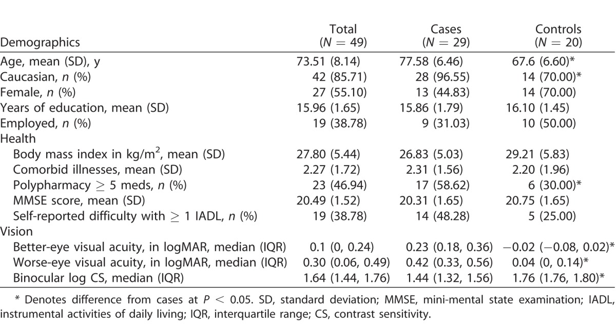
Data from a total of 49 participants comprising 29 AMD cases and 20 glaucoma suspect controls were analyzed. AMD participants were older (mean [SD]: 77.6 [6.46] vs. 67.6 [6.60] years, P <0.05) than controls and more likely to be white (N = 28, 97% vs. N = 14, 70%, P < 0.05), but did not differ from them with regard to sex, education level, employment status, BMI, number of other illnesses, MMSE score, or IADL score (P > 0.05 for all, Table 1). Frequency of polypharmacy was greater in AMD cases than controls (58.62% vs. 30.00%, P < 0.05). Compared with visually normal controls, AMD cases' worse VA was as follows: median better eye-VA interquartile range (IQR): 0.23 (0.18, 0.36) versus −0.02 (−0.08, 0.02)logMAR, and worse CS was as follows: median binocular log CS (IQR): 0.42 (0.33, 0.56) versus 0.04 (0, 0.14) log units), (P < 0.05 for both). Mean walking speed was 0.995 m/sec (SD: 0.190) in the control group and 0.866 m/sec (SD: 0.142) in the AMD group.
Table 1.
Population Characteristics

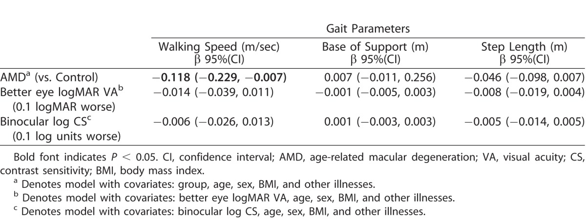
In separate multivariable regression models, all adjusted for age, sex, BMI, and other illnesses, the impact of AMD, VA, and CS on gait variables was analyzed (Table 2). When compared to controls, AMD cases had slower walking speed (β = −0.118 m/sec [95% confidence interval (CI): −0.229, −0.007]), slower stride velocity (β= −0.119 m/sec [95% CI: −0.232, −0.007]), and greater double support time (β = 3.381% [95% CI: 1.006, 5.757]), (P < 0.05 for all). A pattern of association between shorter step lengths AMD status (β = −0.046 m [95% CI: −0.098, 0.007], P = 0.086) that did not reach statistical significance was found. Similarly, a pattern of association between worse VA (per 0.1 logMAR) and greater double support time (β= 0.532 [95% CI: −0.017, 1.081] %, P = 0.057) that did not reach statistical significance was seen.
Table 2.
Multivariable Linear Regressions Assessing Associations between Covariates and Gait Characteristics

Table 2.
Extended

Multivariable linear regression models used to assess the relationship between inter-stride CV values of gait characteristics and AMD status, VA, or CS showed no associations between those variables (P > 0.05 for all; Table 3).
Table 3.
Multivariable Linear Regressions Assessing Associations between Covariates and Coefficients of Variations of Gait Characteristics

Table 3.
Extended

Discussion
In this pilot study population, AMD status was associated with slower walking speed and stride velocity, and greater double support time. However, it is possible that these findings may be largely related to the older age of the AMD group compared with the controls. While neither VA nor CS, evaluated as continuous measures, were associated with statistically significant differences in gait parameters, the model evaluating the relationship between VA, and greater double support time showed a pattern of an association. AMD subjects did not show any differences in inter-stride variability of gait patterns in comparison with controls.
Our results are in accord with previous studies by Spaulding et al.7,20 examining gait in AMD that found that AMD was associated with slower stride velocity. Another study performed by Wood et al.13 found that reduced CS in AMD was associated with slower walking velocity, and increased double-support time. While we report similar gait adaptations in our AMD cohort, our findings differ in that CS or other covariates in our study did not explain our results.
These changes noted in walking patterns, such as decreased walking speed32,33 and greater double support time34 have been previously associated with an increased risk of falling. It has also been postulated that these changes in gait parameters are indicative of stabilizing gait adaptations related to increased caution expressed by patients secondary to a fear of falling.35 In fact, prior literature suggests that a slower walking velocity is a compensatory mechanism adopted secondary to an effort to increase postural stability in the elderly.13,36 Our results suggest that AMD subjects have certain gait characteristics that may contribute to mobility issues; however, we did not specifically test the role of past falls, fear of falling, or assess falls prospectively.
This study adds to the limited body of existing literature on gait parameters in AMD and is one of only three studies providing detailed three-dimensional kinematic gait analysis. Additionally, this is the first study to examine stride-to-stride CV in gait characteristics in AMD. However, this study has some limitations.
First, we tested gait under simple walking conditions alone. Research assessing the effect of AMD on walking under more challenging settings, such as extreme ambient lighting, courses with obstructions, and uneven terrain, such as the those studies conducted by Spaulding et al.7,20 will provide more robust data helping understand gait under “real world” settings.
A second concern is the unequal age and sex distributions between our groups, with the control group being close to a decade younger and having a larger proportion of females than the AMD group. While we controlled for age and sex in our analysis, a more balanced distribution in these demographics between groups may have provided more optimal precision to our effect measures. Additionally, because AMD cases were substantially older than the controls, our results should be interpreted with caution, as it is possible that our findings might not be due to AMD itself, but rather the considerably older age of the AMD group. Limited resources precluded recruitment of more appropriate controls outside our clinic but it might be worthwhile for future studies to attempt wider recruitment of more elderly, age-matched controls.
Finally, the study might have benefited from additional testing and collection of information on history of falls, handedness/footedness, and VFs. Information on foot dominance was not obtained and we uniformly analyzed right leg data. Using data from the dominant limb that is preferentially used for performance of mobilization tasks might be a better indicator of gait than data from the same limb in all subjects. Furthermore, because we did not perform VF testing, we could not investigate the effects of a central scotoma on gait. Similarly, past falls and/or fear of falling experienced by our participants might have been important factors to consider as they may have influenced their gait making those who are fearful adopt more cautious gait patterns.
In summary, we conclude that patients with AMD have slower walking speeds and stride velocities, and spend a greater proportion of time with both feet on the ground while walking, compared with controls, and that these gait characteristics could potentially result in mobility difficulties and increased fall risk in AMD. Maintenance of the ability of independent and safe ambulation is important to physical and psychosocial well being, and our research supports the need for further evaluation of gait and variability in gait in AMD in studies with larger populations, and a longitudinal design allowing the examination of adaptation of gait as it develops. It would also be useful to examine if the adoption of any stabilizing adaptations actually results in a lower risk of falls.
Acknowledgments
Supported by grants from Research to Prevent Blindness.
Disclosure: V. Varadaraj, None; A. Mihailovic, None; R. Ehrenkranz, None; S. Lesche, None; P.Y. Ramulu, None; B.K. Swenor, None
References
- 1. Lord SR,, Smith ST,, Menant JC. Vision and falls in older people: risk factors and intervention strategies. Clin Geriatr Med. 2010; 26: 569–581. [DOI] [PubMed] [Google Scholar]
- 2. Lord SR,, Dayhew J. Visual risk factors for falls in older people. J Am Geriatr Soc. 2001; 49: 508–515. [DOI] [PubMed] [Google Scholar]
- 3. Boptom RQI,, Cumming RG,, Mitchell P,, et al. Visual impairment and falls in older adults: the Blue Mountains Eye Study. J Am Geriatr Soc. 1998; 46: 58–64. [DOI] [PubMed] [Google Scholar]
- 4. Lord SR,, Lloyd DG,, Li SK. Sensori-motor function, gait patterns and falls in community-dwelling women. Age Ageing. 1996; 25: 292–299. [DOI] [PubMed] [Google Scholar]
- 5. Morrison A,, Fan T, Sen SS, et al. Epidemiology of falls and osteoporotic fractures: a systematic review. Clinicoecon Outcomes Res. 2013; 5: 9–18. [DOI] [PMC free article] [PubMed] [Google Scholar]
- 6. National Eye Institute (NEI). 2010 U.S. Age-Specific Prevalence Rates for AMD by Age and Race/Ethnicity. Avaialble at: https://nei.nih.gov/eyedata/amd#1. Accessed December 24, 2016. [Google Scholar]
- 7. Spaulding SJ,, Patla AE,, Elliott DB,, et al. Waterloo Vision and Mobility Study: gait adaptations to altered surfaces in individuals with age-related maculopathy. Opt Vis Sci. 1994; 71: 770–777. [DOI] [PubMed] [Google Scholar]
- 8. Hassan SE,, Lovie-Kitchin JE,, Woods RL. Vision and mobility performance of subjects with age-related macular degeneration. Opt Vis Sci. 2002; 79: 697–707. [DOI] [PubMed] [Google Scholar]
- 9. Wood JM,, Lacherez P,, Black AA,, et al. Risk of falls, injurious falls, and other injuries resulting from visual impairment among older adults with age-related macular degeneration. Invest Ophthalmol Vis Sci. 2011; 52: 5088–5092. [DOI] [PubMed] [Google Scholar]
- 10. Szabo S,, Janssen P,, Khan K,, et al. Neovascular AMD: an overlooked risk factor for injurious falls. Osteoporos Int. 2010; 21: 855–862. [DOI] [PubMed] [Google Scholar]
- 11. Patino CM,, Mckean-Cowdin R,, Azen SP,, et al. Central and peripheral visual impairment and the risk of falls and falls with injury. Ophthalmology. 2010; 117;199–206, e191. [DOI] [PMC free article] [PubMed] [Google Scholar]
- 12. Radvay X,, Duhoux S,, Koenig-Supiot F,, et al. Balance training and visual rehabilitation of age-related macular degeneration patients. J Vestib Res. 2007; 17: 183–193. [PubMed] [Google Scholar]
- 13. Wood JM,, Lacherez PF,, Black AA,, et al. Postural stability and gait among older adults with age-related maculopathy. Invest Ophthalmol Vis Sci. 2009; 50: 482–487. [DOI] [PubMed] [Google Scholar]
- 14. Hassell J,, Lamoureux E,, Keeffe J. Impact of age related macular degeneration on quality of life. Br J Ophthalmol. 2006; 90: 593–596. [DOI] [PMC free article] [PubMed] [Google Scholar]
- 15. Sengupta S,, Nguyen AM,, Van Landingham SW,, et al. Evaluation of real-world mobility in age-related macular degeneration. BMC Ophthalmol. 2015; 15: 1. [DOI] [PMC free article] [PubMed] [Google Scholar]
- 16. Garber CE,, Greaney ML,, Riebe D,, et al. Physical and mental health-related correlates of physical function in community dwelling older adults: a cross sectional study. BMC Geriatr. 2010; 10: 6. [DOI] [PMC free article] [PubMed] [Google Scholar]
- 17. Wilcox D,, Burdett R. Contrast sensitivity function and mobility in elderly patients with macular degeneration. J Am Optometric Aassoc. 1989; 60: 504–507. [PubMed] [Google Scholar]
- 18. Brown B,, Brabyn L,, Welch L,, et al. Contribution of vision variables to mobility in age-related maculopathy patients. Opt Vis Sci. 1986; 63: 733–739. [DOI] [PubMed] [Google Scholar]
- 19. Kuyk T,, Elliott JL. Visual factors and mobility in persons with age-related macular degeneration. J Rehab Res Devel. 1999; 36: 303–312. [PubMed] [Google Scholar]
- 20. Spaulding S,, Patla A,, Flanagan J,, et al. Waterloo Vision and Mobility Study: normal gait characteristics during dark and light adaptation in individuals with age-related maculopathy. Gait Posture. 1995; 3: 227–235. [Google Scholar]
- 21. Hausdorff JM,, Rios DA,, Edelberg HK. Gait variability and fall risk in community-living older adults: a 1-year prospective study. Arch Phys Med Rehab. 2001; 82: 1050–1056. [DOI] [PubMed] [Google Scholar]
- 22. Tinetti ME,, Speechley M,, Ginter SF. Risk factors for falls among elderly persons living in the community. N Engl J Med. 1998; 319; 1701–1707. [DOI] [PubMed] [Google Scholar]
- 23. Wolfson L,, Whipple R,, Amerman P,, et al. Gait assessment in the elderly: a gait abnormality rating scale and its relation to falls. J Gerontol. 1990; 45: M12–M19. [DOI] [PubMed] [Google Scholar]
- 24. Bilney B,, Morris M,, Webster K. Concurrent related validity of the GAITRite® walkway system for quantification of the spatial and temporal parameters of gait. Gait Posture 2003; 17; 68–74. [DOI] [PubMed] [Google Scholar]
- 25. Bailey I,, Bullimore M,, Raasch T,, et al. Clinical grading and the effects of scaling. Invest Ophthalmol Vis Sci. 1991; 32; 422–432. [PubMed] [Google Scholar]
- 26. Arditi A. Improving the design of the letter contrast sensitivity test. Invest Ophthalmol Vis Sci. 2005; 46: 2225–2229. [DOI] [PubMed] [Google Scholar]
- 27. Ramulu PY,, Maul E,, Hochberg C,, et al. Real-world assessment of physical activity in glaucoma using an accelerometer. Ophthalmology. 2012; 119: 1159–1166. [DOI] [PMC free article] [PubMed] [Google Scholar]
- 28. Busse A,, Sonntag A,, Bischkopf J,, et al. Adaptation of dementia screening for vision-impaired older persons: administration of the Mini-Mental State Examination (MMSE). J Clin Epidemiol. 2002; 55: 909–915. [DOI] [PubMed] [Google Scholar]
- 29. Katz S. Assessing self-maintenance: activities of daily living, mobility, and instrumental activities of daily living. J Am Geriatr Soc. 1983; 31: 721–727. [DOI] [PubMed] [Google Scholar]
- 30. Linjakumpu T,, Hartikainen S,, Klaukka T,, et al. Use of medications and polypharmacy are increasing among the elderly. J Clin Epidemiol. 2002; 55: 809–817. [DOI] [PubMed] [Google Scholar]
- 31. Jörgensen T,, Johansson S,, Kennerfalk A,, et al. Prescription drug use, diagnoses, and healthcare utilization among the elderly. Ann Pharmacother. 2001; 35: 1004–1009. [DOI] [PubMed] [Google Scholar]
- 32. Van Kan GA,, Rolland Y,, Andrieu S,, et al. Gait speed at usual pace as a predictor of adverse outcomes in community-dwelling older people an International Academy on Nutrition and Aging (IANA) Task Force. J Nutr Health Aging 2009; 13: 881–889. [DOI] [PubMed] [Google Scholar]
- 33. Cummings SR,, Nevitt MC. Falls. N Engl J Med. 1994; 331: 872–873. [DOI] [PubMed] [Google Scholar]
- 34. Hill K,, Schwarz J,, Flicker L,, et al. Falls among healthy, community-dwelling, older women: a prospective study of frequency, circumstances, consequences and prediction accuracy. Aust N Z J Public Health. 1999; 23: 41–48. [DOI] [PubMed] [Google Scholar]
- 35. Maki BE. Gait changes in older adults: predictors of falls or indicators of fear? J Am Geriatr Soc. 1997; 45: 313–320. [DOI] [PubMed] [Google Scholar]
- 36. Woollacott MH,, Tang P-F. Balance control during walking in the older adult: research and its implications. Phys Ther. 1997; 77: 646–660. [DOI] [PubMed] [Google Scholar]


