

Question
57-year-old woman with an incidentally detected pancreatic mass and multiple liver lesions diagnosed on her annual check-up. She was previously healthy and denied any surgery. At presentation, the laboratory findings and tumor markers were within the normal range. The abdominal ultrasound demonstrated an 8.0 × 6.6 cm heterogeneous mass in the pancreatic tail (Figure A, arrow), and two liver masses measuring up to 5.5 cm (Figure B, arrows). A computed tomography (CT) was subsequently requested.
Figure A.

Figure B.

The CT scan showed a large solid-cystic mass in the tail of the pancreas, with central tiny foci of calcification on the pre-contrast phase (Figure C, arrows). Contrast-enhanced CT demonstrated the liver lesions with peripheral hyperenhancement and hypovascular center (Figures D,E,F arrows). The pancreatic mass presented peripheral and septal progressive enhancement (Figure E, F, arrowheads), and was associated with occlusion of the splenic vein (Figure E, dashed arrow), and consequent gastric varices (Figures D, E, asterisks).
Figure C.

Figure D.

Figure E.
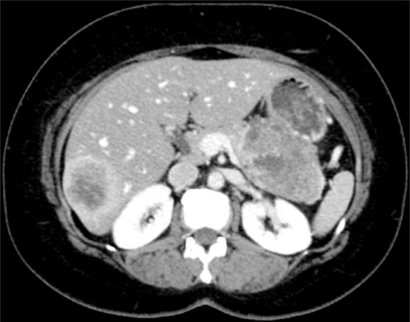
Figure F.

What is the diagnosis?
Answer to the Clinical Challenges and Images in GI
Serous Cystadenocarcinoma of the Pancreas with Liver Metastasis.
An ultrasound-guided biopsy of the larger liver mass was obtained and the diagnosis was microcystic neoplasm with features of serous cystic neoplasm of the pancreas. One month later, the patient underwent distal pancreatectomy, splenectomy, and hepatic metastasectomy. The pancreatectomy showed variably sized small cysts lined by cuboidal cells with small hyperchromatic nuclei and subepithelial capillaries (Figure G, 100× magnification, hematoxylin and eosin). The liver nodules were histologically similar with extensive lymphovascular invasion (Figure H, 100× magnification, hematoxylin and eosin). These findings confirmed the diagnosis of metastatic serous cystadenocarcinoma of the pancreas. One year afterward, the patient is alive with no evidence of recurrent disease (Figure I).
Figure G.

Figure H.

Figure I.

Serous cystadenocarcinoma (SCAc) of the pancreas is a rare but well-established entity first described by George et al in 19891, and since then less than 30 cases have been published2. The WHO classification defines it by the presence of distant metastasis, even if the primary tumor presents a benign histologic appearance3. The origin and evolution of this disorder remain unclear, but even metastatic cases have an excellent prognosis.
Huh et al2 in a recent systematic review showed that these patients were between 42 and 87 years of age and the female to male ratio was 2.9. The most common symptoms or signs were abdominal pain (48%), palpable mass (22%), and weight loss (18%), and 11% of the patients presented an incidentally detected SCAc. There was no significant predilection for location, however, it presents a slightly higher incidence in the body and/or tail of the pancreas (≈ 51%). These tumors are very similar to benign serous cystic neoplasms, except that they tend to be larger (mean diameter of 10.2 cm2), present distant metastasis, and are locally invasive. The most frequent local invasion are adjacent vessels, spleen, peripancreatic fat, stomach, and duodenum3. The most common site of distant metastasis is the liver, however, cases of metastasis to the lung, bone, adrenal glands, peritoneum, and spleen have also been described2.
Acknowledgments
The authors would like to thank Carlie Sigel, Department of Pathology, Memorial Sloan Kettering Cancer Center for her assistance in the histopathological analysis. The authors Natally de Souza Maciel Rocha Horvat, Serena Monti, and Lorenzo Mannelli were equally involved in acquisition of data, analysis and interpretation of data, drafting of the manuscript, critical revision of the manuscript for important intellectual content, statistical analysis, technical, or material support of this study.
Footnotes
The authors disclose no conflict.
References
- 1.George DH, Murphy F, Michalski R, et al. Serous cystadenocarcinoma of the pancreas: a new entity? Am J Surg Pathol. 1989;13(1):61–6. doi: 10.1097/00000478-198901000-00009. [DOI] [PubMed] [Google Scholar]
- 2.Huh J, Byun JH, Hong SM, et al. Malignant pancreatic serous cystic neoplasm: systematic review with a new case. BMC Gastroenterology. 2016;16(1):97. doi: 10.1186/s12876-016-0518-0. [DOI] [PMC free article] [PubMed] [Google Scholar]
- 3.Bosman FT, Carneiro F, Hruban RH, et al. WHO classification of tumours of the digestive system. Geneva: World Health Organization; 2010. [Google Scholar]