Abstract
Adrenal infarction in pregnancy is an extremely rare event. We report a case of a 29-year-old pregnant woman at the twenty-fourth week of gestation that presented with an acute episode of severe localized right upper quadrant pain. Her preliminary blood investigations and abdominal ultrasonography were essentially unremarkable. A diagnosis of right adrenal infarction was subsequently established on the basis of a non-enhanced swollen right adrenal gland on CT scanning of the abdomen with contrast, consistent with the clinical presentation. She was treated with subcutaneous low molecular weight heparin (LMWH) until 2 weeks postpartum. A thrombophilia screen post-partum revealed a significantly elevated factor VIII level and a hypercoagulable state that justified prolonged anticoagulation. This case highlights the importance of a high index of suspicion for adrenal infarction in pregnancy on the clinical grounds of otherwise unexplained acute abdominal pain accompanied by suggestive radiological findings, especially in the presence of thrombophilia.
The adrenal glands are an atypical site for thrombotic events, and in the event of infarction a search for a cause for thrombophilia is warranted. Bilateral adrenal hemorrhagic infarction secondary to a hypercoagulable state such as antiphospholipid syndrome is well characterized, with the usual presentation being primary adrenal insufficiency.1 Unilateral adrenal infarction, with preservation of contralateral adrenal function, may present solely as an acute abdomen during pregnancy, and it may be especially challenging to arrive at a confident diagnosis. This latter condition is extremely rare, and although the pregnant state itself may constitute a thrombogenic factor, cases in association with a hypercoagulable state such as methylene tetra hydro folate reductase C677T gene mutation2 and essential thrombocythemia3 have been reported. Elevated factor VIII has also been observed as a cause of thrombogenesis in association with unilateral adrenal infarction in pregnancy. Our objective in presenting this particular case is to highlight the importance of suspicion of adrenal infarction in a pregnant women with severe abdominal pain.
Case Report
A 29-year-old Kuwaiti pregnant woman (gravida 5, para 4), presented at 24 weeks of gestation to the emergency department with acute onset of severe right-sided abdominal pain of 5-hours duration. The pain was localized and sharp in nature and associated with nausea and vomiting. There was no history of fever, chills, constipation, or urinary symptoms.
Her medical history was unremarkable, she was a non-smoker and had no history of alcohol intake. She had not used oral contraceptives, had 4 previous pregnancies with spontaneous vaginal deliveries at full term, and had no history of miscarriages. She was currently taking oral iron, calcium, and folic acid supplements for her pregnancy and antacids for heartburn on an as-needed basis.
On examination, she was in severe pain, but fully conscious and alert. Her vital signs showed a blood pressure of 132/70 mm Hg, pulse rate 110 per minute and regular, respiratory rate 22 per minute, temperature 37°C, and oxygen saturation (SpO2) 99% on room air. Abdominal examination revealed moderate tenderness over the right upper quadrant with no rebound tenderness. There was no fundal tenderness on uterine palpation, and bowel sounds were present. Chest and cardiovascular examinations were unremarkable.
Laboratory investigations on admission were essentially unremarkable, as follows (normal ranges [NR] are shown in brackets): white blood cell count 10,000 cells/micrL (NR: 4,000-11,000/micrL), neutrophils 58.8% (NR: 40-80%), lymphocytes 33% (NR: 20-40%) hemoglobin level 114 g/L (118-148 g/L); hematocrit 0.34 L/L (0.36-0.44 L/L); mean corpuscular volume 80.4, Femtoliter (82-98 FL); and platelets 448 x 109/L (150-450 x 109/L). Her coagulation profile, renal function test, liver function test, and electrolytes, serum amylase, and lipase were all within normal ranges. Random blood sugar was 5.2 mmol/L, and routine urine microscopy did not show microscopic hematuria, proteinuria, or evidence of infection. Her lipid profile was not markedly deranged: triglycerides of 1.97 mmol/L (0-1.7 mmol/L), high-density lipoprotein cholesterol 0.95 mmol/L (1.29-1.69 mmol/L), and low-density lipoprotein cholesterol 1.53 mmol/L (0-3.4 mmol/L). Serum cortisol (8:00 am) was 560 nmol/L.
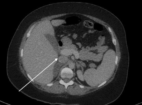
In view of the acute abdomen, ultrasonography of the abdomen was performed and revealed only fatty liver with no evidence of acute cholecystitis or appendicitis. No free fluid was detected in the retro-peritoneum. However, the persistence and severity of symptoms did not correlate well with the unremarkable initial blood investigations and sonographic imaging. Further radiological investigations were therefore performed. A CT abdomen with contrast revealed a swollen hypodense non-enhanced right adrenal gland suggestive of right adrenal infarction (Figure 1).
Figure 1.

Abdominal CT showing right adrenal swollen hypodense lesion (arrow).
In light of this radiological finding, which was consistent with the site of pain and acute clinical presentation, a diagnosis of right adrenal infarction was made. Anticoagulation with subcutaneous low-molecular-weight heparin (LMWH) 0.1 mg/kg twice daily was commenced to prevent propagation of adrenal infarction and a similar occurrence in the contralateral gland. A thrombophilia screen was sought and antinuclear antibodies (ANA), lupus anticoagulant, anti-cardiolipin antibody, and anti-glycoprotein were noted to be negative.
On the second day following admission, the symptoms improved and the patient was discharged home on LMWH. The remainder of her pregnancy was uneventful. In her thirty-eighth week of gestation, she had an uneventful spontaneous vaginal delivery following spontaneous rupture of membranes. Her LMWH was continued for 2 weeks postpartum.
One month after discontinuation of anticoagulation, a second set of thrombophilia screening tests was performed and this revealed significantly elevated plasma factor VIII activity 270% (51-165%). An assessment of other thrombophilic tendencies was unremarkable: anti-thrombin activity 114 (82-130%), protein C activity 94 (64-149%), protein S activity 110 (61-121%), activated protein C resistance 2.7 (>2.2).
After a 6-week interval, factor VIII activity remained persistently elevated at 303.5% (51-165%). Based on high plasma factor VIII activity in association with the history of adrenal infarction it was decided to recommend life-long anticoagulation to limit the recurrence of venous thromboembolic events.
Discussion
This case highlights 2 important clinical issues. Despite its rarity, adrenal infarction should be entertained as one of the differential diagnoses of acute severe abdominal pain during pregnancy. Once the diagnosis of adrenal infarction is established on clinical and radiological grounds a search for a thrombophilic state should be pursued, including, as this case, for elevation of factor VIII.
While bilateral adrenal infarction may present with primary adrenal insufficiency,1 rare cases of unilateral adrenal infarction during pregnancy have been reported to present with acute severe upper abdominal pain,2 which may mimic a wide range of other diagnoses. Where preliminary blood investigations and abdominal sonography are unremarkable, the unexplained nature and severity should prompt further radiological investigation, especially in the known presence of a hypercoagulable state. The diagnosis of adrenal infarction can be established by either CT or MRI scans.3 Initiation of anticoagulation in adrenal infarction should be approached with caution because of the high possibility of adrenal hemorrhage.4 While pregnancy itself is a thrombogenic state, other reported thrombophilic causes in this condition include antiphospholipid syndrome, essential thrombocythemia, and methylenetetrahydrofolate reductase C677T gene mutation.2,4
Thrombotic tendencies rely on multiple factors categorized under Virchow’s triad; namely, endothelial function, vascular flow, and blood composition.5 Factor VIII is a coagulation factor, which, if pathologically elevated (either congenitally or acquired), may cause arterial and venous thrombosis.10
The level of factor VIII varies between individuals, depending on genetic predispositions.7,8 Von Willebrand factor (vWF) and blood group are important determinants of the factor VIII level in plasma, with non-O blood group patients having higher levels of vWF and factor VIII.9 Other factors such as obesity, diabetes mellitus, elevated triglyceride levels, and increase age are also known causes of increased factor VIII plasma levels.10
In multivariate analysis, it has been found that factor VIII levels are an independent risk factor for venous thrombosis, despite blood group type or vWF level.10 The risk of venous thrombosis increases up to three-fold when the level of factor VIII exceeds 150 IU/dL, and each additional increase in factor VIII of 10 IU/dL is associated with a further 10% increase in the risk of thrombosis.10
In conclusion, despite its rarity, this case highlights the importance of a high index of suspicion for the diagnosis of adrenal infarction in pregnancy in the clinical setting of otherwise, unexplained acute abdominal pain accompanied by suggestive radiological findings, especially in the presence of thrombophilia. Factor VIII should be considered as one of the possible causative factors underlying this condition.
Footnotes
References
- 1.Ramon I, Mathian A, Bachelot A, Hervier B, Haroche J, Boutin-Le Thi Huong D, et al. Primary adrenal insufficiency due to bilateral adrenal hemorrhage-adrenal infarction in the antiphospholipid syndrome: long-term outcome of 16 patients. J Clin Endocrinol Metab. 2013;98:3179–3189. doi: 10.1210/jc.2012-4300. [DOI] [PubMed] [Google Scholar]
- 2.Green PA, Ngai IM, Lee TT, Garry DJ. Unilateral adrenal infarction in pregnancy. BMJ Case Rep 2013. 2013 doi: 10.1136/bcr-2013-009997. pii: bcr2013009997. [DOI] [PMC free article] [PubMed] [Google Scholar]
- 3.Riddell AM, Khalili K. Sequential adrenal infarction without MRI-detectable hemorrhage in primary antiphospholipid-antibody syndrome. AJR Am J Roentgenol. 2004;183:220–222. doi: 10.2214/ajr.183.1.1830220. [DOI] [PubMed] [Google Scholar]
- 4.Godfrey RL, Clark J, Field B. Bilateral adrenal haemorrhagic infarction in a patient with antiphospholipid syndrome. BMJ Case Rep 2014. 2014 doi: 10.1136/bcr-2014-207050. pii:bcr2014207050-bcr2014207050. [DOI] [PMC free article] [PubMed] [Google Scholar]
- 5.Palta S, Saroa R, Palta A. Overview of the coagulation system. Indian J Anaesth. 2014;58:515–523. doi: 10.4103/0019-5049.144643. [DOI] [PMC free article] [PubMed] [Google Scholar]
- 6.Kraaijenhagen RA, Anker PS, Koopman MM, Reitsma PH, Prins MH, van den Ende A, et al. High plasma concentration of factor VIIIc is a major risk factor for venous thromboembolism. Thromb Haemost. 2000;83:5–9. [PubMed] [Google Scholar]
- 7.Kerr CB, Preston AE, Barr A, Biggs R. Further studies on the inheritance of factor VIII. Br J Haematol. 1966;12:212–233. doi: 10.1111/j.1365-2141.1966.tb05627.x. [DOI] [PubMed] [Google Scholar]
- 8.Veltkamp JJ, Mayo O, Motulsky AG, Fraser GR. Blood Coagulation Factors I, II, V, VII, VIII, IX, X, XI and XII in Twins. Hum Hered. 1972;22:102–117. [Google Scholar]
- 9.Gill JC, Endres-Brooks J, Bauer PJ, Marks WJ, Jr, Montgomery RR. The effect of ABO blood group on the diagnosis of von Willebrand disease. Blood. 1987;69:1691–1695. [PubMed] [Google Scholar]
- 10.Conlan MG, Folsom AR, Finch A, Davis CE, Sorlie P, Marcucci G, et al. Associations of factor VIII and von Willebrand factor with age, race, sex, and risk factors for atherosclerosis. The Atherosclerosis Risk in Communities (ARIC) Study. Thromb Haemost. 1993;70:380–385. [PubMed] [Google Scholar]


