Acute myeloid leukemia (AML) is an aggressive, genetically diverse hematopoietic stem cell (HSC) malignancy with a generally poor prognosis: most patients die from the disease or from successive but futile rounds of chemotherapy. The FMS-like tyrosine kinase 3 (FLT3) gene is frequently altered in AML; an internal tandem duplication (FLT3-ITD) is seen in approximately 35% of patients, with an amino acid substitution in the tyrosine kinase domain detected in approximately 10%.1 FLT3-ITD mutations are associated with an extremely poor prognosis in AML.
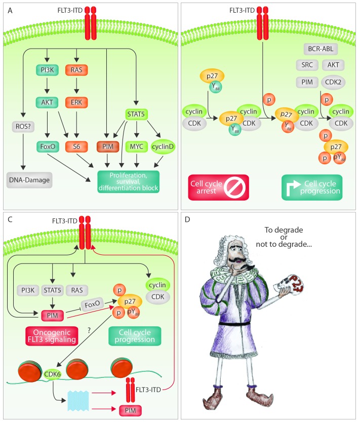
The specific pathways elicited by constitutive FLT3 activation include RAS/ERK/MAPK and PI3K/AKT (Figure 1A). In contrast to the wild-type, FLT3-ITD is a potent activator of STAT5 (Figure 1A); the acquisition of FLT3-ITD ensures leukemic stem cell (LSC) survival by upregulation of MCL-1 via constitutive STAT5 activation.2 The ITD mutations not only constitutively trigger FLT3 kinase activity, but also promote aberrant receptor functions, thereby influencing myeloid differentiation.3 The induction of PIM1, c-MYC and cyclin D3 is likely to contribute to the altered genetic programs downstream of FLT3-ITD necessary for leukemic transformation.4 The clinical importance of FLT3 has stimulated the development of FLT3 tyrosine kinase inhibitors (FLT3-TKI). Unfortunately, the initial high hopes have not been fulfilled due to the rapid development of resistance.5,6 A detailed understanding of the molecular pathways involved in FLT3-ITD signaling may pave the way for improving targeted therapies.
Figure 1.
Vicious feed-forward loop in FLT3-driven acute myeloid leukemia (AML). (A) Schematic presentation of signaling pathways initiated by FLT3-ITD mutations. (B) p27, an inhibitor of cyclin-dependent kinases (CDK), is a key regulator of cell cycle progression. In a pathological condition, many oncogenic kinases phosphorylate p27 on tyrosine 88. The enhanced pY88-p27 leads to a conformational change that allows for further phosphorylation, thereby leading to its proteasomal degradation. This mechanism liberates CDKs from p27-mediated inhibition and stimulates cell-cycle progression. (C) In the proposed model, FLT3-ITD receptors constitutively activate pro-survival pathways through STAT5-dependent and -independent mechanisms. FLT3 and PIM1 inhibit p27 function by direct phosphorylation and/or by transcriptional repression, leading to enhanced CDK6 kinase activity, which in turn promotes the transcription and activity of FLT3 and PIM1. (D) p27 may represent a therapeutic target. The big question in the “cure” of AML remains to be adressed: to degrade or not to degrade p27.
In this issue of Haematologica, Peschel and colleagues reveal p27 to be a direct substrate of FLT3. p27 is an inhibitor of cell cycle progression and is abundant in quiescent cells. p27 is rapidly degraded to enable cells to enter the S-phase. Wild-type and FLT3-ITD bind to p27 and phosphorylate it on tyrosine 88, a residue linked to oncogenic transformation of tumor cells. Y88 phosphorylation is a prerequisite for p27 phosphorylation on T187 by the CDK2-cyclin complex, which results in its ubiquitin-dependent degradation via the E3 ligase SCFSKP2. FLT3-TKI treatment significantly reduces pY88-p27 in FLT3-ITD cells, thereby increasing the level of p27 protein and causing cell cycle arrest. Peschel et al. detect p27-Y88 phosphorylation in primary AML blast cells at levels comparable to those observed in FLT3-ITD+ cell lines and hypothesize that reduced levels of p27 upon Y88 phosphorylation and/or an increased localization to the cytoplasm might be useful indicators of disease outcome.
Although there is a considerable body of evidence to show that p27 can predict responsiveness in solid tumors,7 the findings in AML are conflicting: while all primary wild-type FLT3 samples have reduced pY88-p27 upon TKI exposure, the levels in ITD+ material may be either increased or decreased. The difference most likely arises from compensatory phosphorylation by Src family kinases. p27 is known to be targeted by oncogenic tyrosine kinases other than FLT3 (see Figure 1B). In chronic myeloid leukemia, BCR-ABL alters p27 functions by two means:8,9 i) a kinase-dependent pathway activates SCFSKP2 and promotes degradation of nuclear p27, thereby undermining its CDK inhibitory activity; and ii) a kinase-independent pathway increases the cytoplasmic level of p27, thereby preventing apoptosis by mechanisms that remain elusive. The prediction of therapeutic response thus represents a challenge.
p27 has functions in addition to its role as cell cycle inhibitor and inducer of anti-apoptotic responses. There is recent evidence for an involvement in transcriptional regulation and cell motility.10 p27 has been implicated in differentiation:11 it provokes an erythroid differentiation response and its suppression decreases myeloid differentiation. AML is characterized by a perturbed differentiation and FLT3-ITD activation leads to inhibition of many myeloid transcription factors, such as myeloid Pu.1 and C/EBPα.3 It remains to be determined whether p27 contributes to the pro-survival signals and the maturation arrest downstream of FLT3-ITD.
p27 is a predominantly nuclear protein that inhibits certain CDKs, although it is able to shuttle to the cytoplasm. As a cell cycle inhibitor, nuclear p27 is a candidate tumor suppressor but the homozygous loss or silencing of the p27 locus is exceedingly rare.12 The complete deletion of p27 causes spontaneous tumorigenesis, predominantly in the pituitary. A decrease in p27 levels due to p27 degradation occurs in roughly half of carcinomas and correlates with aggressive, high-grade tumors and a poor prognosis. However, when mislocated in the cytoplasm, p27 has been reported to show oncogenic activity. Mice expressing a cytoplasmic p27 mutant lacking the nuclear CDK inhibitory function have a higher rate of spontaneous tumors in many organs, such as lung, retina, pituitary, ovary, adrenals and spleen. A low nuclear:cytoplasmic p27 ratio in solid tumors is an adverse prognostic marker. Although the mechanisms by which p27 exerts its oncogenic effects remain enigmatic, cytoplasmic p27 may represent a therapeutic target.
In AML, the cytoplasmic abundance of p27 is regulated by the PIM family members, which are serine/threonine kinases. PIM kinases phosphorylate p27 at T157 and T198 to induce nuclear export and proteasome-dependent degradation. They also repress p27 transcription by phosphorylation and inactivation of forkhead transcription factors FoxO1a and FoxO3a.13 A similar mechanism is employed by mutant FLT3, which induces FoxO3a inactivation and thereby suppresses p27 expression.14 In addition, FLT3 induces PIM1 expression through STAT5 activation and PIM2 through an unknown STAT5-independent mechanism.15 FLT3-ITD thus uses two routes to ‘silence’ p27: direct phosphorylation on Y88 and activation of the STAT5-PIM-FoxO3A pathway.
Encouraged by clinical trials of small-molecule CDK inhibitors in AML therapy, the authors propose that preventing p27-Y88 phosphorylation by FLT3-TKI might represent an alternative strategy to inactivate CDKs, thereby inducing G1 arrest. This strategy comes with a caveat: when the LSCs are in a non-cycling stage (dormant), they are more resistant to therapy and may develop TKI-induced resistance. In practice, therapeutic failure in AML is due more to the emergence of treatment-resistant clones than to treatment-related mortality. One may speculate that FLT3 inhibitors stabilize p27, which in turn restores cell cycle arrest and dormancy, and enhances the risk of developing resistance. A potential strategy to circumventing resistance might be the simultaneous application of a TKI with a specific p27 degrader using novel technologies based on proteolysis-targeting chimeras.16 Support for this idea comes from a report showing that deficiency of both p27 and p57 in HSCs induced cycling of dormant cells by activating CDK4/CDK6.17
A recent study found that FLT3-TKI and the CDK4/6 inhibitor palbociclib act synergistically in FLT3-ITD mutant cells.18 The rationale for this combination is that CDK6 inhibition has effects that go beyond cell cycle control, which would be induced by the TKI-mediated stabilization of p27 without palbociclib. The synergistic effects are mediated by an inhibition of cell-cycle progression in combination with the loss of CDK6-mediated transcription of FLT3, PIM1 and other potential targets.18 As PIM kinases phosphorylate and stabilize FLT3 in vitro,19 the combined treatment disrupts a vicious cycle and feed-forward loop.
The same feed-forward loop may explain why leukemic cells become more dependent on FLT3-ITD signaling in the majority of patients, while the ratio of FLT3-ITD mutant alleles is higher after relapse. The finding is consistent with a model in which FLT3-ITD triggers its own expression via PIM1 and CDK6, thereby promoting hyperproliferation of leukemic cells. When p27 function is impaired by FLT3 and PIM1, CDK6 kinase activity is enhanced, which stimulates the transcription and activity of FLT3 and PIM1. Leukemic cells with mutated FLT3-ITD alleles presumably have a selective advantage, which results in the expansion of mutant FLT3 clones. In parallel, FLT3 prevents apoptosis by activating STAT5, which stimulates production of c-MYC, cyclin D and PIM. It also blocks differentiation, at least partially, via p27 downregulation, an aspect that requires further investigation (Figure 1C).
Peschel et al. now add a further layer of complexity to our understanding. In AML cells, p27 partially co-localizes with FLT3 in extended perinuclear structures. The co-localization is accompanied by enhanced p27 Y88-phosphorylation. Cytoplasmic p27 is susceptible to SCFSKP2-triggered proteolysis but is potentially able to exert proto-oncogenic functions. Although the stabilization of p27 is expected to have a net tumor suppressive effect, the simultaneous increase in the cytoplasmic level of p27 might have unintended consequences that offset the benefits of restoring nuclear p27. Whether AML therapy should aim to stabilize p27 or to degrade it is still unclear (Figure 1D). The importance of learning whether to degrade or not to degrade cannot be overstated. We hope that drug synergy screens will soon provide an answer.
Supplementary Material
Acknowledgments
We are grateful to Philipp Jodl and Peter Alexander Martinek for their excellent technical help in generating the figures. We are deeply indebted to Graham Tebb for editing the manuscript. This work was supported by the Austrian Science Fund (FWF) Grant SFB F47 (to VS) and by an ERC-advanced grant (to VS).
References
- 1.Hawley TS, Fong AZ, Griesser H, Lyman SD, Hawley RG. Leukemic predisposition of mice transplanted with gene-modified hematopoietic precursors expressing flt3 ligand. Blood. 1998;92(6):2003–2011. [PubMed] [Google Scholar]
- 2.Yoshimoto G, Miyamoto T, Jabbarzadeh-Tabrizi S, et al. FLT3-ITD up-regulates MCL-1 to promote survival of stem cells in acute myeloid leukemia via FLT3-ITD-specific STAT5 activation. Blood. 2009;114(24):5034–5043. [DOI] [PMC free article] [PubMed] [Google Scholar]
- 3.Mizuki M, Schwable J, Steur C, et al. Suppression of myeloid transcription factors and induction of STAT response genes by AML-specific Flt3 mutations. Blood. 2003;101(8):3164–3173. [DOI] [PubMed] [Google Scholar]
- 4.Li L, Piloto O, Kim K-T, et al. FLT3/ITD expression increases expansion, survival and entry into cell cycle of human haematopoietic stem/progenitor cells. Br J Haematol. 2007;137(1):64–75. [DOI] [PubMed] [Google Scholar]
- 5.Smith CC, Wang Q, Chin C-S, et al. Validation of ITD mutations in FLT3 as a therapeutic target in human acute myeloid leukaemia. Nature. 2012;485(7397):260–263. [DOI] [PMC free article] [PubMed] [Google Scholar]
- 6.Wander SA, Levis MJ, Fathi AT. The evolving role of FLT3 inhibitors in acute myeloid leukemia: quizartinib and beyond. Ther Adv Hematol. 2014;5(3):65–77. [DOI] [PMC free article] [PubMed] [Google Scholar]
- 7.Chu IM, Hengst L, Slingerland JM. The Cdk inhibitor p27 in human cancer: prognostic potential and relevance to anticancer therapy. Nat Rev Cancer. 2008;8(4):253–267. [DOI] [PubMed] [Google Scholar]
- 8.Chu S, McDonald T, Bhatia R. Role of BCR-ABL-Y177-mediated p27kip1 phosphorylation and cytoplasmic localization in enhanced proliferation of chronic myeloid leukemia progenitors. Leukemia. 2010;24(4):779–787. [DOI] [PMC free article] [PubMed] [Google Scholar]
- 9.Agarwal A, Mackenzie RJ, Besson A, et al. BCR-ABL1 promotes leukemia by converting p27 into a cytoplasmic oncoprotein. Blood. 2014;124(22):3260–3273. [DOI] [PMC free article] [PubMed] [Google Scholar]
- 10.Coqueret O. New roles for p21 and p27 cell-cycle inhibitors: a function for each cell compartment¿ Trends Cell Biol. 2003;13(2):65–70. [DOI] [PubMed] [Google Scholar]
- 11.Munoz-Alonso MJ, Acosta JC, Richard C, et al. p21Cip1 and p27Kip1 Induce Distinct Cell Cycle Effects and Differentiation Programs in Myeloid Leukemia Cells. J Biol Chem. 2005;280(18):18120–18129. [DOI] [PubMed] [Google Scholar]
- 12.Fero ML, Randel E, Gurley KE, Roberts JM, Kemp CJ. The murine gene p27Kip1 is haplo-insufficient for tumour suppression. Nature. 1998;396(6707):177–180. [DOI] [PMC free article] [PubMed] [Google Scholar]
- 13.Morishita D, Katayama R, Sekimizu K, Tsuruo T, Fujita N. Pim kinases promote cell cycle progression by phosphorylating and down-regulating p27Kip1 at the transcriptional and posttranscriptional levels. Cancer Res. 2008;68(13):5076–5085. [DOI] [PubMed] [Google Scholar]
- 14.Scheijen B, Ngo HT, Kang H, Griffin JD. FLT3 receptors with internal tandem duplications promote cell viability and proliferation by signaling through Foxo proteins. Oncogene. 2004;23(19):3338–3349. [DOI] [PubMed] [Google Scholar]
- 15.Green AS, Maciel TT, Hospital M-A, et al. Pim kinases modulate resistance to FLT3 tyrosine kinase inhibitors in FLT3-ITD acute myeloid leukemia. Sci Adv. 2015;1(8):e1500221–e1500221. [DOI] [PMC free article] [PubMed] [Google Scholar]
- 16.Lai AC, Crews CM. Induced protein degradation: an emerging drug discovery paradigm. Nat Rev Drug Discov. 2017;16(2):101–114. [DOI] [PMC free article] [PubMed] [Google Scholar]
- 17.Zou P, Yoshihara H, Hosokawa K, et al. p57(Kip2) and p27(Kip1) cooperate to maintain hematopoietic stem cell quiescence through interactions with Hsc70. Cell Stem Cell. 2011;9(3):247–261. [DOI] [PubMed] [Google Scholar]
- 18.Uras IZ, Walter GJ, Scheicher R, et al. Palbociclib treatment of FLT3-ITD+ AML cells uncovers a kinase-dependent transcriptional regulation of FLT3 and PIM1 by CDK6. Blood. 2016;127(23):2890–2902. [DOI] [PMC free article] [PubMed] [Google Scholar]
- 19.Natarajan K, Xie Y, Burcu M, et al. Pim-1 kinase phosphorylates and stabilizes 130 kDa FLT3 and promotes aberrant STAT5 signaling in acute myeloid leukemia with FLT3 internal tandem duplication. PLoS One. 2013;8(9):e74653. [DOI] [PMC free article] [PubMed] [Google Scholar]
Associated Data
This section collects any data citations, data availability statements, or supplementary materials included in this article.


