Abstract
Aims/Hypothesis:
We sought to establish whether the increased incidence of diabetes associated with Down syndrome was due to a congenital deficit in β cells.
Methods:
The pancreas was obtained at autopsy from nondiabetic subjects with Down syndrome (n = 29) and age-matched nondiabetic control subjects without Down syndrome (n = 28). The pancreas sections were evaluated for the fractional β-cell area.
Results:
No difference was found in the fractional β-cell area between the subjects with Down syndrome and the control subjects.
Conclusions/Interpretations:
The increased incidence and prevalence of diabetes in individuals with Down syndrome is not due to an underlying congenital deficiency of β cells.
Keywords: Down syndrome, β cell, diabetes
Precis: The increased incidence of diabetes in those with Down syndrome is not due to a congenital deficiency of β cells.
Down syndrome, caused by trisomy 21, is a common congenital condition, occurring in approximately 1 in 700 live births. Currently, 250,000 to 400,000 individuals have Down syndrome in the United States, with >5 million cases worldwide. In addition to causing a broad range of developmental anomalies [1, 2], Down syndrome is associated with an increased incidence of autoimmune diseases, including thyroid disorders [3, 4] and celiac disease [5–7], with a well-documented increased risk and prevalence of type 1 diabetes [8–11]. In addition, metabolic syndrome and type 2 diabetes occur at an increased frequency at a relatively early age in those with Down syndrome [12].
Development of an individual’s complement of β cells begins during embryonic life and undergoes a rapid postnatal expansion, largely accomplished through replication of the existing β cells [13, 14]. It has been suggested that a risk factor for the development of diabetes might be a failure to establish a sufficient β-cell mass during infancy [15]. β-Cell replication requires nuclear translocation of the transcription factor nuclear factor of activated T cells (NFAT) [16]. Nuclear translocation of NFAT is activated by the phosphatase calcineurin and inhibited by NFAT kinases, which includes dual-specificity tyrosine phosphorylation-regulated kinase 1A [16, 17]. The Down syndrome critical region (DSCR) of chromosome 21 encodes a calcineurin inhibitor, Down syndrome critical region gene 1 (DSCR1), and the NFAT kinase dual-specificity tyrosine phosphorylation-regulated kinase 1A [16]. The intriguing hypothesis was posed that the presumptive increased generation of these 2 inhibitors of β-cell replication might suppress β-cell replication during infancy, leading to a deficient β-cell mass and, thus, the increased risk of diabetes [16].
To address that hypothesis in humans, in the present study, we evaluated the β-cell area in pancreas specimens obtained at autopsy from child and adult nondiabetic individuals with Down syndrome compared with unaffected controls. Our findings reject the hypothesis that there is a deficiency of β cells in nondiabetic subjects with Down syndrome.
1. Materials and Methods
A. Autopsy Cases
Human pancreatic tissue was obtained at autopsy from 29 nondiabetic individuals with documented Down syndrome during life and 28 nondiabetic, age-matched control individuals without Down syndrome (Tables 1 and 2). The subjects were identified by retrospective analysis of the Mayo Clinic autopsy database. For inclusion in the present study, a full autopsy had to have been performed within 24 hours of death and a sample of pancreatic tissue of adequate size and quality stored. Subjects were excluded if the pancreas integrity had been compromised by either autolysis or acute pancreatitis. None of the subjects selected for inclusion in the present study had had a history of diabetes or any other disease known to affect the pancreas. The subject characteristics and diagnoses leading to death are presented in Tables 1 and 2. The institutional review board of the Mayo Clinic and the University of California, Los Angeles, approved the present study. Fasting blood glucose values in health were unavailable from the included subjects. Our presumption that neither the subjects with Down syndrome nor the control subjects had diabetes was determined by an absence of a history of diabetes in the previous medical records and an absence of diabetes documented in their final illness.
Table 1.
Clinical Characteristics of Subjects With Down Syndrome
| Pt. No. | Sex | Age | Age (y) | BCA (%) | Cause of Death |
|---|---|---|---|---|---|
| Age <5 y | |||||
| 1 | Male | Newborn | 0.00 | 1.57 | Fetal distress |
| 2 | Female | 4 d | 0.01 | 5.02 | Aspiration pneumonia |
| 3 | Female | 12 d | 0.03 | 3.33 | Congenital heart disease |
| 4 | Female | 3 mo | 0.25 | 1.49 | Congenital heart disease |
| 5 | Male | 4 mo | 0.33 | 1.93 | Congenital heart disease |
| 6 | Male | 4 mo | 0.33 | 1.93 | Congenital heart disease |
| 7 | Female | 4 mo | 0.33 | 1.10 | Congenital heart disease |
| 8 | Female | 5 mo | 0.42 | 1.70 | Congenital heart disease |
| 9 | Female | 6 mo | 0.50 | 3.24 | Congenital heart disease |
| 10 | Female | 7 mo | 0.58 | 5.56 | Congenital heart disease |
| 11 | Female | 9 mo | 0.75 | 2.07 | Congenital heart disease |
| 12 | Female | 9 mo | 0.75 | 2.38 | Congenital heart disease |
| 13 | Female | 17 mo | 1.42 | 3.43 | Congenital heart disease |
| 14 | Male | 22 mo | 1.83 | 2.16 | Chronic bronchiolitis |
| Mean | 0.54 | 2.64 | |||
| SEM | 0.14 | 0.36 | |||
| Age 5–15 y | NA | ||||
| 1 | Male | 10 | 2.48 | Congenital heart disease | |
| 2 | Female | 5 | 1.07 | Congenital heart disease | |
| 3 | Male | 9 | 1.86 | Congenital heart disease | |
| 4 | Male | 9 | 2.68 | Congenital heart disease | |
| 5 | Male | 13 | 2.00 | Respiratory insufficiency | |
| Mean | 9.20 | 2.02 | |||
| SEM | 1.28 | 0.28 | |||
| >15 y | NA | ||||
| 1 | Female | 19 | 1.53 | Congenital heart disease | |
| 2 | Female | 33 | 1.22 | Congenital heart disease | |
| 3 | Male | 34 | 2.29 | Right ventricular hypertrophy | |
| 4 | Male | 38 | 3.61 | Congenital heart disease | |
| 5 | Female | 47 | 0.79 | Respiratory failure | |
| 6 | Female | 48 | 1.14 | Bronchopneumonia | |
| 7 | Female | 56 | 1.39 | Hemorrhage | |
| 8 | Male | 57 | 2.70 | Bronchopneumonia | |
| 9 | Male | 62 | 0.96 | Bronchopneumonia | |
| 10 | Female | 62 | 0.88 | Bronchopneumonia | |
| Mean | 45.60 | 1.65 | |||
| SEM | 4.52 | 0.29 |
Abbreviations: BCA, beta cell area; NA, not applicable; Pt. No., patient number; SEM, standard error of the mean.
Table 2.
Clinical Characteristics of Control Subjects
| Pt. No. | Sex | Age | Age (y) | BCA (%) | Cause of Death |
|---|---|---|---|---|---|
| Age <5 y | |||||
| 1 | Female | 7 d | 0.02 | 2.92 | Pneumothorax |
| 2 | Female | 18 d | 0.05 | 2.94 | Duodenal atresia |
| 3 | Female | 2.5 mo | 0.21 | 2.75 | Sudden infant death syndrome |
| 4 | Female | 3 mo | 0.25 | 4.54 | Respiratory failure |
| 5 | Male | 4 mo | 0.33 | 4.38 | Congenital heart disease |
| 6 | Male | 4 mo | 0.33 | 3.99 | Congenital heart disease |
| 7 | Female | 7 mo | 0.58 | 4.71 | Congenital heart disease |
| 8 | Female | 8 mo | 0.67 | 2.07 | Congenital heart disease |
| 9 | Female | 9 mo | 0.75 | 1.07 | Congenital heart disease |
| 10 | Female | 11 mo | 0.92 | 1.82 | Congenital heart disease |
| 11 | Female | 14 mo | 1.17 | 2.02 | Bronchopneumonia |
| 12 | Male | 18 mo | 1.50 | 1.16 | Acute nonlymphoblastic leukemia |
| Mean | 0.57 | 2.86 | |||
| SEM | 0.13 | 0.37 | |||
| Age 5–15 y | NA | ||||
| 1 | Female | 5 | 1.67 | Congenital heart disease | |
| 2 | Male | 9 | 1.34 | Congenital heart disease | |
| 3 | Male | 9 | 3.87 | Acute lymphoblastic leukemia | |
| 4 | Male | 12 | 0.93 | Acute encephalopathy | |
| 5 | Male | 13 | 1.39 | Hepatitis | |
| Mean | 9.60 | 1.84 | |||
| SEM | 1.40 | 0.52 | |||
| Age >15 y | NA | ||||
| 1 | Female | 19 | 2.39 | Bronchopneumonia | |
| 2 | Female | 19 | 1.29 | Accidental poisoning | |
| 3 | Female | 33 | 3.76 | Lymphoma | |
| 4 | Male | 33 | 1.03 | Respiratory arrest | |
| 5 | Male | 34 | 2.17 | Teratocarcinoma | |
| 6 | Male | 38 | 1.57 | Cardiac arrhythmia | |
| 7 | Female | 47 | 1.31 | Respiratory arrest | |
| 8 | Male | 48 | 2.78 | Acute myocardial infarction | |
| 9 | Female | 48 | 2.14 | Breast adenocarcinoma | |
| 10 | Female | 62 | 1.81 | Bronchopneumonia | |
| 11 | Male | 64 | 1.67 | Chronic ischemic heart disease | |
| Mean | 40.45 | 1.99 | |||
| SEM | 4.53 | 0.24 |
Abbreviations: BCA, beta cell area; NA, not applicable; Pt. No., patient number; SEM, standard error of the mean.
Because of the substantial changes in the fractional β-cell area during the rapid growth phase of childhood [16], the subjects were grouped into 3 age brackets: <5 years (Down syndrome, n = 14; control, n = 12), 5 to 15 years (Down syndrome, n = 5; control, n = 5), and >15 years (Down syndrome, n = 10; control, n = 11).
B. Pancreatic Tissue Processing
All autopsies were performed at the Mayo Clinic, where a sample of the tail of the pancreas measuring approximately 2.0 × 1.0 × 0.5 cm in size was resected and, together with a sample of spleen, was fixed in formaldehyde before being embedded in paraffin. Next, 5-µm sections were obtained from these tissue blocks and stained for insulin (peroxidase staining) and hematoxylin for light microscopy. For immunohistochemical staining, the primary antibody used was guinea pig anti-insulin (1:200; Dako Laboratories, Carpinteria, CA; Research Resource Identification, AB_2617169).
C. Morphometric Analysis
The pancreatic fractional β-cell area was determined by imaging the entire pancreatic section at ×40 magnification (4× objective). The ratio of the β-cell area to the exocrine pancreatic area was digitally quantified, as previously described [18], using Image Pro Plus software (Image Pro Plus, version 4.5.1; Media Cybernetics, Silver Springs, MD). By digitally excluding the interlobular connective tissue, large blood vessels, and adipocytes, the analysis concentrated on the ratio of the pancreatic islets to the acinar tissue. Two independent observers (A.E.B. and W.S.) performed the analysis. If the interobserver measurements in a sample differed by >5%, the sample was reevaluated.
D. Statistical Analysis
The data are presented as the mean ± standard error. The statistical calculations were performed using GraphPad Prism, version 5 (GraphPad Software, San Diego, CA).
2. Results
A. Age
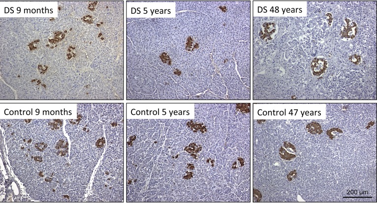
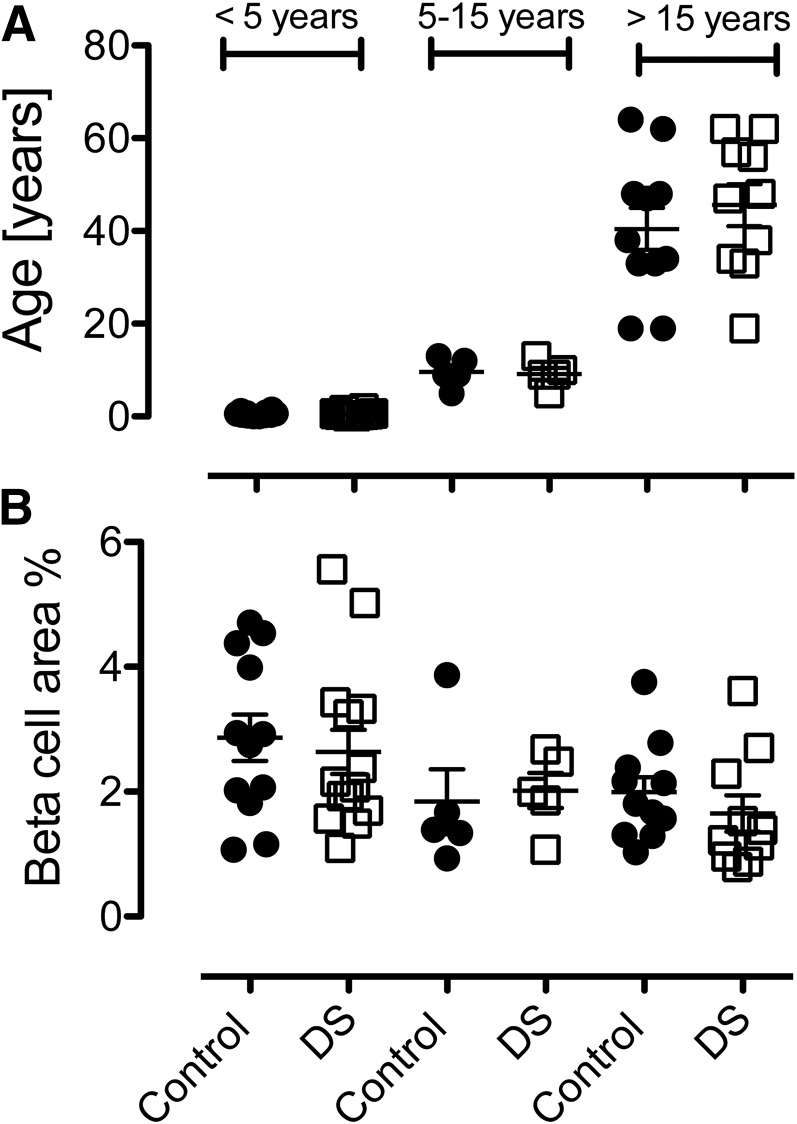
In each of the 3 defined age brackets, the ages of the Down syndrome group were matched with the ages of the control group [Down syndrome vs control, age <5 years, 0.54 ± 0.14 vs 0.57 ± 0.13 years; age 5 to 15 years, 9.2 ± 1.3 vs 9.6 ± 1.4 years; age >15 years, 45.6 ± 4.5 vs 40.5 ± 4.5 years; Fig. 1 and Fig. 2(A)].
Figure 1.
Representative images from pancreatic sections of (upper panels) individuals with Down syndrome (DS) and (lower panels) age-matched control subjects. Insulin-positive β cells are shown in brown (3,3′-diaminobenzidine) with hematoxylin counterstain. In early childhood (left panels), the islet density was greater, with more small clusters of insulin-positive cells compared with later childhood (middle panels) or adulthood (right panels). No difference was found in the β-cell mass between subjects with and without DS. Images were taken at ×200 magnification (20× objective). Scale bar = 200 µm.
Figure 2.
Age and fractional β-cell area percentage for nondiabetic subjects with Down syndrome (DS) and age-matched nondiabetic controls. (A) In each of the 3 defined age brackets, no difference was found between the ages of the Down syndrome group and the ages of the control group (Down syndrome vs control, age <5 years, 0.54 ± 0.14 vs 0.57 ± 0.13 years; age 5 to 15 years, 9.20 ± 1.28 vs 9.60 ± 1.40 years; age >15 years, 45.60 ± 4.52 vs 40.45 ± 4.53 years). (B) No difference was found in the fractional β-cell area between the subjects with Down syndrome and the control subjects in the 3 defined age brackets studied (Down syndrome vs control, age <5 years, 2.64% ± 0.36% vs 2.86% ± 0.37%; age 5 to 15 years, 2.02% ± 0.28% vs 1.84% ± 0.52%; age >15 years, 1.65% ± 0.29% vs 1.99% ± 0.24%).
B. Fractional β-Cell Area
No difference was found in the fractional β-cell area between the subjects with Down syndrome and the control subjects in the 3 defined age brackets studied [Down syndrome vs control, age <5 years, 2.64% ± 0.36% vs 2.86% ± 0.37%; age 5 to 15 years, 2.02% ± 0.28% vs 1.84% ± 0.52%; age >15 years, 1.65% ± 0.29% vs 1.99% ± 0.24%; Fig. 1 and Fig. 2(B)].
3. Discussion
We examined pancreas across a range of individuals with Down syndrome to test the hypothesis that the pancreatic β-cell area would be decreased and thus predispose these subjects to development of type 2 diabetes.
The intriguing hypothesis proposed by Shen et al. [16] was that the β-cell mass in those with Down syndrome would be deficient owing to suppression of the usual high β-cell replication in infancy through an increased dosage of genes in the DSCR. An alternative cause of the decreased β-cell mass in Down syndrome could be the low birth weight commonly present with this syndrome [19]. A low birth weight predicts an increased risk of type 2 diabetes [14], and a low β-cell mass arises after induced placental dysfunction in animal studies [20, 21]. However, the fractional pancreatic β-cell area was normal in children with Down syndrome.
Just as with all studies, the present study had limitations that should be considered. The sample size was small; thus, small differences could have been overlooked. None of the pancreata examined in the present study came from individuals with diabetes. It is plausible that abnormally decreased β-cell growth is present only in the subset of individuals with an increased risk of diabetes. We did not have the whole pancreas available to evaluate nor, therefore, the pancreas mass. It is possible that if the pancreata were much smaller in those with Down syndrome, the total β-cell mass would be decreased, despite a normal β-cell fractional area. We were unable to find any reports of pancreatic size in those with Down syndrome. We were unable to assess the β-cell replication, because the Ki67 marker was unreliable in these sections of pancreas from blocks stored long term.
However, other potential contributors to the increased predisposition to the development of diabetes in the setting of Down syndrome exist. Functional β-cell defects can arise as a consequence of excessive 21 gene dosage [22]. Individuals with Down syndrome might be more susceptible to enteroviral infections, which might predispose these individuals to the development of type 1 diabetes [23]. Epigenetic dysregulation of β cells in those with Down syndrome has also been proposed [24]. The high prevalence of obesity in those with Down syndrome is a predisposing factor for type 2 diabetes and, through endoplasmic stress, might increase the risk of the development of type 2 diabetes [25].
4. Conclusion
The pancreata from this nondiabetic group of subjects with Down syndrome did not have a deficit in the fractional β-cell area. Rather than a low number of β cells as the underlying trigger, the increased risk of type 1 diabetes in those with Down syndrome likely relates to defects in the immune system that increases the frequency of other related autoimmune disorders such as celiac disease and Hashimoto thyroid disease. Furthermore, the increased risk for the development of type 2 diabetes is presumably related to the increased incidence of central adiposity and metabolic syndrome.
Acknowledgments
We appreciate the editorial assistance of Bonnie Lui from the Hillblom Islet Research Center at the University of California, Los Angeles.
These experiments were funded by the National Institutes of Health/ National Institute of Diabetes and Digestive and Kidney Diseases (grant DK077967).
Author contributions: A.E.B. performed the studies and assisted in study design and interpretation and writing the manuscript. W.S. and M.C. assisted in performing the studies and study interpretation. R.A.R. assisted in study design and study interpretation. P.C.B. contributed to the study design, study interpretation, and preparation of the manuscript.
Disclosure Summary: The authors have nothing to disclose.
Footnotes
- DSCR
- Down syndrome critical region
- DSCR1
- Down syndrome critical region gene 1
- NFAT
- nuclear factor of activated T cells.
References and Notes
- 1.Steele J, Stratford B. The United Kingdom population with Down syndrome: present and future projections. Am J Ment Retard. 1995;99(6):664–682. [PubMed] [Google Scholar]
- 2.Korenberg JR, Chen XN, Schipper R, Sun Z, Gonsky R, Gerwehr S, Carpenter N, Daumer C, Dignan P, Disteche C. Down syndrome phenotypes: the consequences of chromosomal imbalance. Proc Natl Acad Sci USA. 1994;91(11):4997–5001. [DOI] [PMC free article] [PubMed] [Google Scholar]
- 3.Kennedy RL, Jones TH, Cuckle HS. Down’s syndrome and the thyroid. Clin Endocrinol (Oxf). 1992;37(6):471–476. [DOI] [PubMed] [Google Scholar]
- 4.Karlsson B, Gustafsson J, Hedov G, Ivarsson SA, Annerén G. Thyroid dysfunction in Down’s syndrome: relation to age and thyroid autoimmunity. Arch Dis Child. 1998;79(3):242–245. [DOI] [PMC free article] [PubMed] [Google Scholar]
- 5.Book L, Hart A, Black J, Feolo M, Zone JJ, Neuhausen SL. Prevalence and clinical characteristics of celiac disease in Downs syndrome in a US study. Am J Med Genet. 2001;98(1):70–74. [PubMed] [Google Scholar]
- 6.Gale L, Wimalaratna H, Brotodiharjo A, Duggan JM. Down’s syndrome is strongly associated with coeliac disease. Gut. 1997;40(4):492–496. [DOI] [PMC free article] [PubMed] [Google Scholar]
- 7.George EK, Mearin ML, Bouquet J, von Blomberg BM, Stapel SO, van Elburg RM, de Graaf EA. High frequency of celiac disease in Down syndrome. J Pediatr. 1996;128(4):555–557. [DOI] [PubMed] [Google Scholar]
- 8.Anwar AJ, Walker JD, Frier BM. Type 1 diabetes mellitus and Down’s syndrome: prevalence, management and diabetic complications. Diabet Med. 1998;15(2):160–163. [DOI] [PubMed] [Google Scholar]
- 9.Van Goor JC, Massa GG, Hirasing R. Increased incidence and prevalence of diabetes mellitus in Down’s syndrome. Arch Dis Child. 1997;77(2):186. [DOI] [PMC free article] [PubMed] [Google Scholar]
- 10.Farquhar JW. Early-onset diabetes in the general and the Down’s syndrome population. Lancet. 1969;2(7615):323–324. [DOI] [PubMed] [Google Scholar]
- 11.Gillespie KM, Dix RJ, Williams AJ, Newton R, Robinson ZF, Bingley PJ, Gale EA, Shield JP. Islet autoimmunity in children with Down’s syndrome. Diabetes. 2006;55(11):3185–3188. [DOI] [PubMed] [Google Scholar]
- 12.Alexander M, Petri H, Ding Y, Wandel C, Khwaja O, Foskett N. Morbidity and medication in a large population of individuals with Down syndrome compared to the general population. Dev Med Child Neurol. 2016;58(3):246–254. [DOI] [PubMed] [Google Scholar]
- 13.Meier JJ, Butler AE, Saisho Y, Monchamp T, Galasso R, Bhushan A, Rizza RA, Butler PC. Beta-cell replication is the primary mechanism subserving the postnatal expansion of beta-cell mass in humans. Diabetes. 2008;57(6):1584–1594. [DOI] [PMC free article] [PubMed] [Google Scholar]
- 14.Georgia S, Bhushan A. Beta cell replication is the primary mechanism for maintaining postnatal beta cell mass. J Clin Invest. 2004;114(7):963–968. [DOI] [PMC free article] [PubMed] [Google Scholar]
- 15.Meier JJ. Linking the genetics of type 2 diabetes with low birth weight: a role for prenatal islet maldevelopment? Diabetes. 2009;58(6):1255–1256. [DOI] [PMC free article] [PubMed] [Google Scholar]
- 16.Shen W, Taylor B, Jin Q, Nguyen-Tran V, Meeusen S, Zhang YQ, Kamireddy A, Swafford A, Powers AF, Walker J, Lamb J, Bursalaya B, DiDonato M, Harb G, Qiu M, Filippi CM, Deaton L, Turk CN, Suarez-Pinzon WL, Liu Y, Hao X, Mo T, Yan S, Li J, Herman AE, Hering BJ, Wu T, Martin Seidel H, McNamara P, Glynne R, Laffitte B. Inhibition of DYRK1A and GSK3B induces human β-cell proliferation. Nat Commun. 2015;6:8372. [DOI] [PMC free article] [PubMed] [Google Scholar]
- 17.Arron JR, Winslow MM, Polleri A, Chang CP, Wu H, Gao X, Neilson JR, Chen L, Heit JJ, Kim SK, Yamasaki N, Miyakawa T, Francke U, Graef IA, Crabtree GR. NFAT dysregulation by increased dosage of DSCR1 and DYRK1A on chromosome 21. Nature. 2006;441(7093):595–600. [DOI] [PubMed] [Google Scholar]
- 18.Butler AE, Janson J, Bonner-Weir S, Ritzel R, Rizza RA, Butler PC. Beta-cell deficit and increased beta-cell apoptosis in humans with type 2 diabetes. Diabetes. 2003;52(1):102–110. [DOI] [PubMed] [Google Scholar]
- 19.Toledo C, Alembik Y, Aguirre Jaime A, Stoll C. Growth curves of children with Down syndrome. Ann Genet. 1999;42(2):81–90. [PubMed] [Google Scholar]
- 20.Limesand SW, Jensen J, Hutton JC, Hay WW Jr. Diminished beta-cell replication contributes to reduced beta-cell mass in fetal sheep with intrauterine growth restriction. Am J Physiol Regul Integr Comp Physiol. 2005;288(5):R1297–R1305. [DOI] [PubMed] [Google Scholar]
- 21.Simmons RA, Templeton LJ, Gertz SJ. Intrauterine growth retardation leads to the development of type 2 diabetes in the rat. Diabetes. 2001;50(10):2279–2286. [DOI] [PubMed] [Google Scholar]
- 22.Meier JJ, Bonadonna RC. Role of reduced β-cell mass versus impaired β-cell function in the pathogenesis of type 2 diabetes. Diabetes Care. 2013;36(Suppl 2):S113–S119. [DOI] [PMC free article] [PubMed] [Google Scholar]
- 23.Jun HS, Yoon JW. A new look at viruses in type 1 diabetes. Diabetes Metab Res Rev. 2003;19(1):8–31. [DOI] [PubMed] [Google Scholar]
- 24.Dang MN, Buzzetti R, Pozzilli P. Epigenetics in autoimmune diseases with focus on type 1 diabetes. Diabetes Metab Res Rev. 2013;29(1):8–18. [DOI] [PubMed] [Google Scholar]
- 25.Halban PA, Polonsky KS, Bowden DW, Hawkins MA, Ling C, Mather KJ, Powers AC, Rhodes CJ, Sussel L, Weir GC. β-Cell failure in type 2 diabetes: postulated mechanisms and prospects for prevention and treatment. Diabetes Care. 2014;37(6):1751–1758. [DOI] [PMC free article] [PubMed] [Google Scholar]


