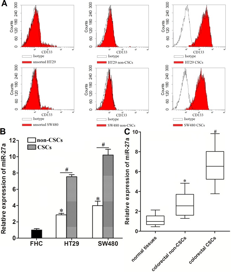
Figure 1. Overexpresion of miR-27a in colorectal cancer stem cells.
(A) Flow cytometry analysis was performed to detect the populations of CSCs and non-CSCs in HT29 and SW48 cell lines. (B) Expression of miR-27a in FHC, HT29 and SW480 CSCs and non-CSCs was measured by using qRT-PCR analysis. *P < 0.05 vs. FHC cells. #P < 0.05. (C) Expression of miR-27a in normal tissues, colorectal CSCs and non-CSCs was measured by using qRT-PCR analysis. *P < 0.05 vs. normal tissues. #P < 0.05 vs. colorectal non-CSCs.

