Supplemental Digital Content is Available in the Text.
Key Words: Descemet membrane endothelial keratoplasty, endothelial keratoplasty, chandelier illuminator, shallow anterior chamber, bullous keratopathy
Abstract
Purpose:
To describe a simple technique that uses posterior chandelier illumination during Descemet membrane endothelial keratoplasty in cases of severe bullous keratopathy (BK).
Methods:
Five eyes of 4 patients with advanced BK undergoing Descemet membrane endothelial keratoplasty were retrospectively analyzed. The pupil of the host eye was not treated with mydriatic or miotic agents. The chandelier illuminator was inserted transconjunctivally into the vitreous cavity from the pars plana.
Results:
In all eyes, BK was secondary to laser iridotomy, which was performed for prevention or treatment of angle closure glaucoma. The implanted graft was clearly confirmed in the anterior chamber using activated chandelier illumination. The graft was immediately attached to the host cornea, with eventual healing of BK in all eyes. No complication involving insertion or removal of the 25-gauge trocar and the chandelier illuminator was observed. No vision-threatening complication was observed in any of the 5 eyes.
Conclusions:
The chandelier illuminator provided good visibility of the anterior chamber and enhanced the safety of surgery by preventing formation of an inverted graft.
Descemet membrane endothelial keratoplasty (DMEK) is one of the most useful forms of corneal transplantation for the treatment of corneal endothelial dysfunction.1,2 DMEK is widely used because it allows rapid visual recovery.3,4 Although it results in an excellent visual outcome and low immunological rejection, it is initially difficult to achieve proficiency in graft preparation, orientation, insertion, and unfolding.5–10
Graft orientation is crucial for successful DMEK because an inverted graft results in failure.11 To assist in proper graft orientation, methods such as graft staining,12 marking,13 and stamping,14 as well as the use of intraoperative optical coherence tomography15,16 have been introduced. However, adequate visualization of the DMEK graft is necessary for correct graft orientation because the graft is not detectable once the color has faded, especially in eyes with severe corneal edema.
DMEK is the most suitable treatment for eyes in the early stage of Fuchs corneal endothelial dystrophy with mild corneal stromal edema. A normal anterior chamber volume and an intact lens iris diaphragm with no dense stromal opacity are important conditions for DMEK surgery.17,18 However, because of advanced corneal epithelial and stromal edema resulting from long-standing bullous keratopathy (BK), eyes with an opaque cornea are not suitable for DMEK because the thin membrane graft cannot be recognized within the anterior chamber during surgery. BK results in a pathological change in the corneal stroma after the onset of stromal edema.7,8,19,20 In such eyes, most surgeons select Descemet stripping automated endothelial keratoplasty or penetrating keratoplasty. Burkhart et al reported a slit-beam technique during DMEK and deep anterior lamellar keratoplasty with a thick cornea.21 This technique improved the visibility in the anterior chamber, making it possible to identify the orientation of the graft in corneas with a thickness exceeding 1 mm.
In this report, we describe a simple technique that uses posterior chandelier illumination during DMEK in cases of severe BK. The chandelier illuminator is inserted transconjunctivally into the vitreous cavity from the pars plana. This novel technique does not require an expensive instrument, such as intraoperative optical coherence tomography, and is easily learned by DMEK surgeons.
TECHNIQUES
Materials and Methods
Five eyes of 4 cases (1 man and 3 women; average age, 76 ± 4.3 years) with advanced BK secondary to laser iridotomy, who underwent DMEK surgery between February 2015 and December 2015 at Yokohama Minami Kyosai Hospital, were enrolled in the study. The medical records of all patients were analyzed retrospectively. All eyes underwent DMEK surgery by the same procedure and surgeon (T.H.). In addition to the usual ophthalmic examinations, measurements of the best spectacle-corrected visual acuity, corneal topography, central corneal thickness, and 6-month postoperative endothelial cell density (ECD) were performed. The study protocol was approved by the Ethical Review Board of Yokohama Minami Kyosai Hospital and adhered to the tenets of the Declaration of Helsinki.
The results were statistically analyzed using SPSS for Windows statistical software (SPSS, Chicago, IL). The Wilcoxon signed-rank test was used for statistical analyses. P ≤ 0.05 indicated statistical significance.
Surgical Procedures
All surgery was performed under retrobulbar anesthesia and a Nadbath facial nerve block. The pupil of the host eye was not treated with mydriatic or miotic agents. The Descemet membrane graft was prepared under sterile conditions shortly before the start of surgery. A donor disc was held on a vacuum punch (Moria Japan, Tokyo, Japan) and stained with 0.06% trypan blue dye. Descemet membrane was peeled gently from the stroma, and 4 small asymmetric semicircular marks indicating the graft orientation were made on the edge of the donor graft. This technique was a slight modification of that previously reported by Kruse et al.22
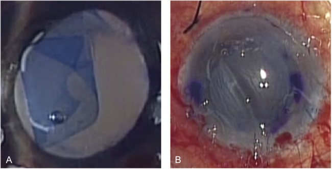
A 25-gauge trocar was inserted 3.5-mm posterior from the corneal limbus. If the vitreous pressure was high, core vitrectomy was performed. A chandelier illuminator (TotalView Chandelier for a cannula system, 25 gauge/0.5 mm; DORC, Zuidland, the Netherlands) was inserted into the vitreous cavity. After creating 2 paracenteses in the corneal limbus, a 2.8-mm-wide corneoscleral tunnel was created at 12-o'clock. Peripheral iridotomy was performed at 6-o'clock using 25-gauge microscissors. An anterior chamber maintainer was inserted through 1 corneal side port, and Descemet membrane was stripped under air using a reverse Sinskey Hook. The DMEK donor graft was inserted using an intraocular lens inserter (WJ-60M; Santen, Osaka, Japan), and all incisions were sutured using 10-0 nylon (Mani, Tochigi, Japan). Then, the chandelier illuminator was activated, and the donor graft was unfolded using a “no-touch technique” under retroillumination (Fig. 1A and see Video 1, Supplemental Digital Content 1, http://links.lww.com/ICO/A530).23 Air was injected underneath the unfolded graft, and the graft was attached to the host corneal stroma (Fig. 1B). After 15 minutes, when the graft was confirmed to be firmly attached to the host cornea, the chandelier illuminator and 25-gauge trocar were removed. At the end of surgery, 0.4 mg betamethasone (Rinderon; Shionogi, Osaka, Japan) was injected subconjunctivally, and levofloxacin eye drops (Cravit; Santen) were instilled. Postoperative medications included 1.5% levofloxacin (Cravit; Santen), betamethasone (Sanbetason; Santen), and 2% rebamipide ophthalmic solution (Mucosta; Otsuka, Tokyo, Japan), 4 times daily for 3 months and tapered thereafter.
FIGURE 1.

DMEK procedure. A, Retroillumination provided good visibility of the anterior chamber. The rolled form of the stained DMEK graft is clearly visible. B, The graft position, form, and folding were difficult to recognize without retroillumination.
RESULTS
The implanted graft was clearly observed during surgery under activated chandelier illumination from the pars plana and was immediately attached to the host cornea, with eventual healing of BK in all eyes. No complication involving insertion or removal of the 25-gauge trocar or the chandelier illuminator was observed.
The best-corrected visual acuity, expressed as the logarithm of the minimal angle of resolution, was 1.44 ± 0.43 preoperatively but improved to 0.17 ± 0.12 at 6 months after surgery (P = 0.007). Central corneal thickness was 883 ± 60 μm (range: 788–955 μm) preoperatively but decreased to 529 ± 103 μm at 6 months after surgery (P = 0.001). ECD was 2355 ± 177 cells/mm2 (range: 2214–2704 cells/mm2) preoperatively but decreased to 1296 ± 376 cells/mm2 at 6 months after surgery (P = 0.002). The endothelial cell loss rate was 45.7 ± 11.4%. No vision-threatening complication was observed in any of the 5 eyes.
DISCUSSION
Chandelier illumination is used frequently for cataract surgery in eyes with advanced BK or with corneal stromal opacity resulting from various causes. Diffuse retroillumination from the vitreous cavity helps to visualize the structures of the anterior eye, providing better visualization than a normal surgical microscope. Oshima et al reported use of chandelier illumination for cataract surgery with an opaque cornea.24 Inoue et al also reported use of chandelier illumination in the anterior chamber during Descemet stripping automated endothelial keratoplasty surgery in severe cases of corneal haze,25 and DMEK assisted by transcorneal illumination was reported by Jacob et al or Kobayashi et al.26,27
We used chandelier illumination during DMEK surgery through the pars plana approach. The thin Descemet membrane graft stained with trypan blue was clearly recognized, even in the presence of a cloudy cornea. Moreover, our technique facilitated DMEK with both hands free. Although the patient we present in the video had paralytic mydriasis before DMEK surgery, we confirmed the improved anterior chamber visibility with a 2- to 3-mm-diameter pupil.
There are some concerns regarding this method. When using chandelier illumination, the pupil should not be miotic, for better visibility of the graft. First, the DMEK procedure is believed to be easier in eyes with a miotic pupil because the anterior chamber is shallower when the pupil is miotic,28 making unfolding of the graft easier. Second, although the endothelial cells of the inserted graft may be protected by the iris stroma in miotic pupils, they may be damaged by contact with the intraocular lens in a pupil without miotic agents. However, our results show that ECD loss was comparable to that published in previous reports,9,15,16 indicating that the pupil without miotic agents did not damage corneal endothelial cells. Third, a concern was whether visualization of the anterior structures was sufficient without the use of mydriatic agents, but the results show that there was sufficient visualization during DMEK surgery with an undilated pupil. Fourth, chandelier illumination is not cheap because a chandelier illuminator is usually used only once. This technique should therefore not be used for all DMEK surgery. However, it is advantageous, especially for very advanced DMEK cases with an opaque cornea.
In conclusion, our novel technique enabled DMEK surgery that resulted in improved visual function, with a lower incidence of incorrect graft insertion, even in advanced corneal haze cases.
Footnotes
The authors have no funding or conflicts of interest to disclose.
T. Shimizu and T. Hayashi contributed equally to this work.
Supplemental digital content is available for this article. Direct URL citations appear in the printed text and are provided in the HTML and PDF versions of this article on the journal's Web site (www.corneajrnl.com).
REFERENCES
- 1.Melles GRJ. Posterior lamellar keratoplasty: DLEK to DSEK to DMEK. Cornea. 2006;25:879–881. [DOI] [PubMed] [Google Scholar]
- 2.Melles GRJ, Ong TS, Ververs B, et al. Descemet membrane endothelial keratoplasty (DMEK). Cornea. 2006;25:987–990. [DOI] [PubMed] [Google Scholar]
- 3.Rodrigues EB, Costa EF, Penha FM, et al. The use of vital dyes in ocular surgery. Surv Ophthalmol. 2009;54:576–617. [DOI] [PubMed] [Google Scholar]
- 4.Guerra FP, Anshu A, Price MO, et al. Descemet's membrane endothelial keratoplasty: prospective study of 1-year visual outcomes, graft survival, and endothelial cell loss. Ophthalmology. 2011;118:2368–2373. [DOI] [PubMed] [Google Scholar]
- 5.Ham L, Dapena I, van Luijk C, et al. Descemet membrane endothelial keratoplasty (DMEK) for Fuchs endothelial dystrophy: review of the first 50 consecutive cases. Eye Lond Engl. 2009;23:1990–1998. [DOI] [PubMed] [Google Scholar]
- 6.Monnereau C, Quilendrino R, Dapena I, et al. Multicenter study of Descemet membrane endothelial keratoplasty: first case series of 18 surgeons. JAMA Ophthalmol. 2014;132:1192–1198. [DOI] [PubMed] [Google Scholar]
- 7.Phillips PM, Phillips LJ, Muthappan V, et al. Experienced DSAEK Surgeon's transition to DMEK: outcomes comparing the last 100 DSAEK surgeries with the first 100 DMEK surgeries exclusively using previously published techniques. Cornea. 2017;36:275–279. [DOI] [PubMed] [Google Scholar]
- 8.Ang M, Mehta JS, Newman SD, et al. Descemet membrane endothelial keratoplasty: preliminary results of a donor insertion pull-through technique using a donor mat device. Am J Ophthalmol. 2016;171:27–34. [DOI] [PubMed] [Google Scholar]
- 9.Arnalich-Montiel F, Pérez-Sarriegui A, Casado A. Impact of introducing 2 simple technique modifications on the Descemet membrane endothelial keratoplasty learning curve. Eur J Ophthalmol. 2016;27:16–20. [DOI] [PubMed] [Google Scholar]
- 10.Debellemanière G, Guilbert E, Courtin R, et al. Impact of surgical learning curve in Descemet membrane endothelial keratoplasty on visual acuity gain. Cornea. 2017;36:1–6. [DOI] [PubMed] [Google Scholar]
- 11.Dirisamer M, van Dijk K, Dapena I, et al. Prevention and management of graft detachment in Descemet membrane endothelial keratoplasty. Arch Ophthalmol. 1960;2012:280–291. [DOI] [PubMed] [Google Scholar]
- 12.Majmudar PA, Johnson L. Enhancing DMEK success by identifying optimal levels of trypan blue dye application to donor corneal tissue. Cornea. 2017;36:217–221. [DOI] [PubMed] [Google Scholar]
- 13.Bachmann BO, Laaser K, Cursiefen C, et al. A method to confirm correct orientation of Descemet membrane during Descemet membrane endothelial keratoplasty. Am J Ophthalmol. 2010;149:922–925.e2. [DOI] [PubMed] [Google Scholar]
- 14.Veldman PB, Dye PK, Holiman JD, et al. Stamping an S on DMEK donor tissue to prevent upside-down grafts: laboratory validation and detailed preparation technique description. Cornea. 2015;34:1175–1178. [DOI] [PubMed] [Google Scholar]
- 15.Steven P, Le Blanc C, Velten K, et al. Optimizing Descemet membrane endothelial keratoplasty using intraoperative optical coherence tomography. JAMA Ophthalmol. 2013;131:1135–1142. [DOI] [PubMed] [Google Scholar]
- 16.Saad A, Guilbert E, Grise-Dulac A, et al. Intraoperative OCT-assisted DMEK: 14 consecutive cases. Cornea. 2015;34:802–807. [DOI] [PubMed] [Google Scholar]
- 17.Weller JM, Tourtas T, Kruse FE. Feasibility and outcome of Descemet membrane endothelial keratoplasty in complex anterior segment and vitreous disease. Cornea. 2015;34:1351–1357. [DOI] [PubMed] [Google Scholar]
- 18.Yoeruek E, Rubino G, Bayyoud T, et al. Descemet membrane endothelial keratoplasty in vitrectomized eyes: clinical results. Cornea. 2015;34:1–5. [DOI] [PubMed] [Google Scholar]
- 19.Morishige N, Yamada N, Zhang X, et al. Abnormalities of stromal structure in the bullous keratopathy cornea identified by second harmonic generation imaging microscopy. Investig Opthalmology Vis Sci. 2012;53:4998. [DOI] [PubMed] [Google Scholar]
- 20.Morishige N, Yamada N, Teranishi S, et al. Detection of subepithelial fibrosis associated with corneal stromal edema by second harmonic generation imaging microscopy. Investig Opthalmology Vis Sci. 2009;50:3145. [DOI] [PubMed] [Google Scholar]
- 21.Burkhart ZN, Feng MT, Price MO, et al. Handheld slit beam techniques to facilitate DMEK and DALK. Cornea. 2013;32:722–724. [DOI] [PubMed] [Google Scholar]
- 22.Kruse FE, Laaser K, Cursiefen C, et al. A stepwise approach to donor preparation and insertion increases safety and outcome of Descemet membrane endothelial keratoplasty. Cornea. 2011;30:580–587. [DOI] [PubMed] [Google Scholar]
- 23.Dapena I, Moutsouris K, Droutsas K, et al. Standardized “no-touch” technique for Descemet membrane endothelial keratoplasty. Arch Ophthalmol. 2011;129:88–94. [DOI] [PubMed] [Google Scholar]
- 24.Oshima Y, Shima C, Maeda N, et al. Chandelier retroillumination-assisted torsional oscillation for cataract surgery in patients with severe corneal opacity. J Cataract Refract Surg. 2007;33:2018–2022. [DOI] [PubMed] [Google Scholar]
- 25.Inoue T. Chandelier illumination for use during Descemet stripping automated endothelial keratoplasty in patients with advanced bullous keratopathy. Cornea. 2011;30:S50–S53. [DOI] [PubMed] [Google Scholar]
- 26.Jacob S, Agarwal A, Agarwal A, et al. Endoilluminator-assisted transcorneal illumination for Descemet membrane endothelial keratoplasty: enhanced intraoperative visualization of the graft in corneal decompensation secondary to pseudophakic bullous keratopathy. J Cataract Refract Surg. 2014;40:1332–1336. [DOI] [PubMed] [Google Scholar]
- 27.Kobayashi A, Yokogawa H, Yamazaki N, et al. The use of endoillumination probe-assisted Descemet membrane endothelial keratoplasty for bullous keratopathy secondary to argon laser iridotomy. Clin Ophthalmol. 2015;9:91–93. [DOI] [PMC free article] [PubMed] [Google Scholar]
- 28.Wilkie J, Drance SM, Schulzer M. The effects of miotics on anterior-chamber depth. Am J Ophthalmol. 1969;68:78–83. [DOI] [PubMed] [Google Scholar]


