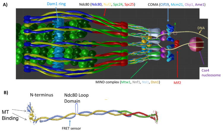
Figure 1.
Structural organization of the major kinetochore constituents at the microtubule plus-end. 3-D visualization of the metaphase budding yeast kinetochore-microtubule attachment as predicted by the protein localization data assuming a symmetric arrangement of kinetochore protein complexes around the cylindrical microtubule lattice. Ndc80c shown as blue/yellow and red/green coiled-coils in A and B with more detail. A: The MIND complex (Mtw1, Nnf1, Nsl1 and Dsn1) (gray/green ovoids and yellow/blue balls) provide the linkage from Ndc80c to COMA. COMA (Ctf19, Okp1, Mcm21, and Ame1) dk and lt blue triangles and purple spheres. Mif2 is depicted in red, the Cse4 containing nucleosome in shades of purple wrapped by a DNA double strand (yellow fiber). B: Structural view of Ndc80c, including the Ndc80 loop domain and the site of insertion of the FRET biosensor [53].

