Abstract
Background/Aims
The purpose was to determine whether lifestyle interventions have different effects on regional fat in women with normal vs. impaired glucose tolerance (NGT vs. IGT).
Methods
Changes in glucose metabolism (2-hr OGTT), android to gynoid fat mass ratio (DXA), visceral to subcutaneous abdominal fat area ratio (computed tomography), and abdominal to gluteal subcutaneous fat cell weight (FCW; adipose tissue biopsies) were determined in 60 overweight postmenopausal women (45–80 years) following 6 months of weight loss alone (WL; N=28) or with aerobic exercise (AEX+WL; N=32).
Results
The interventions led to ~8% decrease in weight, but only the AEX+WL group improved fitness (↑11% in VO2max) and reduced android to gynoid fat mass ratio (↓5%) (P’s<0.05). Both NGT and IGT groups reduced visceral and subcutaneous abdominal fat areas and abdominal and gluteal FCWs, which related to improvements in HOMA-IR (r’s=0.34–0.42) and 2-hr glucose (r’s=0.34–0.35), respectively (P’s<0.05). The decline in FCW was 2× greater in women with IGT following WL (P<0.05). The ratios of abdominal to gluteal FCW did not change following either intervention in women.
Conclusions
The mechanisms by which WL with and without exercise impact regional fat loss should be explored as reductions in abdominal fat area and subcutaneous FCW appear to influence glucose metabolism.
Introduction
Central (android) obesity is associated with an increased risk for metabolic dysfunction compared to gluteal/femoral (gynoid) obesity [1]. Metabolically unhealthy men and women with impaired glucose tolerance (IGT) tend to have a greater body mass index (BMI) and waist to hip ratio (WHR) than normal glucose tolerant (NGT) adults [2]. Obese persons with larger abdominal compared to gluteal fat cells have higher fasting insulin and glucose levels [3, 4], indicating that the accumulation of fat in the android region places obese individuals at higher metabolic risk. Fat located within the android region may be located both inside (visceral) and outside (subcutaneous) of the abdominal cavity. Insulin sensitivity by a hyperinsulinemic-euglycemic clamp is related to both subcutaneous and visceral abdominal fat [5], but there is evidence that subcutaneous abdominal fat retains significance after adjusting for visceral fat [6], suggesting that the location of fat within the android region also affects metabolic risk. Understanding the interrelationships among regional fat distribution, obesity, and risk for type 2 diabetes mellitus (T2DM) is especially relevant in obese postmenopausal women since menopause is associated with a shift of fat deposition from gynoid and toward android adiposity and this shift increases risk for T2DM [7].
Weight loss-induced reductions in abdominal fat cell size [8, 9] are associated with declines in upper body fat mass [10] and improvements in insulin sensitivity by a hyperinsulinemic-euglycemic clamp [11]. However, we showed that the addition of aerobic exercise to weight loss results in greater reductions in 2-hr insulin than weight loss alone [12]. Moreover, the addition of exercise to weight loss is associated with the preferential reduction in subcutaneous abdominal fat cell weight (FCW) compared to weight loss alone, but both weight loss with and without aerobic exercise reduce gluteal fat cell size equivalently [13]. Thus, literature indicates that the ratio of android to gynoid fat cell size increases following weight loss alone, but does not change with the addition of exercise [13]. Conversely, despite evidence that visceral abdominal fat change is inversely related to increases in VO2max, preferential loss of subcutaneous, visceral, or the ratio of subcutaneous to visceral abdominal fat is not observed when comparing the effects of weight loss with and without exercise [14], and reductions in visceral and subcutaneous abdominal fat following both interventions appear to result in glucose metabolic improvements (i.e. improvements in fasting plasma glucose and insulin, glucose tolerance or insulin sensitivity) [12, 15, 16].
The degree of glucose metabolic improvements during weight loss with and without aerobic exercise may vary depending upon baseline glucose tolerance status. Improvements in glucose metabolism are greater in adults with T2DM and IGT compared to those with NGT following either weight loss alone [17, 18] or when aerobic exercise is combined with weight loss [12, 19, 20]. However, how baseline glucose tolerance affects the changes in the distribution of fat, which may influence glucose metabolism, following these lifestyle interventions has not been compared in postmenopausal women. Therefore, this study examines the hypothesis that in overweight and obese postmenopausal women with IGT, weight loss alone, but more so with the addition of aerobic exercise, will result in greater reductions in upper than lower body fat (i.e. greater reductions in WHR, android to gynoid FM ratio, and abdominal to gluteal FCW ratio), as well as greater reductions in visceral than subcutaneous abdominal fat area, than in women with NGT. Further, we explore whether greater reductions in the fat distribution ratios are associated with greater improvements in glucose metabolism.
Materials and Methods
Study Overview
Sedentary (<20 min of aerobic exercise 2×/week), overweight and obese, postmenopausal (age 45–80 years) women were recruited from the Baltimore area. A medical history, physical examination, resting 12-lead electrocardiogram, and fasting blood profile were obtained to exclude those with unstable medical conditions. Subjects with evidence of unstable hypertension and hypertriglyceridemia, heart disease, cancer, liver, renal or hematological disease, orthopedic limitations, or medical conditions deemed to impact participation were excluded. All women signed University of Maryland Institutional Review Board approved informed consent forms.
Participants were part of a larger clinical trial [12] examining the effects of weight loss alone (WL) and weight loss with aerobic exercise (AEX+WL) on insulin sensitivity and skeletal muscle metabolism (N=96). Women without diabetes who completed dual energy x-ray absorptiometry (DXA) and computed tomography (CT) scans, oral glucose tolerance tests (OGTT), and adipose tissue biopsies pre and post intervention (N=60) were used for the current analysis. Some of the results have been previously published [12], but changes in the ratios of regional body fat and FCW are unique to this manuscript. VO2max was measured by indirect calorimetry during a graded exercise test on a treadmill as previously described [12]. Subjects met with a Registered Dietitian (RD) weekly for approximately four-six weeks to learn a heart healthy diet (i.e. <30% of diet as total fat, <10% of diet as saturated fat, <2,400 mg sodium, with more fruits, vegetables, and complex carbohydrates) prior to completing baseline testing in order to minimize the effects of diet composition on metabolism [21]. Then, all subjects met weekly for six months with the RD to learn techniques for consuming a hypocaloric (250–350 kcal/d deficit), heart healthy diet designed to promote ~1.0–1.5 kg weight loss per month. In addition, women in the AEX+WL group exercised three days per week for six months using treadmills and elliptical trainers. Training programs were gradually progressed in duration and intensity until the participant was able to exercise at >85% heart rate reserve for 45 minutes. The average adherence to exercise and weight loss classes was approximately 86%. Following the interventions, all subjects were weight stabilized (±2%) for 10 days prior to post-testing.
Body Composition
Height and body weight were measured using a stadiometer and electric scale to calculate body mass index (weight [kg]/height [m2]). A total body DXA scan (DPX-IQ; Lunar Corp., Madison, Wisconsin, USA) was performed to determine total body fat-free mass (lean tissue mass + bone mineral content), fat mass, and % body fat, as well as regional fat mass in the android and gynoid regions. Standard definitions of android and gynoid regions, as defined by the Lunar software, were used. Briefly, the android region is the area around the waist between the mid-point of the lumbar spine and the top of the pelvis and the gynoid region is between the head of the femur and mid-thigh [22]. CT scans were performed with a PQ 6000 scanner (Marconi Medical Systems, Cleveland, OH) to quantify subcutaneous and visceral abdominal fat areas using a single 5-mm scan was taken at the L4–L5 region while the subject was supine, with arms stretched overhead. CT data are expressed as cross-sectional area of tissue (cm2), where adipose tissue is considered −190 to −30 Hounsfield units (HU) [12].
Glucose Metabolism
Blood was collected after 12 hrs of fasting and at 30 minute intervals for 2 hrs after subjects ingested 75 g of glucose during an oral glucose tolerance test (OGTT) to determine glucose tolerance status [23] and total glucose and insulin area under the curve (by trapezoidal method [24]). Plasma glucose concentrations were measured using the glucose oxidase method (2300 STAT Plus; YSI, Yellow Springs, OH). Immunoreactive insulin was measured by radioimmunoassay (Linco Research Inc., St. Charles, MO). Intra- and interassay coefficients of variation of pooled control sera average 5 and 9%, respectively. Baseline values were used to estimate insulin resistance via the homeostatic model assessment (HOMA-IR). HOMA-IR was calculated as [(fasting insulin (μU/ml) × fasting glucose [mmol/l])/22.5] [25]. Post-intervention OGTTs were performed 36–48 h after the last bout of exercise. Glucose metabolic improvements were considered improvements in any of the following: fasting plasma glucose and insulin, HOMA-IR, and glucose tolerance or insulin response during the OGTT.
Fat Cell Weight
Fat aspirations from both the abdominal (ABD) and gluteal (GLT) regions were performed. Participants consumed two days of a metabolically stable diet prior to the fat aspiration. After subjects underwent a 12 hr overnight fast, subcutaneous adipose tissue was aspirated under local anesthesia (0.5% xylocaine) from both the ABD and GLT regions using a 10 mm mini-cannula and fat cells were isolated by collagenase digestion (1 mg/mL) and fat cell weights of at least 300 cells per site, with a diameter between 25 and 250 μm, were calculated (FCW=0.915/106 × π/6 d3, where d is the diameter in microns), as previously described [26, 27]. In the AEX+WL group, the post biopsies were performed within 24–36 hrs of the last exercise session.
Statistics
At baseline, between group comparisons of IGT vs. NGT were performed using independent Student’s t-tests. A χ2 test was used to determine whether the prevalence of African American and Caucasian women differed between groups. Three factorial ANOVAs (time*intervention*IGT status) with Bonferroni post hoc tests were used to determine differences in the effect of the intervention (WL versus AEX+WL) on fat distribution variables by glucose tolerance status (IGT vs. NGT). Pearson and partial correlations were used to assess relationships between key variables. Statistical significance was set at a two-tailed P<0.05. Data were analyzed using SPSS (PAWS Statistics, Version 18, Chicago, IL). Results are expressed as mean ± SEM.
Results
Baseline Comparisons of Data by Glucose Tolerance Status
Women with IGT were of comparable body weight, BMI, and % total body fat as those with NGT, but were older and had an 18% lower relative VO2max (Ps<0.01; Table 1). Race distribution did not differ by glucose tolerance status. As anticipated, HOMA-IR, 2-hr glucose and insulin, and glucose and insulin AUC were higher in women with IGT (P’s<0.05; Table 2). Women with IGT had higher waist circumference, android fat mass, and visceral fat area, which resulted in a higher waist to hip, android to gynoid fat mass, and visceral to subcutaneous abdominal fat ratios (P’s<0.05). Although abdominal FCW also was higher in women with IGT (P<0.05), the ratio of abdominal to gluteal FCW was similar in women with IGT vs. NGT.
Table 1.
Baseline subject characteristics stratified by glucose tolerance status
| Normal Glucose Tolerance (N=35) |
Impaired Glucose Tolerance (N=25) |
|
|---|---|---|
| Race (% Caucasian) | 69% | 56% |
| Age (years) | 58 ± 1 | 63 ± 1** |
| Weight (kg) | 86 ± 2 | 91 ± 3 |
| BMI (kg/m2) | 32 ± 1 | 35 ± 1 |
| Waist circumference (cm) | 94 ± 2 | 103 ± 3** |
| Hip circumference (cm) | 117 ± 2 | 122 ± 3 |
| Waist to hip ratio | 0.80 ± 0.01 | 0.85 ± 0.01** |
| Body fat (%) | 47 ± 1 | 49 ± 1 |
| Total body fat mass (kg) | 41 ± 2 | 45 ± 2 |
| Total body fat-free mass (kg) | 46 ± 1 | 47 ± 1 |
| Android fat mass (kg) | 3.4 ± 0.2 | 4.0 ± 0.2* |
| Gynoid fat mass (kg) | 7.6 ± 0.3 | 7.8 ± 0.4 |
| Android/gynoid fat mass | 0.44 ± 0.01 | 0.49 ± 0.01** |
| Visceral abdominal fat area (cm2) | 137 ± 11 | 175 ± 16 |
| Subcutaneous abdominal fat area (cm2) | 446 ± 28 | 426 ± 31 |
| Visceral/subcutaneous abdominal fat ratio | 0.31 ± 0.03 | 0.44 ± 0.05** |
| Abdominal FCW (μg triglyceride/cell) | 0.56 ± 0.02 | 0.61 ± 0.03** |
| Gluteal FCW (μg triglyceride/cell) | 0.62 ± 0.02 | 0.66 ± 0.02 |
| Abdominal/gluteal FCW | 0.91 ± 0.02 | 0.95 ± 0.03 |
| Absolute VO2max (L/min) | 1.7 ± 0.1 | 1.5 ± 0.1 |
| Relative VO2max (mL/kg/min) | 20.3 ± 0.8 | 16.7 ± 0.9** |
Significantly different from NGT:
P<0.05,
P<0.01
Table 2.
Glucose and insulin responses to an OGTT in subjects classified as having normal vs. impaired glucose tolerance
| Normal Glucose Tolerance |
Impaired Glucose Tolerance |
|
|---|---|---|
| Fasting glucose (mmol/L) | 5.3 ± 0.1 | 5.5 ± 0.1 |
| Fasting insulin (pmol/L) | 74 ± 5 | 110 ± 10** |
| HOMA-IR | 2.9 ± 0.2 | 4.5 ± 0.5** |
| 2-hr glucose (mmol/L) | 5.9 ± 0.2 | 9.0 ± 0.2** |
| 2-hr insulin (pmol/L) | 408 ± 51 | 903 ± 134** |
| Glucose AUC (mmol/L/120 min) | 824 ± 20 | 1 052 ± 24** |
| Insulin AUC (pmol/L/120 min) | 54 305 ± 3 938 | 73 648 ± 10 006* |
Significantly different from NGT:
P<0.05,
P<0.01
After controlling for baseline age and VO2max, a greater ratio of upper to lower body fat was associated with worse glucose metabolic profiles (Table 3). These relationships appear to be driven by upper body fat, as upper body fat was a stronger predictor of HOMA-IR and 2-hr glucose than the lower body equivalent (HOMA-IR: waist vs. hip circumference: r=0.58 [P<0.01] vs. r=0.38 [P<0.01]; android vs. gynoid fat mass: r=0.48 [P<0.01] vs. r=0.35 [P<0.01], and abdominal vs. gluteal FCW: r=0.38 [P<0.05] vs. r=0.24 [P=NS]; 2-hr glucose: waist vs. hip circumference: r=0.36 [P<0.01] vs. r=0.25 [P=NS]; android vs. gynoid fat mass: r=0.37 [P<0.05] vs. r=0.31 [P=NS], and abdominal vs. gluteal FCW: r=0.30 [P<0.05] vs. r=0.24 [P=NS]). Further, greater visceral to subcutaneous abdominal fat area ratio was associated with worse glucose metabolic profiles (Table 3), with visceral fat area being a stronger predictor of glucose intolerance than subcutaneous abdominal fat area (HOMA-IR: r=0.68 [P<0.01] vs. r=0.38 [P<0.01]; 2-hr glucose: r=0.32 [P<0.01] vs. r=0.04 [P=NS]).
Table 3.
Relationships of baseline body fat distribution ratios to baseline and changes in glucose tolerance
| Pearson Coefficients | Baseline waist/hip circumference | Baseline android/gynoid fat mass | Baseline abdominal/gluteal FCW | Baseline visceral/subcutaneous fat areas | |
|---|---|---|---|---|---|
| Fasting glucose (mmol/L) | Baseline | 0.04 | 0.28* | 0.10 | 0.48** |
| Change | −0.09 | −0.20 | −0.17 | −0.38* | |
| Fasting insulin (pmol/L) | Baseline | 0.49** | 0.32* | 0.30* | 0.46** |
| Change | −0.35** | −0.27* | −0.24* | −0.32* | |
| HOMA-IR | Baseline | 0.45** | 0.34* | 0.29* | 0.52** |
| Change | −0.34* | −0.31* | −0.25* | −0.45** | |
| 2-hr glucose (mmol/L) | Baseline | 0.25 | 0.28* | −0.08 | 0.40** |
| Change | −0.12 | −0.22 | −0.08 | −0.39** | |
| 2-hr insulin (pmol/L) | Baseline | 0.33* | 0.37** | 0.18 | 0.20 |
| Change | −0.13 | −0.37* | −0.20 | −0.36 | |
| Glucose AUC (mmol/L/120 min) | Baseline | 0.15 | 0.28* | 0.34** | 0.39* |
| Change | 0.01 | −0.15 | 0.07 | −0.32* | |
| Insulin AUC (pmol/L/120 min) | Baseline | 0.31* | 0.36** | 0.20 | 0.14 |
| Change | −0.07 | −0.22 | 0.09 | 0.14 | |
Controlled for age and VO2max.
P<0.05;
P<0.01.
Effects of Weight Loss with and without Exercise on Regional Fat Distribution and FCW (Table 4)
Table 4.
Effects of the WL and AEX+WL interventions stratified by glucose tolerance status
| WL | AEX+WL | Intervention *Group | Intervention | Group | |||
|---|---|---|---|---|---|---|---|
| NGT (N=15) | IGT (N=13) | NGT (N=20) | IGT (N=12) | ||||
| ΔBody weight (kg) | −6.5 ± 0.7‡ | −7.7 ± 0.9‡ | −7.2 ± 0.7‡ | −7.4 ± 1.4‡ | NS | NS | NS |
| BMI (kg/m2) | −2.4 ± 0.3‡ | −2.9 ± 0.3‡ | −2.7 ± 0.2‡ | −2.7 ± 0.5‡ | NS | NS | NS |
| ΔWaist circumference (cm) | −5.9 ± 1.6‡ | −5.9 ± 1.4‡ | −4.4 ± 1.1‡ | −4.4 ± 1.7† | NS | NS | NS |
| ΔHip circumference (cm) | −5.7 ± 0.9‡ | −4.6 ± 1.5‡ | −5.2 ± 1.3‡ | −7.0 ± 1.4‡ | NS | NS | NS |
| ΔWHR | −0.01 ± 0.01 | −0.01 ± 0.01 | 0.01 ± 0.01 | 0.00 ± 0.02 | NS | NS | NS |
| ΔBody fat (%) | −2.1 ± 0.4‡ | −3.0 ± 0.6‡ | −4.6 ± 0.7‡ | −3.1 ± 1.1‡ | NS | NS | NS |
| ΔTotal body fat mass (kg) | −4.9 ± 0.6‡ | −6.5 ± 0.7‡ | −6.4 ± 0.7‡ | −5.8 ± 1.5‡ | NS | NS | NS |
| ΔTotal body fat-free mass (kg) | −1.6 ± 0.5‡ | −2.4 ± 1.0† | −0.6 ± 0.3 | −1.0 ± 1.0 | NS | <0.05 | NS |
| ΔAndroid fat mass (kg) | −0.42 ± 0.10‡ | −0.65 ± 0.10‡ | −0.61 ± 0.07‡ | −0.55 ± 0.14‡ | NS | NS | NS |
| ΔGynoid fat mass (kg) | −0.97 ± 0.16‡ | −1.10 ± 0.13‡ | −1.29 ± 0.17‡ | −1.03 ± 0.21‡ | NS | NS | NS |
| ΔAndroid/gynoid fat mass | 0.002 ± 0.010 | −0.007 ± 0.009 | −0.020 ± 0.006‡ | −0.014 ± 0.010 | NS | <0.05 | NS |
| ΔVisceral abdominal fat area (cm2) | −20.3 ± 11.9 | −23.5 ± 8.1† | −20.3 ± 5.9‡ | −25.6 ± 15.5 | NS | NS | NS |
| ΔSubcutaneous abdominal fat area (cm2) | 16.6 ± 51.1 | −61.70 ± 29.0 | −77.2 ± 15.1‡ | −68.4 ± 18.5† | NS | NS | NS |
| ΔVisceral/subcutaneous abdominal fat area | −0.09 0.09 | 0.05 0.07 | −0.01 0.02 | −0.03 0.02 | NS | NS | NS |
| ΔAbsolute VO2max (L/min) | −0.10 ± 0.06 | −0.03 ± 0.04 | 0.21 ± 0.04‡ | 0.19 ± 0.06‡ | NS | <0.01 | NS |
Significant change from baseline:
P<0.05,
P<0.01.
Intervention represents WL vs. AEX+WL; Group represents NGT vs. IGT.
Similar to our prior report [12], weight change was comparable across groups (~8%), but only those in the AEX+WL groups improved VO2max (WL vs. AEX+WL: −3 vs. +11%; P<0.01) and maintained FFM (−4 vs. −1%; P<0.05), and these improvements were similar between NGT and IGT groups within each intervention. Although there were no time*intervention*IGT status interactions for changes in glucose metabolism, there was a significant group*time effect, which showed that women with IGT had greater reductions in fasting insulin (IGT vs. NGT: −26% vs. −16%), 2-hr glucose (−18% vs. +5%), insulin AUC (−44% vs. −16%), glucose AUC (−10% vs. −2%), and HOMA-IR (−32% vs. −21%) than women with NGT, independent of intervention (P’s<0.05; data available in supplementary table).
A decline in waist and hip circumference, android and gynoid fat mass, abdominal and gluteal FCW, and visceral and subcutaneous abdominal fat area was observed (P’s<0.05) in each group following both interventions. The changes in ABD and GLT FCW were ~2-fold greater in women with IGT who underwent WL alone compared to all other groups (P’s<0.01) (Figure 1). This remained true even after adjusting for changes in body fat. The declines in upper and lower body circumference and FCW and abdominal fat areas were similar by region, as there were no changes in waist to hip circumference, abdominal to gluteal FCW, or visceral to subcutaneous abdominal fat area ratios in either IGT or NGT groups. However, the decline in android to gynoid fat mass ratio was significant in women following AEX+WL (Figure 2A), but not WL alone (WL vs. AEX+WL: −1 vs. −5%; P<0.05; Figure 2B), regardless of IGT status.
Figure 1.
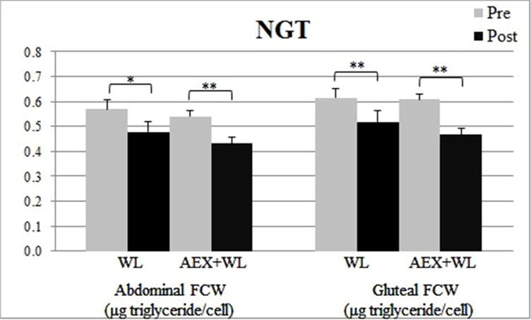
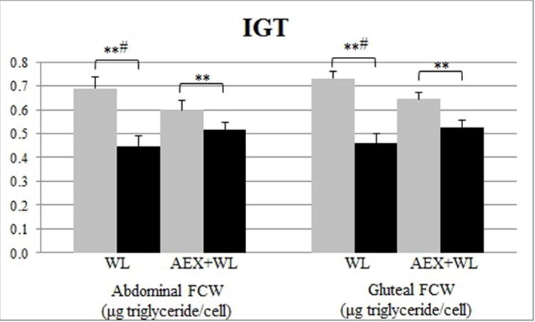
Changes in fat cell weight (FCW) with following weight loss with (AEX+WL) and without aerobic exercise (WL) in those with normal (NGT: Figure 1A) and impaired (IGT: Figure 1B) glucose tolerance. *P<0.05, **P<0.01: significant change from prebaseline. #P<0.05: the change is significantly different from all other groups (IGT following AEX+WL and NGT following WL and AEX+WL).
Figure 2.
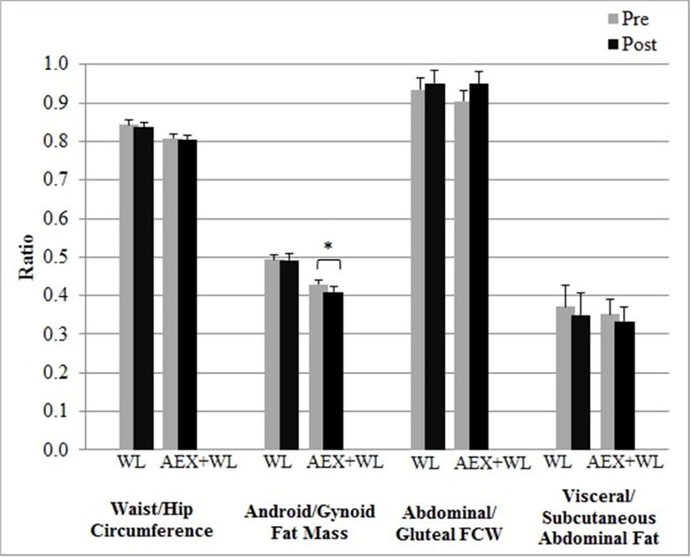
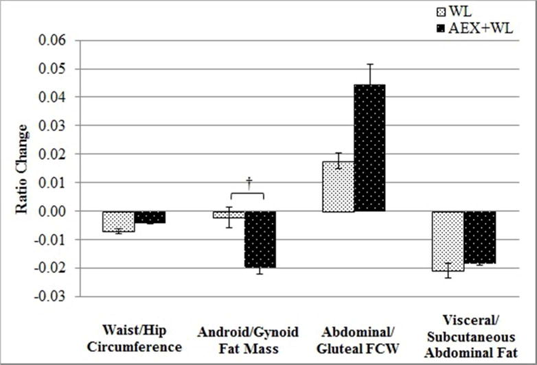
Bar graphs representing the changes in the ratios of body fat distribution (raw data: Figure 2A and change data: Figure 2B). Normal and impaired glucose tolerance groups were combined as no “glucose tolerance status” group differences were observed. *P<0.05: significantly different than pre. †P<0.05: significantly different than WL.
Relationships of Changes in Regional Fat Distribution and FCW to Glucose Metabolism after the Interventions
Glucose metabolic improvements (i.e. fasting and 2-hr glucose and insulin and HOMA-IR) negatively related to lower baseline android to gynoid fat mass and visceral to subcutaneous abdominal fat area ratios, but not WHR or ABD to GLT FCW ratio (Table 3). However, the changes in these glucose and insulin associated variables did not correlate with changes in waist or hip circumference, WHR, android or gynoid fat mass, or the ratio of android to gynoid fat mass in the total group. After adjusting for changes in body fat, reductions in 2-hr glucose and glucose AUC were associated with declines in ABD FCW (2-hr glucose: r=0.35 [Figure 3A]; glucose AUC: r=0.28) and GLT FCW (2-hr glucose: r=0.34 [Figure 3B]; glucose AUC: r=0.31) (P’s<0.05), but not the ratio of ABD to GLT FCW. Reductions in fasting glucose and HOMA-IR were associated with declines in visceral (fasting glucose: r=0.31; HOMA-IR: r=0.42) and subcutaneous (fasting glucose: r=0.36; HOMA-IR: r=0.34) abdominal fat areas (P’s<0.05), but not the ratio of visceral to subcutaneous abdominal fat area.
Figure 3.
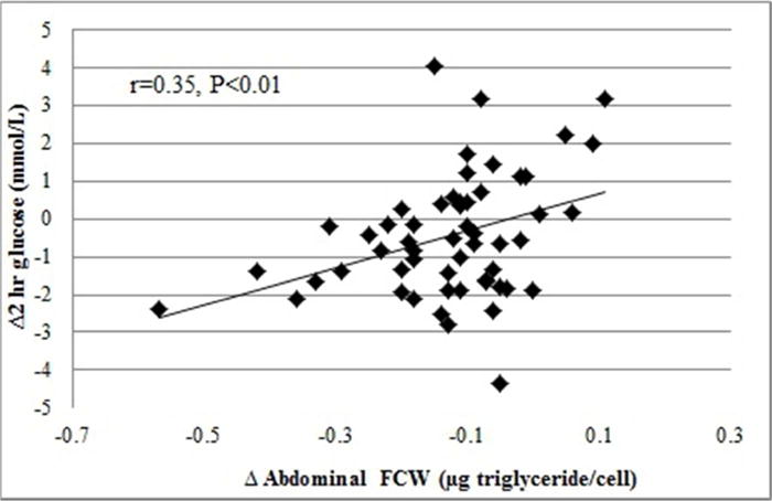
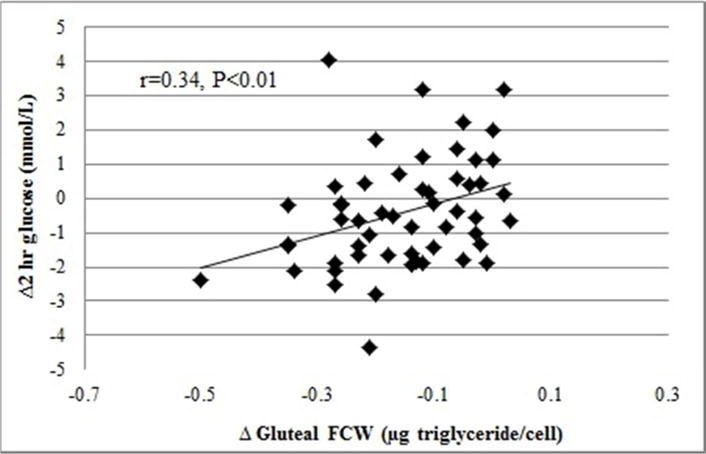
Relationship of changes in abdominal (Figure 3A) and gluteal (Figure 3B) FCW to changes in 2-hour glucose in all participants (WL and AEX+WL combined).
Discussion
Despite observing a greater decline in android to gynoid fat mass ratio in the women that underwent aerobic exercise in addition to weight loss, we did not find that a greater change in upper vs. lower body fat or visceral to subcutaneous fat area is a mediator of glucose metabolism following weight loss with and without aerobic exercise. Rather, we find that greater reductions in FCW and abdominal fat area (absolute changes, not the ratios) are associated with improvements in glucose tolerance following WL and AEX+WL. The relationship is similar for the abdominal and gluteal region and the visceral and subcutaneous abdominal region, despite evidence that ABD FCW and visceral fat area seem to be stronger predictors of glucose intolerance and insulin resistance then GLT FCW [6, 28] and subcutaneous fat area [29], respectively. We find that reductions in FCW appear to depend upon intervention and glucose tolerance status, as women with baseline glucose intolerance undergoing WL have the greatest reductions in FCW, suggesting a link between fat cell metabolism and insulin resistance. Thus, in overweight and obese, older women, interventions that lead to the greatest reductions in FCW and abdominal fat area seem to have the greatest impact on glucose metabolism. These data suggest that women who have the highest accumulation of central body fat have the ability to make the greatest glucose metabolic improvements and that it is the overall loss of fat and not necessarily that from a particular region that affects improvements in glucose metabolism.
Our results show that, in overweight and obese postmenopausal women, the gynoid region contains ~2× more fat than the android region (~7.5 vs. ~3.5 kg). The android region makes up only 7–9% of total body fat and contains <50% of its fat in the visceral region. These data, in addition to our finding that upper body fat is a stronger predictor of baseline glucose metabolic dysfunction (HOMA-IR and 2-hr glucose) than lower body fat, indicate the robust influence of central obesity on metabolic dysfunction and suggest that declines in upper body fat may have greater benefit to glucose metabolism than lower body fat. To the best of our knowledge, this is the only study examining the change in the ratio of android to gynoid fat mass by DXA following weight loss. Previous studies show that WHR does not change following weight loss in women, even when metabolic improvements (i.e. increases in VO2max and reductions in fasting lipid and glucose profiles) are observed [13, 30]. However, other studies show declines in WHR following weight loss [31–33] and weight loss combined with exercise [34, 35]. We show that women participating in AEX+WL reduce their abdominal to gluteal fat mass ratio and that this change is greater than in women undergoing WL alone; however, the change in the ratio of android to gynoid fat mass did not correlate with the improvements in glucose metabolism. Thus, it appears that although android to gynoid fat mass is reduced with the addition of aerobic exercise to weight loss, it is not the mechanism for improvements in glucose tolerance.
While many studies examine how changes in visceral and subcutaneous abdominal fat relate to metabolic improvements following WL and AEX+WL [36–38], surprisingly few examine this relationship utilizing the change in the ratio, with mixed outcomes observed [39–41]. It appears that the loss of fat from each depot may be influenced by gender [42] and the amount of weight lost [36]. A systematic review reports that visceral fat may be providing energy at times of acute negative energy balance [36]; therefore, our gradual weight loss may not have been a sufficient stimuli to require breakdown of visceral fat for energy utilization beyond that required from subcutaneous tissue. This review also did not find an overall effect of exercise with and without weight loss on the ratio of visceral to subcutaneous abdominal fat (when expressed as % change) [36], suggesting that weight loss is of greater influence than exercise.
The results of the few studies that examine the effects of WL with and without exercise on the ratio of abdominal to gluteal FCW are equivocal. In women, WL alone seems to either decrease [43] or increase [13] the ratio of abdominal to gluteal FCW, whereas our findings and those of You et al. [13] show that the addition of AEX to WL is associated with no change in the ratio. We suspect that this heterogeneity is due to differences in subject characteristics and interventions, which include menopausal status, presence of central obesity, weight loss achieved, and differences in exercise intensities. Paracrine responses to weight loss may affect regional lipid accumulation, including those regulating triglyceride accumulation (i.e. lipoprotein lipase activity) and lipolysis (i.e. hormone sensitive lipase) [44]. This may further be modulated by exercise, as it appears that endurance trained women have preferential lipid mobilization from subcutaneous abdominal compared to femoral adipose tissue stores [45]. Unfortunately, this study was limited to subcutaneous FCW assessment, but it is suggested that visceral adipocytes may be more sensitive to weight reduction because visceral adipocytes appear more metabolically active and sensitive to lipolysis than subcutaneous adipocytes [46]. A more comprehensive molecular examination of the effects of weight loss and exercise on adipocyte metabolism would help clarify these issues.
In summary, these results suggest that it is reductions in abdominal fat area and subcutaneous FCW, but not the ratios of visceral to subcutaneous fat areas or upper to lower body fat, that have the greatest influence on glucose metabolism. Future studies should focus on the mechanisms by which weight loss with and without exercise training impact fat cell lipid storage capacity to improve glucose metabolism in postmenopausal women.
Supplementary Material
Acknowledgments
Our appreciation is extended to the women who participated in this study. We are grateful to the medical team, exercise physiologists, and registered dietitians of the University of Maryland Division of Gerontology and Geriatric Medicine and Baltimore Veterans Affairs Geriatric Research, Education and Clinical Center (GRECC) for their assistance to this project. This work was funded by the Department of Veterans Affairs Career Development Award Number IK2 RX-000944 (MCS) and IK2 RX-001788–01 (OA) and Senior Research Career Scientist (ASR) from the United States (U.S) Department of Veterans Affairs Rehabilitation R&D (Rehab RD) Service, the National Institute on Aging (NIA) grants R01-AG19310 (ASR), R01-AG20116 (APG), Claude D. Pepper Older Americans Independence Center P30-AG028747, NIDDK Mid-Atlantic Nutrition Obesity Research Center (NIH P30 DK072488), GCRC of the University of Maryland, Baltimore (5M01RR016500), and Baltimore VA GRECC and Research Service.
Footnotes
Conflict of Interest
All authors have nothing to disclose.
References
- 1.Shuster A, Patlas M, Pinthus JH, Mourtzakis M. The clinical importance of visceral adiposity: a critical review of methods for visceral adipose tissue analysis. Br J Radiol. 2012;85(1009):1–10. doi: 10.1259/bjr/38447238. [DOI] [PMC free article] [PubMed] [Google Scholar]
- 2.Sekikawa A, Eguchi H, Igarashi K, Tominaga M, Abe T, Fukuyama H, Kato T. Waist to hip ratio, body mass index, and glucose intolerance from Funagata population-based diabetes survey in Japan. Tohoku J Exp Med. 1999;189(1):11–20. doi: 10.1620/tjem.189.11. [DOI] [PubMed] [Google Scholar]
- 3.Krotkiewski M, Bjorntorp P, Sjostrom L, Smith U. Impact of obesity on metabolism in men and women. Importance of regional adipose tissue distribution. J Clin Invest. 1983;72(3):1150–62. doi: 10.1172/JCI111040. [DOI] [PMC free article] [PubMed] [Google Scholar]
- 4.Weyer C, Foley JE, Bogardus C, Tataranni PA, Pratley RE. Enlarged subcutaneous abdominal adipocyte size, but not obesity itself, predicts type II diabetes independent of insulin resistance. Diabetologia. 2000;43(12):1498–506. doi: 10.1007/s001250051560. [DOI] [PubMed] [Google Scholar]
- 5.Ryan AS. Insulin resistance with aging: effects of diet and exercise. Sports Med. 2000;30(5):327–46. doi: 10.2165/00007256-200030050-00002. [DOI] [PubMed] [Google Scholar]
- 6.Goodpaster BH, Thaete FL, Simoneau JA, Kelley DE. Subcutaneous abdominal fat and thigh muscle composition predict insulin sensitivity independently of visceral fat. Diabetes. 1997;46(10):1579–85. doi: 10.2337/diacare.46.10.1579. [DOI] [PubMed] [Google Scholar]
- 7.Davis SR, Castelo-Branco C, Chedraui P, Lumsden MA, Nappi RE, Shah D, Villaseca P, Writing Group of the International Menopause Society for World Menopause D Understanding weight gain at menopause. Climacteric. 2012;15(5):419–29. doi: 10.3109/13697137.2012.707385. [DOI] [PubMed] [Google Scholar]
- 8.Gurr MI, Jung RT, Robinson MP, James WP. Adipose tissue cellularity in man: the relationship between fat cell size and number, the mass and distribution of body fat and the history of weight gain and loss. Int J Obes. 1982;6(5):419–36. [PubMed] [Google Scholar]
- 9.Lofgren P, Andersson I, Adolfsson B, Leijonhufvud BM, Hertel K, Hoffstedt J, Arner P. Long-term prospective and controlled studies demonstrate adipose tissue hypercellularity and relative leptin deficiency in the postobese state. J Clin Endocrinol Metab. 2005;90(11):6207–13. doi: 10.1210/jc.2005-0596. [DOI] [PubMed] [Google Scholar]
- 10.Singh P, Somers VK, Romero-Corral A, Sert-Kuniyoshi FH, Pusalavidyasagar S, Davison DE, Jensen MD. Effects of weight gain and weight loss on regional fat distribution. Am J Clin Nutr. 2012;96(2):229–33. doi: 10.3945/ajcn.111.033829. [DOI] [PMC free article] [PubMed] [Google Scholar]
- 11.Andersson DP, Eriksson Hogling D, Thorell A, Toft E, Qvisth V, Naslund E, Thorne A, Wiren M, Lofgren P, Hoffstedt J, Dahlman I, Mejhert N, Ryden M, Arner E, Arner P. Changes in subcutaneous fat cell volume and insulin sensitivity after weight loss. Diabetes Care. 2014;37(7):1831–6. doi: 10.2337/dc13-2395. [DOI] [PubMed] [Google Scholar]
- 12.Ryan AS, Ortmeyer HK, Sorkin JD. Exercise with calorie restriction improves insulin sensitivity and glycogen synthase activity in obese postmenopausal women with impaired glucose tolerance. Am J Physiol Endocrinol Metab. 2012;302(1):E145–52. doi: 10.1152/ajpendo.00618.2010. [DOI] [PMC free article] [PubMed] [Google Scholar]
- 13.You T, Murphy KM, Lyles MF, Demons JL, Lenchik L, Nicklas BJ. Addition of aerobic exercise to dietary weight loss preferentially reduces abdominal adipocyte size. Int J Obes (Lond) 2006;30(8):1211–6. doi: 10.1038/sj.ijo.0803245. [DOI] [PubMed] [Google Scholar]
- 14.Nicklas BJ, Wang X, You T, Lyles MF, Demons J, Easter L, Berry MJ, Lenchik L, Carr JJ. Effect of exercise intensity on abdominal fat loss during calorie restriction in overweight and obese postmenopausal women: a randomized, controlled trial. Am J Clin Nutr. 2009;89(4):1043–52. doi: 10.3945/ajcn.2008.26938. [DOI] [PMC free article] [PubMed] [Google Scholar]
- 15.Rossi AP, Fantin F, Zamboni GA, Mazzali G, Zoico E, Bambace C, Antonioli A, Pozzi Mucelli R, Zamboni M. Effect of moderate weight loss on hepatic, pancreatic and visceral lipids in obese subjects. Nutr Diabetes. 2012;2:e32. doi: 10.1038/nutd.2012.5. [DOI] [PMC free article] [PubMed] [Google Scholar]
- 16.Lesser IA, Dick TJ, Guenette JA, Hoogbruin A, Mackey DC, Singer J, Lear SA. The association between cardiorespiratory fitness and abdominal adiposity in postmenopausal, physically inactive South Asian women. Prev Med Rep. 2015;2:783–7. doi: 10.1016/j.pmedr.2015.09.007. [DOI] [PMC free article] [PubMed] [Google Scholar]
- 17.Kim MJ, Maachi M, Debard C, Loizon E, Clement K, Bruckert E, Hainque B, Capeau J, Vidal H, Bastard JP. Increased adiponectin receptor-1 expression in adipose tissue of impaired glucose-tolerant obese subjects during weight loss. Eur J Endocrinol. 2006;155(1):161–5. doi: 10.1530/eje.1.02194. [DOI] [PubMed] [Google Scholar]
- 18.Steven S, Hollingsworth KG, Small PK, Woodcock SA, Pucci A, Aribisala B, Al-Mrabeh A, Daly AK, Batterham RL, Taylor R. Weight Loss Decreases Excess Pancreatic Triacylglycerol Specifically in Type 2 Diabetes. Diabetes Care. 2016;39(1):158–65. doi: 10.2337/dc15-0750. [DOI] [PubMed] [Google Scholar]
- 19.Jenkins NT, Hagberg JM. Aerobic training effects on glucose tolerance in prediabetic and normoglycemic humans. Med Sci Sports Exerc. 2011;43(12):2231–40. doi: 10.1249/MSS.0b013e318223b5f9. [DOI] [PubMed] [Google Scholar]
- 20.Malin SK, Kirwan JP. Fasting hyperglycaemia blunts the reversal of impaired glucose tolerance after exercise training in obese older adults. Diabetes Obes Metab. 2012;14(9):835–41. doi: 10.1111/j.1463-1326.2012.01608.x. [DOI] [PMC free article] [PubMed] [Google Scholar]
- 21.Dietary guidelines for healthy American adults. A statement for physicians and health professionals by the Nutrition Committee, American Heart Association. Circulation. 1988;77(3):721A–724A. [PubMed] [Google Scholar]
- 22.National Health and Nutrition Examination Survey. Dual Energy X-Ray Absorptiometry - Android/Gynoid Measurements. 2013 03/10/2015]; Available from: http://wwwn.cdc.gov/nchs/nhanes/2005–2006/DXXAG_D.htm.
- 23.Kerner W, Bruckel J. Definition, classification and diagnosis of diabetes mellitus. Exp Clin Endocrinol Diabetes. 2014;122(7):384–6. doi: 10.1055/s-0034-1366278. [DOI] [PubMed] [Google Scholar]
- 24.Purves RD. Optimum numerical integration methods for estimation of area-under-the-curve (AUC) and area-under-the-moment-curve (AUMC) J Pharmacokinet Biopharm. 1992;20(3):211–26. doi: 10.1007/BF01062525. [DOI] [PubMed] [Google Scholar]
- 25.Matthews DR, Hosker JP, Rudenski AS, Naylor BA, Treacher DF, Turner RC. Homeostasis model assessment: insulin resistance and beta-cell function from fasting plasma glucose and insulin concentrations in man. Diabetologia. 1985;28(7):412–9. doi: 10.1007/BF00280883. [DOI] [PubMed] [Google Scholar]
- 26.Lavau M, Susini C, Knittle J, Blanchet-Hirst S, Greenwood MR. A reliable photomicrographic method to determining fat cell size and number: application to dietary obesity. Proc Soc Exp Biol Med. 1977;156(2):251–6. doi: 10.3181/00379727-156-39916. [DOI] [PubMed] [Google Scholar]
- 27.Hirsch J, Gallian E. Methods for the determination of adipose cell size in man and animals. J Lipid Res. 1968;9(1):110–9. [PubMed] [Google Scholar]
- 28.Lundgren M, Svensson M, Lindmark S, Renstrom F, Ruge T, Eriksson JW. Fat cell enlargement is an independent marker of insulin resistance and ‘hyperleptinaemia’. Diabetologia. 2007;50(3):625–33. doi: 10.1007/s00125-006-0572-1. [DOI] [PubMed] [Google Scholar]
- 29.Usui C, Asaka M, Kawano H, Aoyama T, Ishijima T, Sakamoto S, Higuchi M. Visceral fat is a strong predictor of insulin resistance regardless of cardiorespiratory fitness in non-diabetic people. J Nutr Sci Vitaminol (Tokyo) 2010;56(2):109–16. doi: 10.3177/jnsv.56.109. [DOI] [PubMed] [Google Scholar]
- 30.Wing RR, Jeffery RW, Burton LR, Thorson C, Kuller LH, Folsom AR. Change in waisthip ratio with weight loss and its association with change in cardiovascular risk factors. Am J Clin Nutr. 1992;55(6):1086–92. doi: 10.1093/ajcn/55.6.1086. [DOI] [PubMed] [Google Scholar]
- 31.Pasquali R, Antenucci D, Casimirri F, Venturoli S, Paradisi R, Fabbri R, Balestra V, Melchionda N, Barbara L. Clinical and hormonal characteristics of obese amenorrheic hyperandrogenic women before and after weight loss. J Clin Endocrinol Metab. 1989;68(1):173–9. doi: 10.1210/jcem-68-1-173. [DOI] [PubMed] [Google Scholar]
- 32.Hainer V, Stich V, Kunesova M, Parizkova J, Zak A, Wernischova V, Hrabak P. Effect of 4-wk treatment of obesity by very-low-calorie diet on anthropometric, metabolic, and hormonal indexes. Am J Clin Nutr. 1992;56(1 Suppl):281S–282S. doi: 10.1093/ajcn/56.1.281S. [DOI] [PubMed] [Google Scholar]
- 33.Park HS, Lee KU. Postmenopausal women lose less visceral adipose tissue during a weight reduction program. Menopause. 2003;10(3):222–7. doi: 10.1097/00042192-200310030-00009. [DOI] [PubMed] [Google Scholar]
- 34.Maiorana A, O’Driscoll G, Goodman C, Taylor R, Green D. Combined aerobic and resistance exercise improves glycemic control and fitness in type 2 diabetes. Diabetes Res Clin Pract. 2002;56(2):115–23. doi: 10.1016/s0168-8227(01)00368-0. [DOI] [PubMed] [Google Scholar]
- 35.Nalini M, Moradi B, Esmaeilzadeh M, Maleki M. Does the effect of supervised cardiac rehabilitation programs on body fat distribution remained long time? J Cardiovasc Thorac Res. 2013;5(4):133–8. doi: 10.5681/jcvtr.2013.029. [DOI] [PMC free article] [PubMed] [Google Scholar]
- 36.Chaston TB, Dixon JB. Factors associated with percent change in visceral versus subcutaneous abdominal fat during weight loss: findings from a systematic review. Int J Obes (Lond) 2008;32(4):619–28. doi: 10.1038/sj.ijo.0803761. [DOI] [PubMed] [Google Scholar]
- 37.Busetto L. Visceral obesity and the metabolic syndrome: effects of weight loss. Nutr Metab Cardiovasc Dis. 2001;11(3):195–204. [PubMed] [Google Scholar]
- 38.Ohkawara K, Tanaka S, Miyachi M, Ishikawa-Takata K, Tabata I. A dose-response relation between aerobic exercise and visceral fat reduction: systematic review of clinical trials. Int J Obes (Lond) 2007;31(12):1786–97. doi: 10.1038/sj.ijo.0803683. [DOI] [PubMed] [Google Scholar]
- 39.Kanai H, Tokunaga K, Fujioka S, Yamashita S, Kameda-Takemura KK, Matsuzawa Y. Decrease in intra-abdominal visceral fat may reduce blood pressure in obese hypertensive women. Hypertension. 1996;27(1):125–9. doi: 10.1161/01.hyp.27.1.125. [DOI] [PubMed] [Google Scholar]
- 40.Fujioka S, Matsuzawa Y, Tokunaga K, Kawamoto T, Kobatake T, Keno Y, Kotani K, Yoshida S, Tarui S. Improvement of glucose and lipid metabolism associated with selective reduction of intra-abdominal visceral fat in premenopausal women with visceral fat obesity. Int J Obes. 1991;15(12):853–9. [PubMed] [Google Scholar]
- 41.Park HS, Lee K. Greater beneficial effects of visceral fat reduction compared with subcutaneous fat reduction on parameters of the metabolic syndrome: a study of weight reduction programmes in subjects with visceral and subcutaneous obesity. Diabet Med. 2005;22(3):266–72. doi: 10.1111/j.1464-5491.2004.01395.x. [DOI] [PubMed] [Google Scholar]
- 42.Gallagher D, Heshka S, Kelley DE, Thornton J, Boxt L, Pi-Sunyer FX, Patricio J, Mancino J, Clark JM, Group MRIASGoLAR Changes in adipose tissue depots and metabolic markers following a 1-year diet and exercise intervention in overweight and obese patients with type 2 diabetes. Diabetes Care. 2014;37(12):3325–32. doi: 10.2337/dc14-1585. [DOI] [PMC free article] [PubMed] [Google Scholar]
- 43.Presta E, Leibel RL, Hirsch J. Regional changes in adrenergic receptor status during hypocaloric intake do not predict changes in adipocyte size or body shape. Metabolism. 1990;39(3):307–15. doi: 10.1016/0026-0495(90)90052-e. [DOI] [PubMed] [Google Scholar]
- 44.Berman DM, Nicklas BJ, Ryan AS, Rogus EM, Dennis KE, Goldberg AP. Regulation of lipolysis and lipoprotein lipase after weight loss in obese, postmenopausal women. Obes Res. 2004;12(1):32–9. doi: 10.1038/oby.2004.6. [DOI] [PubMed] [Google Scholar]
- 45.Mauriege P, Prud’Homme D, Marcotte M, Yoshioka M, Tremblay A, Despres JP. Regional differences in adipose tissue metabolism between sedentary and endurance- trained women. Am J Physiol. 1997;273(3 Pt 1):E497–506. doi: 10.1152/ajpendo.1997.273.3.E497. [DOI] [PubMed] [Google Scholar]
- 46.Bjorntorp P. Regional obesity. In: Bjorntorp P, Brodoff BN, editors. Obesity. JB Lippincott Co; Philadelphia, PA: 1992. pp. 579–586. [Google Scholar]
Associated Data
This section collects any data citations, data availability statements, or supplementary materials included in this article.


