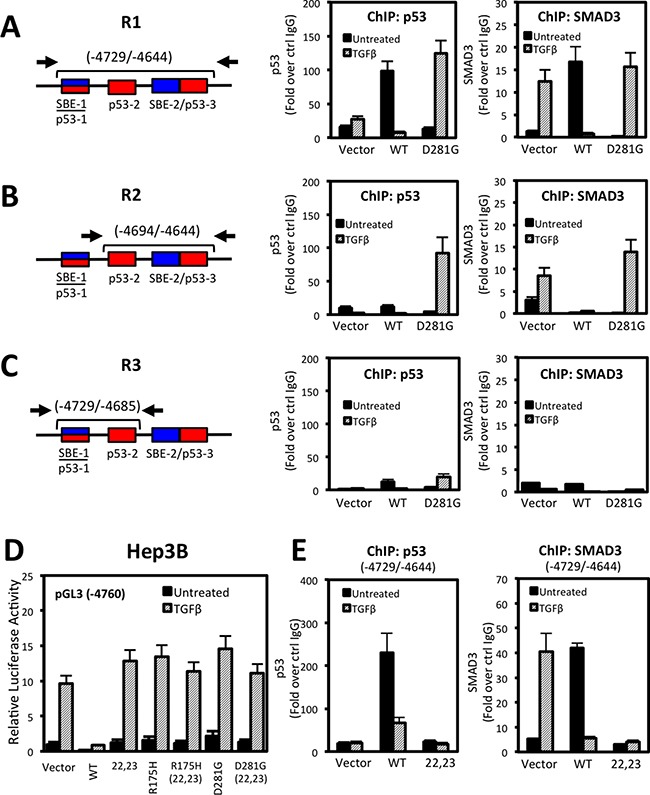
Figure 7. p53 and SMAD3 associate with NOX4 SBE and p53RE sequences in a TGFβ-dependent manner.

(A–C) Schematic showing the NOX4 promoter and the p53 and SMAD target regions tested in ChIP assays (left). See Materials and Methods for primer sequences. ChIP was performed in Hep3B cells transfected with vector control, p53-WT, or p53-D281G plasmids for 24 hours and subsequently treated with TGFβ (5 ng/ml) for an additional 24 hours. ChIP assays were performed using antibodies that specifically recognize p53, SMAD3, or IgG (negative control). Input DNA and immunoprecipitated DNA were quantified by qPCR. The ChIP-qPCR results are represented as fold enrichment of IP with anti-p53 (middle) or anti-SMAD3 (right) ChIP over input DNA relative to IgG negative control (n = 3). (D) Luciferase assays were performed in Hep3B cells using the pGL3-NOX4 promoter-reporter (-4760). The cells were transfected with control vector, p53-WT, p53-L22Q/W23S (transactivation domain mutant), mut-p53-R175H, p53-D281G, or the triple mut-p53-R175H/L22Q/W23S or p53-D281G/L22Q/W23S. Twenty-four hours after transfection, cells were left untreated or treated with TGFβ (5 ng/ml) for an additional 24 hours. (E) ChIP assay was performed as described in panel A in Hep3B cells that were expressing control, p53-WT, or p53-L22Q/W23S followed by 24 hours stimulation with TGFβ (5 ng/ml) (n = 2, in duplicate).
