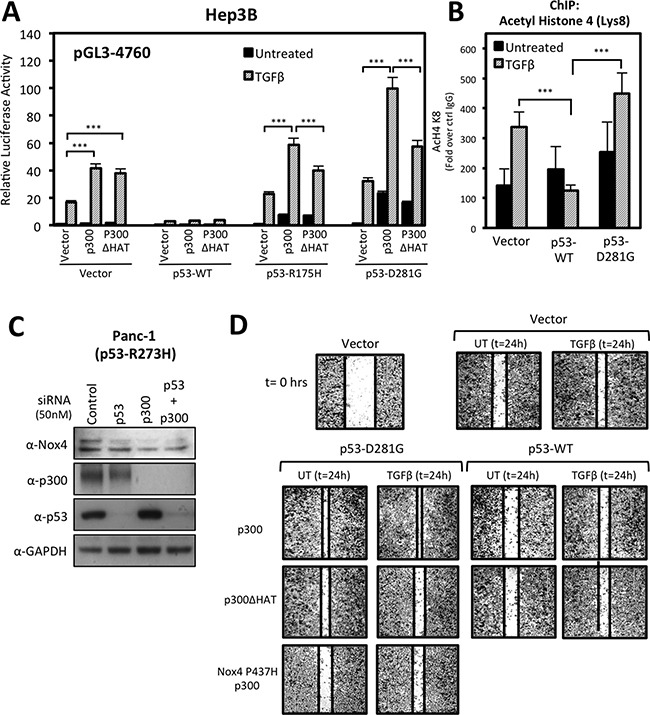
Figure 9. p300 histone acetyltransferase (HAT) activity enhances mutant p53-mediated NOX4 promoter activity and cell migration.

(A) Hep3B cells co-transfected with NOX4 promoter pGL3 (-4760) and either vector control, p53-WT, p53-R175H, or p53-D281G; and either control, p300, or HAT-inactive p300ΔHAT plasmids for 24 hours. The cells were then treated with TGFβ (5 ng/ml) for an additional 24 hours. Total cell lysates were collected following TGFβ treatment and analyzed for luciferase activity (n = 3, in triplicate). (B) ChIP assays were performed in Hep3B cells expressing control, p53-WT, or p53-D281G plasmids and either treated with TGFβ (5 ng/ml) for 24 hours or remained untreated. Histone immunoprecipitation was conducted using ChIP qualified antibodies specific for acetylated lysine-8 histone 4 (H4K8) antibodies, or IgG (negative control). ChIP-qPCR results are represented as fold enrichment of IP: α-H4K8 ChIP over input DNA relative to IgG negative control (n = 2, in triplicate). (C) PANC-1 (p53-R273H) cells were transfected with On-Target SMARTpool p53-specific siRNAs (50 nM), p300-specific siRNAs (50 nM), or non-targeting control siRNAs (50 nM) for 72 h. Fifty micrograms of total cell lysates were analyzed by immunoblotting. (D) H1299 cells transfected with vector control, p53-WT, p53-D281G, p300-WT, p300-ΔHAT, or NOX4-P437H plasmids and seeded into a 96-well tissue culture plate (3.5 × 10⁴ per well) and grown to confluence. Wounds were made using the WoundMaker 96-pin tool, which creates 96 precise and reproducible wounds of 800μm. The cells were then left untreated or were treated with TGFβ (5 ng/ml). Approximately 18-22 hours after wounding and TGFβ treatment, the cells were fixed and stained for imaging. The images presented are representative of four experiments completed in triplicate. Significance values are indicated as ***P-value < 0.001.
