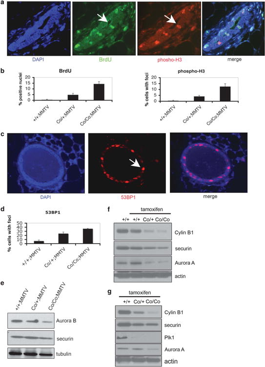
Figure 6.

Wee1 deletion in mammary gland results in mis-coordination of the cell cycle. (a) Mammary gland sections of Wee1+/+;MMTV-Cre, Wee1Co/+;MMTV-Cre and Wee1Co/Co;MMTV-Cre mice were stained with anti-phosphorylated histone-H3 Ser10 (pH3; red) and anti-BrdU antibodies (green). Representative images from Wee1 mutant mammary glands are shown and arrows indicate positive stained cells. Nuclei were counterstained with DAPI. (b) Epithelial cells from tissue sections stained as described before were counted and the percentage of either BrdU (left) or phospho-H3 (right) positive cells is shown. (c) Wee1-defficiency in mammary gland causes DNA damage. Mammary glands from WT, Wee1Co/+;MMTV-Cre and Wee1Co/Co;MMTV-Cre mice were stained with an antibody against 53BP1. Nuclei were counterstained with 4′,6-diamidino-2-phenylindole (DAPI) and arrows indicate positive stained cells from mutant mammary glands. (d) Quantification of the percentage of mammary cells displaying positive staining for 53BP1. (e) Mammary glands from Wee1+/+;MMTV-Cre, Wee1Co/+;MMTV-Cre and Wee1Co/Co;MMTV-Cre mice were immunoblotted with antibodies against Aurora-B and securin. (f) Mammary glands from Wee1+/+;TM-Cre, Wee1Co/+;TM-Cre and Wee1Co/Co;TM-Cre mice either untreated or treaded with tamoxifen were immunoblotted with antibodies against Aurora-A and securin. (g) Liver (upper) and testis (lower) from same mice as described in (f) were checked for levels of mitotic regulators by immunoblot.
