Abstact
Background
To evaluate the left ventricular (LV) longitudinal function changes in rabbits after acute occlusion of the left anterior descending artery (LAD) by two-dimensional speckle tracking imaging (2D–STI).
Methods
Forty-eight New Zealand white rabbits underwent echocardiography examination. EchoPAC was used to measure LV peak systolic longitudinal strain (LS) of the endocardium, middle myocardium, and epicardium, peak longitudinal strain rate (LSr), segmental and global longitudinal rotation (LR) degrees. Ligated the LAD and repeated all measurements after 10 min.
Results
Peak LS and LSr were significantly different between the preoperative and postoperative rabbits among most LV walls (P < 0.05). In apical four-chamber view, there was significant difference in the degrees of rotation of the LV lateral wall in preoperative and postoperative rabbits (P < 0.05). In apical three-chamber view, the rotation degrees of the posterior wall and the LR were significantly lower in the postoperative than in the preoperative (P < 0.001). In apical two-chamber view, the rotation degrees of the inferior wall and the LR were significantly lower in the postoperative (P < 0.05).
Conclusions
Left ventricular function was impaired after acute occlusion of LAD. Segmental rotational degrees and changes in LR could be useful indicators of cardiac function during the early phases of acute myocardial ischemia.
Electronic supplementary material
The online version of this article (doi:10.1186/s12872-017-0655-6) contains supplementary material, which is available to authorized users.
Keywords: Acute myocardial ischemia, Longitudinal, Rotation, Strain, Strain rate
Background
Acute myocardial ischemia (AMI) is commonly associated with heart disease and frequently manifests in the clinic as pericardial discomfort, or chest pain. Diagnosis of AMI is an important factor in the determination of patient prognosis; however, in cases of AMI without myocardial infarction, echocardiogram examination cannot reveal the abnormal segmental wall activity. Coronary angiography is the most commonly used and accurate technique for atherosclerosis diagnosis, but it is invasive and expensive.
Two-dimensional speckle tracking imaging (2D–STI) is a relatively new method of angle-independent quantification of left ventricular (LV) strain, strain rate and LV twist [1–4]. The technique’s namesake “speckles” are the result of the constructive and destructive interference patterns observed in conventional gray-scale ultrasound images. Tracking the unique speckle pattern from one frame to the next allows the investigator or clinician to track myocardial motion [5].
A normal myocardium consists of endocardium, middle myocardium, and epicardium fibers. Current 2D–STI researches on the myocardial function has primarily focus on the global systolic and/or diastolic function, the torsion and torsion velocity changes between the apex and the base of the heart [6–12]. Few studies have reported the use of 2D–STI to assess the endocardium, middle myocardium, and epicardium of the LV function and longitudinal rotation (LR) of the LV. However, some scholars had noted the different layers of the LV can make its function more clearly [13]. There was clockwise LR during the systolic period in the LV of patients with dilated cardiomyopathy or heart failure [14, 15]. More importantly, most of these reports have described the phenomenon in human patients, and few studies have attempted to investigate LR using animal models.
Therefore, the first aim of our research was the measure LV peak systolic longitudinal strain (LS) of the endocardium, middle myocardium, and epicardium, peak longitudinal strain rate (LSr). Second, we phrased a specific hypothesis which is there have global LR in the LV of rabbits. In this study, we sought to investigate whether LR occurs and evaluated early longitudinal function changes in the LV of rabbits after AMI.
Methods
This study conformed to the Guide for the Care and Use of Laboratory Animals and was approved by the Ethics Committee of Changzhou No.2 People’s Hospital Institutional Animal Care and Use Committee. The methods carried out in the experiment were in accordance with the approved guidelines. The ARRIVE guidelines (Additional file 1) were followed in this study.
Animal preparation
Forty-eight New Zealand white rabbits (males: 28 and females: 20), weighing 1.5–2.0 kg, were provided by the Experimental Animal Center of NanJing Medical University, China. All rabbits were lived in an alone cage, that the dimensions were 80 cm for length, 60 cm for width and 60 cm for height. Every day we feed them 3–4 times, and washed the cages 2 times (am: 8:00, and pm: 15:00). All of the rabbits were healthy, and all of them had not examined or treated by veterinarians before or during the research procedures. All rabbits were anesthetized with pentobarbital (1.5%, 2 ml/kg, IP), and ECG leads were connected to each subject. Thoracotomy was performed to expose the heart, and a suture was used to occlude the left anterior descending coronary artery (LAD) for 10 min, following the procedure we just used the Lidocaine to reduce the pain. We used Lidocaine just before thoracotomy and begin to closed thorax. We had taken the subcutaneous injection method. In general, every rabbit used a piece of Lidocaine (5 ml: 0.1 g). Afterwards, the thorax was closed and the rabbits were monitored by ECG to confirm ST segment changes and the establishment of the acute myocardial infarction model.
Conventional two-dimensional Doppler echocardiography
Conventional echocardiography was performed on forty-eight New Zealand white rabbits using the GE Vivid7 Dimension ultrasound machine. Preoperative and postoperative LV end-systolic volume (LVESV), end-diastolic volume (LVEDV), stroke volume (SV) and LV ejection fraction (LVEF) were recorded in the rabbits, and standard high frame rate apical four, three and two-chamber views of three consecutive cardiac cycles were stored for off-line analysis.
Data analysis for LR behavior of LV
We analyzed the apical four, three and two-chamber preoperative and postoperative views using 2D–STI software to obtain the strain, strain rate index and LR (2D–Strain, EchoPAC PC version 113, GE Healthcare, Horten, Norway). LR was defined by the global rotation of the LV cross-section. We sketched the endocardial contours, and the software automatically created regions of interest containing the endocardium, middle myocardium, and epicardium, then adjusted the regions of interest to match the endocardial-myocardial and epicardial-myocardial borders. In the apical four-chamber, three-chamber, two-chamber views, the LV walls were subdivided into six: septum, lateral, anteroseptum, posterior, anterior, inferior walls. Segmental LR and global LR of the LV were assessed from the same echocardiograms using 2D–STI (Fig. 1). All animals were euthanized once data collection was complete, the method of euthanized was injected a certain amount of air (20-50 ml) into the ear vein. All of the rabbits were anesthetized prior to euthanasia via air embolism.
Fig. 1.

Global LR between preoperative and postoperative, the left picture presented the preoperative, and the right picture presented the postoperative
Statistical analysis
All analysis was performed using SPSS 17.0 (SPSS Inc., Chicago, IL, USA). Normal data distribution was assessed using Kolmogorov-Smirnov’s test. If the data distribution was normal, the data was compared using a paired t-test. For variables with non-normal distribution, the nonparametric Mann-Whitney test was used. Data was expressed as the mean ± SD. Differences were considered statistically significant when the p-value was less than 0.05.
Results
Basic information
The values of postoperative LVEF were significantly lower than the values observed in preoperative rabbits. There were no significant differences between the preoperative and postoperative LVEDV, LVESV and SV (Table 1).
Table 1.
The basic information of conventional two-dimensional doppler echocardiography between preoperative and postoperative ()
| HR(bpm) | LVEDV(ml) | LVESV(ml) | SV(ml) | LVEF(%) | |
|---|---|---|---|---|---|
| Preoperative | 276 ± 44 | 1.61 ± 1.36 | 0.52 ± 0.19 | 1.09 ± 1.28 | 63 ± 9 |
| Postoperative | 241 ± 47 | 1.27 ± 0.43 | 0.57 ± 0.23 | 0.70 ± 0.26 | 55 ± 9 |
| P Value | <0.001 | 0.119 | 0.248 | 0.057 | <0.001 |
HR heart rate, LVEDV left ventricular end-diastolic volume, LVESV left ventricular end-systolic volume, SV stroke volume, LVEF left ventricular ejection fraction
Compared LS of the endocardial, middle and epicardial myocardial layers between preoperative and postoperative rabbits
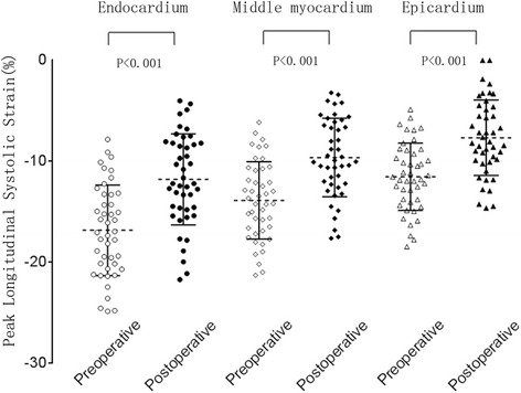
In the preoperative and postoperative rabbits, the pattern of peak systolic LS of the three different myocardial layers was: endocardium > middle myocardium > epicardium. The peak systolic global LS was significantly different between the preoperative and postoperative rabbits. The values of the postoperative were lower than preoperative. The segmental peak systolic LS of the three different myocardial layers had significant difference between the preoperative and postoperative rabbits exclude the epicardial and middle myocardial layers of the septal wall. The values of the postoperative were lower than preoperative. (Tables 2 and 3, Fig. 2)
Table 2.
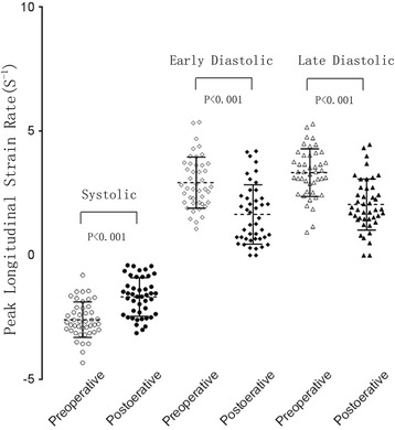
The peak global systolic longitudinal strain of endocardial, middle and epicardial layers, peak global longitudinal strain rate between preoperative and postoperative ()
| Varible | Peak global systolic longitudinal strain(%) | Peak global longitudinal strain rate (S−1) | ||||
|---|---|---|---|---|---|---|
| Endocardial | Middle | Epicardial | Systolic | Early-diastolic | Late-diastolic | |
| Preoperative | −16.84 ± 4.49 | −13.88 ± 3.84 | −11.53 ± 3.34 | −2.59 ± 0.71 | 2.93 ± 1.03 | 3.33 ± 0.96 |
| Postoperative | −11.79 ± 4.51 | −9.63 ± 3.89 | −7.66 ± 3.74 | −1.68 ± 0.77 | 1.65 ± 1.19 | 2.05 ± 1.03 |
| P-value | <0.001 | <0.001 | <0.001 | <0.001 | <0.001 | <0.001 |
Table 3.
The peak segmental systolic longitudinal strain of endocardial, middle and epicardial layers ()
| Segmental LV wall | Endocardial (%) | Middle (%) | Epicardial (%) | |||||||
|---|---|---|---|---|---|---|---|---|---|---|
| Preoperative | Postoperative | P-value | Preoperative | Postoperative | P-value | Preoperative | Postoperative | P-value | ||
| Apical 4-CH view | Lateral | −15.38 ± 7.90 | −8.55 ± 6.58 | <0.001 | −12.64 ± 6.77 | −6.70 ± 5.42 | <0.001 | −10.61 ± 5.99 | −5.32 ± 4.62 | <0.001 |
| Septum | −13.95 ± 7.32 | −10.77 ± 7.19 | 0.026 | −11.27 ± 6.15 | −9.01 ± 6.08 | 0.054 | −9.25 ± 5.33 | −7.71 ± 5.29 | 0.119 | |
| Apical 3-CH view | Anteroseptum | −15.75 ± 7.72 | −8.03 ± 7.10 | <0.001 | −12.28 ± 6.28 | −6.35 ± 5.89 | <0.001 | −9.65 ± 5.26 | −5.13 ± 5.00 | <0.001 |
| Posterior | −17.33 ± 6.88 | −10.97 ± 8.21 | <0.001 | −14.01 ± 6.05 | −8.84 ± 6.87 | <0.001 | −11.46 ± 5.44 | −7.21 ± 5.88 | 0.001 | |
| Apical 2-CH view | Anterior | −15.49 ± 7.07 | −7.51 ± 7.46 | <0.001 | −12.55 ± 6.42 | −5.85 ± 6.16 | <0.001 | −10.31 ± 5.96 | −4.64 ± 5.20 | <0.001 |
| Inferior | −17.34 ± 7.38 | −12.90 ± 8.69 | 0.009 | −13.89 ± 6.26 | −10.40 ± 7.22 | 0.013 | −11.18 ± 5.44 | −8.49 ± 6.15 | 0.020 | |
Fig. 2.

Scatter diagram reveals the peak global systolic longitudinal strain of endocardial, middle and epicardial layers between preoperative and postoperative rabbits
Compared LSr in systolic and diastolic period between preoperative and postoperative rabbits
The global peak LSr in the systolic, early and late diastolic period had significant difference between the preoperative and postoperative rabbits. The values of the postoperative were lower than preoperative. The segmental peak LSr in the systolic, early and late diastolic period had significant difference between the preoperative and postoperative rabbits exclude the systolic and early diastolic of the septal. The values of the postoperative were lower than preoperative. (Tables 2 and 4, Fig. 3)
Table 4.
The peak segmental longitudinal strain rate between preoperative and postoperative ()
| Segmental LV wall | Peak longitudinal systolic strain rate (S−1) | Peak longitudinal early-diastolic strain rate(S−1) | Peak longitudinal late-diastolic strain rate (S−1) | |||||||
|---|---|---|---|---|---|---|---|---|---|---|
| Preoperative | Postoperative | P-value | Preoperative | Postoperative | P-value | Preoperative | Postoperative | P-value | ||
| Apical 4-CH view | Lateral | −3.16 ± 1.19 | −2.16 ± 1.27 | <0.001 | 3.51 ± 2.45 | 1.82 ± 1.77 | 0.001 | 4.61 ± 2.58 | 2.65 ± 2.15 | <0.001 |
| Septum | −2.82 ± 1.27 | −2.40 ± 1.25 | 0.100 | 2.61 ± 1.59 | 2.36 ± 1.76 | 0.417 | 3.45 ± 1.87 | 2.67 ± 1.71 | 0.023 | |
| Apical 3-CH view | Anteroseptum | −3.12 ± 1.32 | −2.11 ± 1.23 | <0.001 | 2.95 ± 1.72 | 1.85 ± 1.49 | 0.004 | 3.04 ± 1.65 | 1.93 ± 1.38 | 0.005 |
| Posterior | −3.53 ± 1.58 | −2.27 ± 1.19 | <0.001 | 3.31 ± 1.94 | 2.20 ± 1.58 | 0.006 | 4.04 ± 1.69 | 2.58 ± 1.47 | <0.001 | |
| Apical 2-CH view | Anterior | −2.97 ± 1.27 | −2.06 ± 1.25 | 0.003 | 3.54 ± 2.15 | 1.50 ± 2.21 | <0.001 | 4.53 ± 2.39 | 2.26 ± 1.72 | <0.001 |
| Inferior | −3.39 ± 1.24 | −2.58 ± 1.43 | 0.001 | 3.35 ± 1.99 | 2.25 ± 1.89 | 0.005 | 4.84 ± 2.46 | 3.30 ± 2.18 | 0.001 | |
Fig. 3.

Scatter diagram reveals the peak global longitudinal strain rate between preoperative and postoperative rabbits
Compared segmental LR with global LR in the LV of preoperative and postoperative rabbits
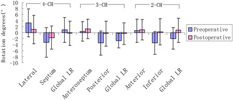
In the apical four-chamber view, there were significant differences in the degrees of rotation of the LV lateral wall in preoperative and postoperative rabbits. In the apical three-chamber view, the rotation degrees of the posterior wall was significantly lower in the postoperative rabbits, and the LR was also significantly lower in the postoperative than in the preoperative rabbits (−2.66° ± 2.34°, −0.33° ± 3.57°, p < 0.001). In the apical two-chamber view, the degrees of rotation about the inferior wall was significantly lower in the postoperative than in the preoperative rabbits, and the LR was also significantly lower in the postoperative rabbits (−1.96° ±3.59°, 0.79° ± 3.98°, p = 0.004). (Table 5, Fig. 4)
Table 5.
The segmental and global longitudinal rotational between preoperative and postoperative (°, )
| Segmental LV wall rotational degrees(°) | Preoperative | Postoperative | P Value | |
|---|---|---|---|---|
| Apical 4-CH view | Lateral | 3.23 ± 4.65 | 1.07 ± 4.74 | 0.043 |
| Septum | −3.27 ± 4.91 | −1.64 ± 3.89 | 0.095 | |
| Global LR | 0.94 ± 4.14 | −0.21 ± 3.99 | 0.136 | |
| Apical 3-CH view | Anteroseptuml | 0.39 ± 3.24 | 1.28 ± 3.21 | 0.199 |
| Posterior | −3.45 ± 4.05 | −0.29 ± 4.16 | <0.001 | |
| Global LR | −2.66 ± 2.34 | −0.33 ± 3.57 | <0.001 | |
| Apical 2-CH view | Anterior | 0.68 ± 3.86 | 0.93 ± 3.37 | 0.725 |
| Inferior | −3.36 ± 3.84 | 0.29 ± 4.41 | <0.001 | |
| Global LR | −1.96 ± 3.59 | 0.79 ± 3.98 | 0.004 | |
Fig. 4.

Segmental wall rotation and the global LR between preoperative and postoperative rabbits, blue means the preoperative, while the magenta means the postoperative
Discussion
In the research, we had ligated the left anterior descending coronary artery to make an acute myocardial infarction model. The main findings of the study were: (1) the peak systolic LS of different myocardial layers, peak LSr in the postoperative rabbits were lower than in the preoperative. (2) There was LR in the cardiac of the postoperative rabbits.
AMI is a common heart disease. In recent years, the incidence of AMI and its associated mortality have increased annually. Cell death occurs after approximately 20 min of severe ischemia, and progresses in a wave front from the subendocardium into the subepicardium of the ischemic bed-at-risk. In ischemia, the lack of oxygen is caused by a reduction in coronary flow and towards cell death and necrosis and or infarction [16]. In order to reduce the mortality rate, we performed a 10 min LAD ligation in rabbits. The acute occlusion of the coronary can damage the systolic and diastolic function of the cardiac. From this research, we knew that, the function of the LV damaged after AMI. After knowing this knowledge, fighting for the time to re-open the occlusion artery is becoming more important.
However, early diagnosis of AMI is difficult. While coronary angiography can accurately evaluate the degree of stenosis of coronary artery, the technique is prohibitively invasive and expensive. 2D–STI is a new technique that is useful for the effective diagnosis of myocardial ischemia, myocardial infarction. In acute myocardial infarction, 2D–STI can easily and accurately determine the infarcted area [17–21]. However, few studies have been performed concerning 2D–STI’s ability to diagnose myocardial ischemia in animal models.
Qian FU et al. [22] evaluated changes in the regional LV myocardial function of rats following acute occlusion of the left anterior descending coronary artery using 2D–STI. They concluded that 2D–STI was a non-invasive technique that can be used to assess the response of regional myocardial function to variations in blood supply in rats following acute occlusion of the LAD. They also concluded 2D–STI could be used as a sensitive and reliable means to follow the process of LV remodeling.
An advantage of rabbits as experimental subjects for myocardial ischemia is that rabbits do not require artificial ventilation during the LAD ligation experiments. According to previous studies, rabbits have minimal coronary collateral circulation. Blood flow to the coronary artery of the rabbit is mainly via the left anterior descending coronary artery, LV branch and the right coronary artery. The LAD perfuses the LV and a small portion of right ventricle wall, and the LV branch supplies a wider portion of the LV, but the right coronary artery only perfuses the right ventricular free wall [23]. LAD ligation in the rabbit is, therefore, a good model of AMI.
In our studies, we experimented with several methods of producing an accurate model of AMI in the rabbit. We observed that ligate the root of the LV branch might lead to sudden death. Ligating the distal end of the LV branch did not produce a good model of AMI, and caused papillary muscle and apical myocardial infarction instead. Therefore, we choose to model of AMI by ligating LAD. This study presented an innovative evaluation of the changes in LV function by detecting the three different myocardial layers and LR in the early stages of AMI in rabbits using the 2D–STI technique. We examined LV function and LR in preoperative rabbits as a self-control to minimize the variability between the groups.
The study reported in this article investigated a rabbit model of AMI induced following LAD ligation. We used this model to calculate and compare the LV function. The values of postoperative LVEF were significantly lower than the values observed in preoperative rabbits, and there were no significant differences in the LVEDV, LVESV and SV between the preoperative and postoperative rabbits. These values indicated that the ligation of LAD in rabbits had a significant influence on LV systolic function.
The pattern of peak systolic LS of the three different myocardial layers was: endocardium > middle myocardium > epicardium. At rest, most left ventricular wall thickening occurs as a result of endocardial thickening; the middle myocardium layer contributes only modestly to thickening; and the contribution of the epicardium is negligible [24]. According to the different orientation of myocardial fibres, the wall stress was in a non-uniform distribution, with decreasing values from endo- to epicardium. The subendocardial longitudinal fibres have a smaller curvature as the mid-wall circumferential fibres, also to the subepicardial fibres; thus, the wall stress will be higher on the longitudinal fibres [20]. The Peak systolic LS and LSr in the infarcted, peri-infarcted and remote myocardial regions were significantly decreased in the postoperative. From this results, we concluded that the longitudinal LV function of postoperative rabbits were impaired in the early phase of AMI.
This study used the LR of the LV to reflect changes in systolic function following AMI in rabbits. Previous studies have reported LR in the LV of dilated cardiomyopathy patients and other heart failure patients; however, before our study it was unknown whether LR was associated with AMI in rabbits. We observed that the degrees of rotation of the LV lateral, posterior and inferior walls were significantly lower in the postoperative rabbits (p < 0.05), and the LR in the apical three and two-champer views were also significantly lower (−2.66° ± 2.34°, −0.33° ± 3.57°, p < 0.001, −1.96° ±3.59°, 0.79° ± 3.98°, p = 0.004). We concluded that the LR changes between the preoperative and postoperative rabbits were related to the anatomy of rabbit coronary.
The LAD of the left coronary artery perfuses the interventricular septum, and when it is occluded, the LR of interventricular septum had no significant differernce between preoperative and postoperative rabbits, but the value is decreased. For the reason, we considered it was related to the collateral circulation, when the LAD was occluded, the collateral circulation maybe rebuilded. Why, then, were the LR of the inferior and posterior walls reduced? Due to the “muscle band”, the inferior and posterior walls are downstream of the interventricular septum, and, therefore, they may be influenced by the upstream muscle. Our results demonstrated that LR may be related to the condition of the surrounding muscle. The origin of the LR is unclear. Gallagher KP et al. [25] performed a research in conscious dogs, and the found that inner and outer wall thickening during systole is not uniform and the nonuniformity is sustained during the greatly altered hemodynamic conditions associated with exercise. After the acute occlusion of the coronary of LAD, according to the distribution of the myocardial fibre, the subendocardium was affected the most. When the cardiac contracted, due to AMI, the original balance of the different myocardium layers was disappeared or changed, may resulting in the changes of LR motion. Also the global LR reversed in the apical 4-ch and 2-ch views maybe explained by the above reason. By examining the LR, it can be determined that early AMI in the LV influenced the function of the heart.
Conclusions
In this study, LV function was impaired in the early phase after acute occlusion of LAD coronary artery. The LS of different myocardial layers could reflect the LV function very conveniently. The LR of LV could be an important indicator of cardiac function in the early phases of AMI. In the clinical practice, the early detection of the cardiac dysfunction after AMI can make these patients have early treatment, and can also evaluate the efficacy of different treatment.
Acknowledgements
The authors would like to thank the department of Echocardiography, ChangZhou No. 2 People’s Hospital Affiliated to NanJing Medical University.
Funding
none
Availability of data and materials
The datasets used and/or analysed during the current study available from the corresponding author on reasonable request.
Abbreviations
- 2D–STI
2-dimensional speckle tracking imaging
- AMI
Acute myocardial ischemia
- LAD
Left anterior descending artery
- LAD
Left anterior descending coronary artery
- LR
Longitudinal rotation
- LS
Longitudinal strain
- LSr
Longitudinal strain rate
- LV
Left ventricular or left ventricle
Additional file
The ARRIVE Guidelines. (PDF 258 kb)
Authors’ contributions
JH, LF and ZNY carried out the study design, data collection and analysis. YFR and XTS designed part of the experiments. JH wrote the manuscript. ZNY revised the manuscript. YFR and XTS performed the statistical analysis. All authors participated to the experiment. All authors have read and approved the manuscript.
Ethics approval and consent to participate
This study conformed to the Guide for the Care and Use of Laboratory Animals and was approved by the Ethics Committee of Changzhou No.2 People’s Hospital Institutional Animal Care and Use Committee. The methods carried out in the experiment were in accordance with the approved guidelines.
Consent for publication
Not applicable.
Competing interests
The authors declare that they have no competing interests.
Publisher’s Note
Springer Nature remains neutral with regard to jurisdictional claims in published maps and institutional affiliations.
Footnotes
Electronic supplementary material
The online version of this article (doi:10.1186/s12872-017-0655-6) contains supplementary material, which is available to authorized users.
Contributor Information
Jun Huang, Email: 305669112@qq.com.
Zi-Ning Yan, Email: docyanzining@163.com.
Li Fan, Email: 1900883927@qq.com.
Yi-Fei Rui, Email: 2318472619@qq.com.
Xiang-Ting Song, Email: docsongxiangting@hotmail.com.
References
- 1.Mizuguchi Y, Oishi Y, Miyoshi H, Iuchi A, Nagase N, Oki T. Concentric left ventricular hypertrophy brings deterioration of systolic longitudinal, circumferential, and radial myocardial deformation in hypertensive patients with preserved left ventricular pump function. J Cardiol. 2010;55:23–33. doi: 10.1016/j.jjcc.2009.07.006. [DOI] [PubMed] [Google Scholar]
- 2.Mizuguchi Y, Oishi Y, Miyoshi H, Iuchi A, Nagase N, Ara N, Oki T. Possible mechanisms of left ventricular torsion evaluated by cardioreparative effects of telmisartan in patients with hypertension. Eur J Echocardiogr. 2010;11:690–697. doi: 10.1093/ejechocard/jeq044. [DOI] [PubMed] [Google Scholar]
- 3.Galderisi M, Lomoriello VS, Santoro A, Esposito R, Olibet M, Raia R, et al. Differences of myocardial systolic deformation and correlates of diastolic function in competitive rowers and young hypertensives: a speckle-tracking echocardiography study. J Am Soc Echocardiogr. 2010;23:1190–1198. doi: 10.1016/j.echo.2010.07.010. [DOI] [PubMed] [Google Scholar]
- 4.van Dalen BM, Soliman OI, Vletter WB, ten Cate FJ, Geleijnse ML. Age-related changes in the biomechanics of left ventricular twist measured by speckle tracking echocardiography. Am J Physiol Heart Circ Physiol. 2008;295:H1705–H1711. doi: 10.1152/ajpheart.00513.2008. [DOI] [PubMed] [Google Scholar]
- 5.Notomi Y, Lysyansky P, Setser RM, Shiota T, Popović ZB, Martin-Miklovic MG, et al. Measurement of ventricular torsion by two-dimensional ultrasound speckle tracking imaging. J Am Coll Cardiol. 2005;45:2034–2041. doi: 10.1016/j.jacc.2005.02.082. [DOI] [PubMed] [Google Scholar]
- 6.Takeuchi M, Nishikage T, Nakai H, Kokumai M, Otani S, Lang RM. The assessment of left ventricular twist in anterior wall myocardial infarction using two-dimensional speckle tracking imaging. J Am Soc Echocardiogr. 2007;20:36–44. doi: 10.1016/j.echo.2006.06.019. [DOI] [PubMed] [Google Scholar]
- 7.Chetboul V, Serres F, Gouni V, Tissier R, Pouchelon JL. Noninvasive assessment of systolic left ventricular torsion by 2-dimensional speckle tracking imaging in the awake dog: repeatability, reproducibility, and comparison with tissue Doppler imaging variables. J Vet Intern Med. 2008;22:342–350. doi: 10.1111/j.1939-1676.2007.0037.x. [DOI] [PubMed] [Google Scholar]
- 8.Helle-Valle T, Crosby J, Edvardsen T, Lyseggen E, Amundsen BH, Smith HJ, et al. New noninvasive method for assessment of left ventricular rotation: speckle tracking echocardiography. Circulation. 2005;12:3149–3156. doi: 10.1161/CIRCULATIONAHA.104.531558. [DOI] [PubMed] [Google Scholar]
- 9.Saito M, Okayama H, Nishimura K, Ogimoto A, Ohtsuka T, Inoue K, et al. Determinants of left ventricular untwisting behaviour in patients with dilated cardiomyopathy: analysis by two-dimensional speckle tracking. Heart. 2009;95:290–296. doi: 10.1136/hrt.2008.145979. [DOI] [PubMed] [Google Scholar]
- 10.Bertini M, Sengupta PP, Nucifora G, Delgado V, Ng AC, Marsan NA, et al. Role of left ventricular twist mechanics in the assessment of cardiac dyssynchrony in heart failure. JACC Cardiovasc Imaging. 2009;2:1425–1435. doi: 10.1016/j.jcmg.2009.09.013. [DOI] [PubMed] [Google Scholar]
- 11.van Dalen BM, Soliman OI, Vletter WB, Kauer F, van der Zwaan HB, ten Cate FJ, et al. Feasibility and reproducibility of left ventricular rotation parameters measured by speckle tracking echocardiography. Eur J Echocardiogr. 2009;10:669–676. doi: 10.1093/ejechocard/jep036. [DOI] [PubMed] [Google Scholar]
- 12.van Dalen BM, Kauer F, Soliman OI, Vletter WB, Michels M, ten Cate FJ, et al. Influence of the pattern of hypertrophy on left ventricular twist in hypertrophic cardiomyopathy. Heart. 2009;95:657–661. doi: 10.1136/hrt.2008.152777. [DOI] [PubMed] [Google Scholar]
- 13.Popović ZB, Grimm RA, Ahmad A, Agler D, Favia M, Dan G, et al. Longitudinal rotation: an unrecognised motion pattern in patients with dilated cardiomyopathy. Heart. 2007;94:1–6. doi: 10.1136/hrt.2007.122192. [DOI] [PubMed] [Google Scholar]
- 14.Huang J, Ni XD, Hu YP, Song ZW, Yang WY, Xu R. Left ventricular longitudinal rotation changes in patients with dilated cardiography detected by two-dimensional speckle tracking imaging. Chin J Cardiol. 2011;39:920–924. [PubMed] [Google Scholar]
- 15.Sjøli B, Ørn S, Grenne B, Vartdal T, Smiseth OA, Edvardsen T, et al. Comparison of left ventricular ejection fraction and left ventricular global strain as determinants of infarct size in patients with acute myocardial infarction. J Am Soc Echocardiogr. 2009;22:1232–1238. doi: 10.1016/j.echo.2009.07.027. [DOI] [PubMed] [Google Scholar]
- 16.Ferrari R, Balla C, Malagù M, Guardigli G, Morciano G, Bertini M, et al. Reperfusion damage - a story of success, failure, and hope. Circ J. 2017;25(81):131–141. doi: 10.1253/circj.CJ-16-1124. [DOI] [PubMed] [Google Scholar]
- 17.Eek C, Grenne B, Brunvand H, Aakhus S, Endresen K, Smiseth OA, et al. Strain echocardiography predicts acute coronary occlusion in patients with non-ST-segment elevation acute coronary syndrome. Eur J Echocardiogr. 2010;11:501–508. doi: 10.1093/ejechocard/jeq008. [DOI] [PubMed] [Google Scholar]
- 18.Jia DL, Ma CY, Liu S, Sun LF. Evaluation of left ventricular twist in acute myocardial infarction patients using speckle tracking imaging. Cell Biochem Biophys. 2011;61:673–678. doi: 10.1007/s12013-011-9223-3. [DOI] [PubMed] [Google Scholar]
- 19.Biaggi P, Carasso S, Garceau P, Greutmann M, Gruner C, Tsang W, et al. Comparison of two different speckle tracking software systems: does the method matter? Echocardiography. 2011;28:539–547. doi: 10.1111/j.1540-8175.2011.01386.x. [DOI] [PubMed] [Google Scholar]
- 20.Dominguez-Rodriguez A, Abreu-Gonzalez P, Arroyo-Ucar E, Avanzas P, Reiter RJ. Global left ventricular longitudinal strain is associated with decreased melatonin levels in patients with acute myocardial infarction: a two-dimensional speckle tracking study. Biomarkers. 2013;18:310–313. doi: 10.3109/1354750X.2013.778330. [DOI] [PubMed] [Google Scholar]
- 21.Pellikka PA, Douglas PS, Miller JG, Abraham TP, Baumann R, Buxton DB, et al. American Society of Echocardiography cardiovascular technology and research summit: a roadmap for 2020. J Am Soc Echocardiogr. 2013;26:325–338. doi: 10.1016/j.echo.2013.02.003. [DOI] [PubMed] [Google Scholar]
- 22.Fu Q, Xie M, Wang J, Wang X, Lv Q, Lu X. Assessment of regional left ventricular myocardial function in rats after acute occlusion of left anterior descending artery by two-dimensional speckle tracking imaging. J Huazhong Univ Sci Technol. 2009;29:786–790. doi: 10.1007/s11596-009-0623-1. [DOI] [PubMed] [Google Scholar]
- 23.Podesser B, Wollenek G, Seitelberger R, Siegel H, Wolner E, Firbas W, et al. Epicardial branches of the coronary arteries and their distribution in the rabbit heart: the rabbit heart as a model of regional ischemia. Anat Rec. 1997;247:521–527. doi: 10.1002/(SICI)1097-0185(199704)247:4<521::AID-AR11>3.0.CO;2-R. [DOI] [PubMed] [Google Scholar]
- 24.Kaul S. There may be more to myocardial viability than meets the eye. Circulation. 1995;15(92):2790–2793. doi: 10.1161/01.CIR.92.10.2790. [DOI] [PubMed] [Google Scholar]
- 25.Gallagher KP, Osakada G, Matsuzaki M, Miller M, Kemper WS, Ross J., Jr Nonuniformity of inner and outer systolic wall thickening in conscious dogs. Am J Phys. 1985;249:H241–H248. doi: 10.1152/ajpheart.1985.249.2.H241. [DOI] [PubMed] [Google Scholar]
Associated Data
This section collects any data citations, data availability statements, or supplementary materials included in this article.
Data Availability Statement
The datasets used and/or analysed during the current study available from the corresponding author on reasonable request.


