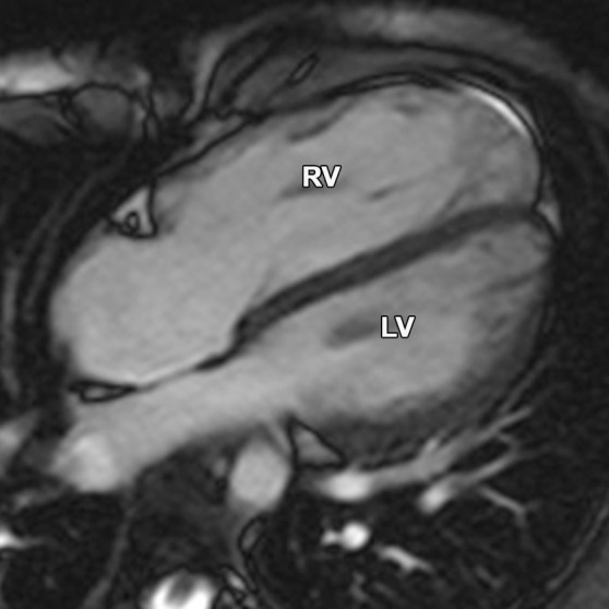
Figure 17a.

Partial anomalous pulmonary venous return compared with ARVC. (a, b) Horizontal long-axis bright-blood SSFP (a) and axial bright-blood (b) MR images of a 16-year-old male adolescent with a history of partial anomalous pulmonary venous return show RV dilatation, with an increase in the transverse chamber diameter. Connection of the pulmonary veins to the superior vena cava (arrow in b), instead of the left atrium, is depicted. (c) Horizontal long-axis MR image of a 36-year-old woman with a history of ARVC shows that the RV size is similar in patients with ARVC and those with partial anomalous pulmonary venous return.
