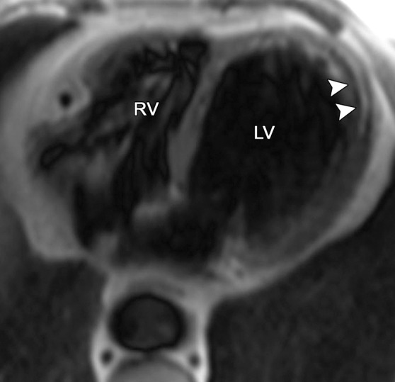
Figure 3b.

Fat deposition from a healed myocardial infarction in a 65-year-old man with a history of myocardial infarction. Axial bright-blood SSFP (a) and T1-weighted fast spin-echo non–fat-suppressed (b) MR images show increased signal intensity in the subendocardial region of the LV apex (arrowheads) secondary to fat deposition in the setting of a chronic myocardial infarction. Etching artifact (arrows in a), a cardiac MR imaging finding that results from the loss of signal in voxels that contain both fat and water protons, surrounds the apical fat on bright-blood MR images.
