

Abstract
Rapid advances in biochemistry and genetics lead to expansion of the various medical instruments for detection and prevention tasks. On the other hand, food safety is an important concern which relates to the public health. One of the most reliable tools to detect bioparticles (i.e., DNA molecules and proteins) and determining the authenticity of food products is the optical ring resonators. By depositing a recipient polymeric layer of target particle on the periphery of an optical ring resonator, it is possible to identify the existence of molecules by calculating the shift in the spectral response of the ring resonators. The main purpose of this paper is to investigate the performance of two structures of optical ring resonators, (i) all-pass and (ii) add-drop resonators for sensing applications. We propose a new configuration for sensing applications by introducing a nanogap in the all-pass ring resonator. The performance of these resonators is studied from sensing point of view. Simulation results, using finite difference time domain paradigm, revealed that the existence of a nanogap in the ring configuration achieves higher amount of sensitivity; thus, this structure is more suitable for biosensing applications.
Keywords: Active layer, DNA molecule, optical resonators, sensitivity
Introduction
Biological sensors, as a bioanalytic tool, play an important role in food sciences and biotechnology. Many investigations are conducted to increase the accuracy, sensitivity, and functionality of these sensors; at the same time, there is a tendency to decrease the size and price of these devices.
In the past few years, optical ring resonators have received a lot of attention as one of the most promising biological sensors. The optical ring resonator measures the target molecules through assessing the deviations in light behavior, which is caused by interaction between electromagnetic wave and biological molecules such as proteins, bacteria, cells, or DNA samples. This change in the behavior of the light is because of interaction between evanescent field of the resonating light inside the resonator and bioparticle that exist in the ambient. Existence of bioparticles in the medium changes the effective refractive index of surrounded medium, which results in deviation of resonance conditions of the resonator. Consequence of such an interaction is resonance wavelength deviation of the resonator that is related to the number for bioparticles in the medium.[1] A sensing mechanism is depicted schematically in Figure 1. To enable the periphery of the resonator to absorb bioparticles, an active polymer layer can be deposited on the boundary of the resonator. This layer is able to mechanically absorb or chemically react with the target bioparticle, resulting in a change in the effective refractive index of the resonating mode.
Figure 1.

Optical ring resonator as a biological sensing platform; (a) ring resonator is placed in an aqueous buffer solution. (a) There are no bioparticles in the buffer solution. (b) There are bioparticles in the medium. The bioparticles captured by the active polymeric layer interact with evanescent field of the light which results in a shift in resonance wavelength of the resonator
In general, ring resonators can be designed and fabricated in two models. First model is optical fiber configuration in which a ring resonator can be realized by connecting both the ends of an optical fiber.[2] Another optical fiber can be used to couple light into the ring.[3] The second model is integrated optical structures (which is created by integrated optics technology). Optical resonators built with this method are repeatable and more reliable. Integrated ring resonators enable parallel detection of multiple bioparticles simultaneously by fabrication of identical ring resonators on the same chip and activation of each of them with a different active layer.[4,5] The latter is the subject of interest in this paper, in which a waveguide is fabricated near the ring resonator and is utilized to inject light to the ring. This happens because of evanescent nature of the propagating light through the waveguide. High-contrast refractive index between resonator and surrounding medium makes it possible to have a higher sensitive as well as more compact resonator. Silicon with high refractive index 3.47 and advanced nanofabrication technology (microelectronics technology) are one of the best choices to develop high sensitivity integrated optical resonators.[6,7,8] In the following, different types of optical resonators are briefly studied.
Microspheres: a sphere usually made of SiO2[9,10] or polymers[11] acts as a cavity to store the energy of light at certain wavelengths. The main advantage of microsphere resonators is very high-quality factor,[9,12] which makes this device an excellent choice for biosensing applications. On the other hand, fabrication of these devices is not repeatable due to lack of a standard fabrication method. In addition, to couple light into the cavity tapered optical fibers is used,[13,14] which makes integration of this kind of microresonators more difficult. As a result, these devices are not suitable to develop commercialized photonic-based biosensors.
Microtoroid resonators: quality factor of microtoroid resonators is lower than microspheres and higher than microdisk/ring resonators. However, recently reliable and repeatable fabrication method for realization of microtoroidal resonators has been reported, but still coupling to the cavity is a challenging part that makes difficult integration and mass production of microtoroids.[15,16,17,18]
Silicon Microdisk/ring resonators: as silicon technology is well developed, thanks to the microelectronics, silicon microdisk/ring resonators are one of the most promising devices to develop integrated photonic chips for biosensing applications. However, these types of resonators are of lower quality than microspheres and microtoroidals; as very high quality factors of micro-disk/ring resonators has been reported recently.[19] In this paper, we design and simulate three different configurations of silicon microresonators. The sensitivity of proposed resonators is compared to each other, and the most sensitive sensor for biological applications is introduced.
In this paper, two structures of optical ring resonators, (i) all-pass and (ii) add-drop resonators are evaluated for sensing applications. In addition, performance of the all-pass ring resonators is improved by introducing a nanogap in its configuration. The rest of the paper is organized as follows: the “Theory of Optical Ring Resonators” section describes the whispering gallery mode basic concepts and formulas. Also the transmission responses of all-pass and add-drop filters are studied in this part. The “Numerical Results and Discussion” section introduces the numerical finite difference time domain (FDTD) simulation for three proposed sensing systems. Finally, the “Conclusion” section concludes the paper.
Theory of Optical Ring Resonators
To make an optical ring resonator, a single-mode waveguide can be bent at its both ends that meet each other. The cavity made by this way can capture and immure the light at certain wavelengths. At resonance wavelength, the propagating light inside the ring, after completing one round trip, constructively interferes with itself. The constructive interference of an electromagnetic wave trapped inside a closed medium can be written as follows:
kd = 2πm (1)
where k = nk0 is the wave number of the light, d is the distance between forward and backward waves in a standing wave or the distance of one round-trip for a propagating wave, and m is the resonance mode. For a ring resonator, someone can replace d with perimeter of the ring and substitute k = 2πneff/ λ0, in which neff is the effective refractive index of the resonating mode and λ0 is the free space wavelength. Applying the aforementioned changes to Equation (1) and rearranging the parameters, following relation is obtained:

where R is radius of the ring. This equation clearly indicates that changing the effective refractive index of the mode due to existence of bioparticles leads to a change in the resonance wavelength.
The principal concept of two different configurations of optical ring resonators that has been presented in the next section is depicted here. On the basis of the number of couplers that interacts with the ring resonator, optical ring resonators could be divided into different categories. The one with one coupler is named all-pass filter, and the resonator that interacts with two waveguides is of add-drop configuration. The spectral response of these two different configurations has been extensively described in literatures.[20]
All-pass Filter
Structure with one waveguide, called all-pass optical filter, is shown in Figure 2. Role of the waveguide is to couple light into the resonator. As it is shown in Figure 1, one input and one output port can be introduced for this filter. The output port usually is named through port. The optical signal injected to the input port propagates through waveguide to reach to the coupling region, which is the area of waveguide with minimum distance with ring resonator. In this section, a part of light evanescently couples to the ring. Amount of coupling depends on the gap spacing between waveguide and ring, as well as matching between propagation constant of propagating mode through the waveguide and resonating mode inside the ring. The coupled light at resonance wavelengths traps and builds up energy inside the ring. At other wavelengths, the light passes through coupling region and reaches to the through port. The transmission response of such a resonator can be expressed by following equation[21]:
Figure 2.

All-pass ring resonator and its typical spectral response

where ϕ is phase shift of the light after one round-trip inside the ring, a is amount of diminution of light after one round-trip, and r is self-coupling coefficient of the coupler.
Add-drop Filters
An add-drop structure includes two waveguides, which is placed on both sides of a ring resonator (see Figure 3). Duty of one waveguide is to inject light to the resonator and the other one is responsible to couple light out of the resonator. As it is shown in Figure 3, four ports can be introduced for this structure. The optical resonance can be observed at two output ports. First one at the other end of the input port is called through port. The transmission response of this port is similar to the through port of the all-pass filter, which means there is a dip at transmission of the through port at resonance wavelength. Second output that has reverse transmission response compared to through port is called drop port. The transmission response of add-drop resonators is expressed by following equations.
Figure 3.

Add-drop ring resonator and its typical spectral response

where r1 and r2 are self-coupling coefficients of the first and second couplers, respectively. The typical transmission response of add-drop filter is presented in Figure 3.
Numerical Results and Discussion
In this section, spectral response of optical ring resonators for three different configurations is numerically studied to introduce optical ring resonators as a good platform for biosensing applications. Simulations are performed with FDTD method.
At first, spectral behavior and sensitivity of an optical ring resonator in all-pass and add-drop configurations are studied. Then, we will introduce a nanogap in the ring resonator to enhance the sensitivity of the resonator. For simulation with FDTD method, the ring resonator is assumed to be made of silicon with refractive index of 3.47. Ambient of the resonator is considered as an aqueous buffer solution with refractive index of 1.31. To investigate the sensitivity of the resonator, refractive index of the ambient is changed from 1.31 to 1.45 as a result of introducing bioparticles to the aquatic surrounding medium[21] (see Figure 1).
Sensitivity Assessment of All-pass Filter
Figure 4(a) shows a schematic representation and parameters of optical ring resonator in all-pass configuration. Parameters of the designed ring resonator for sensing applications are as follows: R (radius of the ring) is 12 μm, WR (width of the ring) is 500 nm, W (width of the waveguide) is 400 nm, and g (gap between ring resonator and waveguide) is 100 nm.
Figure 4.
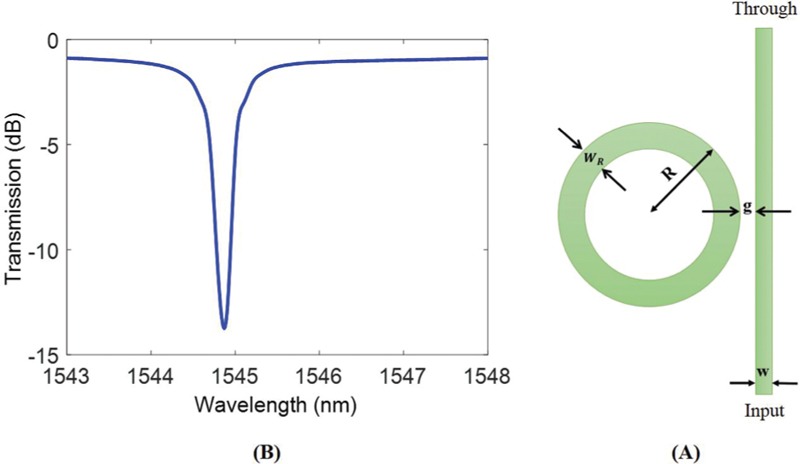
All-pass ring resonator, (a) designed parameters, (b) spectral response
Spectral response of ring resonator calculated numerically using FDTD method is represented in Figure 4(b). The resonance wavelength is 1544.87 nm, in which the injected light to the input port satisfies Equation (2) and traps inside the ring resonator. As a result, there is a dip at the wavelength of passed light to the through port. Quality factor of resonator can be calculated from numerical simulation using the following equation:

where λres and B.W. are resonance wavelength and 3-dB bandwidth of the spectral response, respectively. Calculated quality factor of designed structure is 4291. The other important parameter for a ring-resonator-based sensor is extinction ratio, which is defined as follows:

The extinction value calculated from FDTD simulation is 12.8 dB.
To investigate the sensitivity of the resonator, it is assumed that introducing bioparticle to the aquatic surrounding medium changes ambient refractive index from 1.31 to 1.45 (see Figure 1). Figure 5 represents the shift in resonance wavelength of spectral response due to interaction between evanescent fields of the light with bioparticles. For the designed ring resonator, this interaction leads to 0.88 nm shift in the resonance wavelength.
Figure 5.

Sensitivity of the designed all-pass resonator. It is assumed that by adding bioparticles, refractive index of surrounding medium is changed from 1.31 to 1.45
Sensitivity Assessment of Add-drop Filter
Figure 6(a) shows parameters of the designed add-drop resonator. All dimensions of the resonator are identical with all-pass configuration, except the gap between ring and waveguides reduces to 80 nm to compensate the extra dissipation of optical power because of the second waveguide. Spectral response of this resonator is depicted in Figure 6(b), which includes both through-port and drop-port transmission (see Figure 3). Spectral response shows a dip at 1545.53 nm wavelength with quality factor of 2146 and extinction ratio of 17 dB. Quality factor of the resonator reduces because of additional coupling loss between ring and second waveguide.
Figure 6.

Add-drop ring resonator, (a) designed parameters, (b) spectral response
Now, refractive index of the surrounding medium is increased to 1.45. Similar to all-pass resonator a 0.88 nm wavelength shift is detected due to this variation in the refractive index (Figure 7).
Figure 7.

Sensitivity of the designed Add-drop resonator. It is assumed thatrefractive index of the surrounding medium is changed from 1.31 to 1.45 by adding bioparticles
Sensitivity Assessment of All-pass Filter with a Gap in the Ring
To enhance the sensitivity of ring resonator to the variation of the ambient refractive index, a gap in the loop of the ring is introduced. Figure 8(a) and (b) shows the schematic representation of the ring with a gap and its spectral response, respectively. All dimensions of the ring is identical with the previous design except there is a 400-nm gap in the ring. The gap between ring and waveguide is reduced to 50 nm to couple more light to the resonator and compensate the extra loss due to presence of a gap in the ring. With the new condition, resonance occurs at 1549.85 nm. As it is expected, the extra loss due to presence of a gap in the ring increases 3-dB bandwidth and consequently reduces the quality factor of the resonator. The value of quality factor is calculated 1130 on the basis of Equation (6).
Figure 8.

All-pass ring resonator with a gap in the ring, (a) designed parameters, (b) spectral response
Like resonator without gap, to investigate the sensitivity of the resonator and compare it to the typical all-pass and add-drop ring resonators, refractive index of ambient is changed from 1.31 to 1.45. Figure 9 represents 0.98 nm shift in the resonance wavelength of spectral response which is 10% higher than resonance wavelength shift without gap. This enhancement in sensitivity of the ring resonator can be explained using the fact that by introducing a gap in the ring, not only the evanescent field of the light can interact with bioparticles, but also a part of the propagating mode inside the ring in the introduced gap area strongly interacts with bioparticles. This leads to a jump in the wavelength shift. Table 1 summarizes important specifications of the three different configurations.
Figure 9.

Sensitivity of designed ring resonator with a gap in the ring. It is assumed that the refractive index of surrounding medium has changed from 1.31 to 1.45 as a result of adding bioparticles. Direct interaction between resonating mode and bioparticles in the gap region has caused higher wavelength shift
Table 1.
Summarized specifications of three different configurations for biosensing

Investigation the Effect of Different Sizes of Nanogaps
Sensitivity of the resonator is studied for different sizes of the nanogaps. Figure 10 depicts sensitivity (wavelength shift due to refractive index changes from 1.31 to 1.45) as a function of nanogap size. By increasing the size of the gap from 100 to 700 nm, the wavelength changes increase from 0.89 to 1.23 nm. In a larger gap, larger part of the resonating mode can directly interact with the ambient leads to higher sensitivity to the refractive index variation of the surrounding medium. On the other hand, a large gap in the ring introduces larger loss source, which results in smaller extinction ration (as it can be seen in Figure 11). Small extinction ratio makes the detection of resonance wavelength difficult in optical resonators. This could be compensated by further reducing the separation between resonator and waveguide, but separations smaller than 50 nm is not practical from fabrication point of view. In Figures 10 and 11, it is assumed that the separation between resonator and waveguide is 50 nm for all different gap sizes. As a result, selecting appropriate gap size is a trade-off between required sensitivity and minimum detectable extinction ratio.
Figure 10.
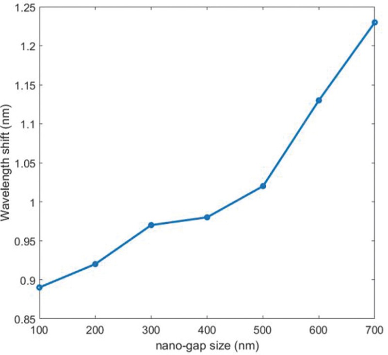
Wavelength shift due to refractive index changes from 1.31 to 1.45 as a function of nanogap size. It is assumed that for different gap sizes the separation between resonator and waveguide is constant
Figure 11.

Extinction ratio as a function of nanogap size. It is assumed that for different gap sizes the separation between resonator and waveguide is constant
Conclusion
In this paper, a new resonator configuration on the basis introduction of a nanogap in the all-pass ring resonator was proposed for sensing applications. Existence of bioparticles in the medium was modeled by increasing the refractive index of the aqueous buffer solution from 1.31 to 1.45. We have compared numerical results of the proposed configuration with typical all-pass and add-drop resonators. It is shown that introducing a nanogap in the ring resonator leads to increased sensitivity. The larger gap size leads to the higher sensitivity achievable by the sensor. It has numerically proved that 40% sensitivity enhancement is achieved through creating a 700 nm gap in a 12-µm-ring resonator. In conclusion, these results reveal that optical ring resonators are excellent choices for biological sensing applications.
Financial support and sponsorship
Financial support by Rasht Branch, Islamic Azad University is gratefully acknowledged.
Conflicts of interest
There are no conflict of interest.
References
- 1.Sun Y, Fan X. Optical ring resonators for biochemical and chemical sensing. Anal Bioanal Chem. 2011;399:205–11. doi: 10.1007/s00216-010-4237-z. [DOI] [PubMed] [Google Scholar]
- 2.Schineeweiss P, Zeiger S, Hoinkes T, Rauschenbeutel A, Volz J. Fiber ring resonator with a nanofiber section for chiral cavity quantum electrodynamics and multimode strong coupling. Opt Lett. 2017;42:85–8. doi: 10.1364/OL.42.000085. [DOI] [PubMed] [Google Scholar]
- 3.Little BE, Laine JP, Haus HA. Analytic theory of coupling from tapered fibers and half-blocks into microsphere resonators. J Lightwave Technol. 1999;17:704–15. [Google Scholar]
- 4.Li X, Zhang Z, Qin S, Qiu M, Su Y. Ultra-compact parallel labelfree biosensors based on concentric micro-ring resonators in silicon-on-insulator. Asia Optical Fiber Communication and optoelectronic Exposition and Conference, AOE. 2008 [Google Scholar]
- 5.Iqbal M, Gleeson MA, Spaugh B, Tabor F, Gunn WG, Hochberg M, Jones TB, Bailey RC, Gunn C. Label-free biosensor arrays based on silicon ring resonators and high-speed optical scanning instrumentation. IEEE J Sel Top Quantum Electron. 2010;16:654–61. [Google Scholar]
- 6.Kim HT, Yu M. Cascaded ring resonator-based temperature sensor with simultaneously enhanced sensitivity and range. Opt Express. 2016;24:9501–10. doi: 10.1364/OE.24.009501. [DOI] [PubMed] [Google Scholar]
- 7.Claes T, Bogaerts W, Bienstman P. Experimental characterization of a silicon photonic biosensor consisting of two cascaded ring resonators based on the Vernier-effect and introduction of a curve fitting method for an improved detection limit. Opt Lett. 2010;18:22747–61. doi: 10.1364/OE.18.022747. [DOI] [PubMed] [Google Scholar]
- 8.Khorasaninejad M, Clarke N, Anantram MP, Saini SS. Optical bio-chemical sensors on SNOW ring resonators. Opt Lett. 2011;19:17575–84. doi: 10.1364/OE.19.017575. [DOI] [PubMed] [Google Scholar]
- 9.Gorodetsky ML, Ilchenko VS. Optical microsphere resonators: optical coupling to high-Q whispering-gallery modes. J Opt Soc Am. 1999;16:147–54. [Google Scholar]
- 10.Gorodetsky ML, Savchenkov AA, Ilchenko VS. Ultimate Q of optical microsphere resonators. Opt Lett. 1996;21:453–5. doi: 10.1364/ol.21.000453. [DOI] [PubMed] [Google Scholar]
- 11.Lin N, Jiang L, Wang S, Chen Q, Xiao H, Lu Y, et al. Simulation and optimization of polymer-coated microsphere resonators in chemical vapor sensing. J Opt Soc Am. 2011;50:5465–72. doi: 10.1364/AO.50.005465. [DOI] [PubMed] [Google Scholar]
- 12.Ilchenko VS, Volikov PS, Velichansky VL, Treussart F, Seguin VL, Raimond JM, et al. Strain-tunable high-Q optical microsphere resonator. Opt Commun. 1998;145:86–90. [Google Scholar]
- 13.Soria S, Berneschi S, Brenci M, Cosi F, Conti GN, Pelli S, et al. Optical microspherical resonators for biomedical sensing. Sensors. 2011;11:785–805. doi: 10.3390/s110100785. [DOI] [PMC free article] [PubMed] [Google Scholar]
- 14.An P, Zheng Y, Yan S, Xue C, Wang W, Liu J. High-Q microsphere resonators for angular velocity sensing in gyroscopes. Appl Phys Lett. 2015;106:063504. [Google Scholar]
- 15.Zhang X, Armani AM. Silica microtoroid resonator sensor with monolithically integrated waveguides. Opt Lett. 2013;21:23592–603. doi: 10.1364/OE.21.023592. [DOI] [PubMed] [Google Scholar]
- 16.Monifi F, Ozdemir SK, Friedlin J, Yang L. Encapsulation of a fiber taper coupled microtoroid resonator in a polymer matrix. IEEE Photonics Technol Lett. 2013;25:1458–61. [Google Scholar]
- 17.Zadeh MH, Vahala K. Free ultra-high-Q microtoroid a tool for designing photonic device. Opt Lett. 2006;15:166–75. doi: 10.1364/oe.15.000166. [DOI] [PubMed] [Google Scholar]
- 18.Armani DK, Kippenberg TJ, Spillane SM, Vahala KJ. Ultra-high-Q toroid microcavity on chip. Nature. 2003;421:925–8. doi: 10.1038/nature01371. [DOI] [PubMed] [Google Scholar]
- 19.Soltani M, Yegnanarayanan S, Adibi A. Ultra-high Q planar silicon microdisk resonators for chip-scale silicon photonics. Opt Lett. 2007;15:4694–4704. doi: 10.1364/oe.15.004694. [DOI] [PubMed] [Google Scholar]
- 20.Schwelb O. Transmission, group delay, and dispersion in single-ring optical resonators and Add/Drop filters—a tutorial overview. J Lightwave Technol. 2004;22:1380–94. [Google Scholar]
- 21.Bogaerts W, Heyn PD, Vaerenbergh TV, Vos KD, Selvaraja SK, Claes T, et al. Silicon microring resonators. Laser Photonics Rev. 2012;6:47–73. [Google Scholar]