

Abstract
Ramsay hunt syndrome is not just a syndrome but it's rather an infectious disease caused by reactivation of latent varicella-zoster virus in geniculate ganglion. This was first explained by J. Ramsay Hunt as a triad of complications like otalgia, mucosal and cutaneous rashes with or without trigeminal facial palsy. The facial palsy can occur with characteristic vesicles along the path of nerve. We present a case of Ramsay Hunt syndrome in a 48-year-old male. The unilateral pattern of facial involvement and presence of vesicles assisted us for early diagnosis, distinguishing the syndrome with diseases mimicking other severe neurological illnesses and prompt treatment.
Keywords: Facial palsy, neuralgia, ramsay hunt syndrome, varicella-zoster virus, vesicullo bullous
Introduction
Sir James Ramsay Hunt, an American physician, in 1907 described Ramsay hunt syndrome (RHS) in a series of patients suffering from viral lesions of the ear, face, oral cavity accompanied by facial palsy, and other neurological disturbances.[1]
This syndrome is also called as geniculate neuralgia or nervus intermediate neuralgia or herpes zoster oticus.[2,3] The syndrome is a rare but severe complication of primary infection of Varicella-Zoster virus (VZV) which causes the most common childhood disease – chicken pox. The virus is assumed to be latent in the dorsal root ganglia and when reactivated due to various factors can lead to herpes zoster.[2]
The disease is thought to be self-limiting and the usual presentation is from 3 months of age to 82 years.[4] RHS is of particular importance to oral physicians and pathologists as it mimics various other vesiculo bullous lesions and hence may go unnoticed or create a perplexing dilemma in diagnosis. Any delay in treatment of this syndrome may lead to permanent neurological damage.
Case Report
A 48-year-old male reported to our department with a chief complaint of multiple eruptions on the right side of face. He also complained of severe pain in oral cavity along with inability to open the mouth. He was afebrile, with pulse and blood pressure within normal limits and no lymphadenopathy. He was diabetic for the past 4 years and under medication for the same. Focal neurological deficit in the form of lower motor neuron facial palsy was noticed. He was referred further for dental evaluation. On physical examination, multiple vesicles along the right side of face involving tragus of ear, auditory canal above ear, lateral side of forehead, lateral margin of eye, lower lip, and right mandibular parasymphyseal region were seen [Figures 1 and 2]. Intraoral examination revealed multiple vesicular eruptions on right buccal mucosa and right half of labial mucosa. Ear, nose, and throat specialist's evaluation revealed tympanic membranes congested in the right ear with blebs. Audiometric analysis was done to rule out any sign of deafness which revealed mild conducive hearing loss [Figure 3]. Ophthalmology evaluation showed no relevant pathological findings. Diagnosis of RHS was made based on the history of varicella infection and the current clinical presentation. Laboratory investigations such as hemogram, urine routine, serum electrolytes, serum glutamic pyruvate transaminase, peripheral blood smear, and blood and urine culture sensitivity results were normal. Oral vesicles were subjected to Tzanck smear which revealed multinucleated giant cells. He was treated without delay and was prescribed with tablet acyclovir 800 mg five times a day, tablet paracetamol 500 mg 6 h, tablet carbamazepine 200 mg thrice daily, and tablet methylcobalamine 1500 mg once daily. He was also advised physiotherapy for facial paralysis.
Figure 1.
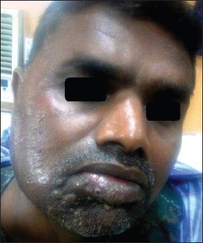
Multiple vesicles on the right side of face involving chin and lower labial mucosa
Figure 2.

Multiple vesicles on the right side of face involving tragus, forehead, and cheek
Figure 3.

Audiometric test revealing mild hearing loss
Discussion
RHS is estimated at an occurrence of 5 per 100,000 and 12% of all facial nerve paralysis.[5] Females are 20% more commonly affected than males.[6] RHS is a relatively rare syndrome with no single definition which is agreed to be gold standard. Hunt classified the disease into four subgroups based on the pathological process taking place in geniculate ganglion as[7] – disease affecting the sensory portion of seventh cranial nerve, disease involving the sensory and motor divisions of seventh cranial nerve, disease involving the sensory and motor divisions of seventh cranial nerve with auditory symptoms, and disease involving the sensory and motor divisions of seventh cranial nerve with auditory and vestibular symptoms.[8]
The pathophysiology of the syndrome lies in VZV reactivation in geniculate ganglia and subsequent neural inflammation, pressure and possible destruction of facial nerve in temporal bone causing facial palsy along with migration of VZV from the ganglia to skin around the ear or oropharynx producing vesicular lesions.[1] Hunt also summarized that gasserian, geniculate, petrous, accessory, jugular, plexiform, and second and third cervical dorsal root ganglia comprised a chain, in which inflammation of a single ganglion could extend to nearby ganglia resulting in a cranial polyneuropathy.[1]
There are also reported literature cases of reactivation of VZV following dental procedures such as incision of gingiva and fitting of a crown, treatment of carious teeth, extraction of a tooth, root canal therapy, and adjustment of dentures in immunocompromised patients.[9] Other underlying systemic illnesses, particularly hematologic malignancies such as leukemia and lymphoma, may be associated with VZV reactivation.[10]
Common symptoms of RHS include prodromal signs of pain, fever, and fatigue for 1–3 days of duration. These signs are then followed by eruption of vesicles in tympanic membrane, external auditory canal with or without tongue involvement. The active stage is characterized by emergence of a rash progressing from erythematous papules and edema to vesicles and finally to pustules within 1–7 days which dry and crust and are exfoliated over 2–3 weeks, leaving erythematous macular lesions that may scar. Diagnostic difficulties may be encountered when vesicular rash does not occur and it is defined as zoster sine herpete.[11] The present case developed similar signs and symptoms of pain, multiple vesicle formation on the right side of face involving tragus of the ear, auditory canal above ear, lateral side of forehead, lateral margin of eye, lower lip, mandibular parasymphyseal region, buccal mucosa, and right half of labial mucosa along with facial paralysis. Generally, facial involvement develops within 1–2 weeks after the rash appears. Symptoms of middle ear involvement present itself as vertigo, nystagmus, tinnitus, and hearing loss.[5] In the present case, there was mild hearing loss instigating the involvement of auditory apparatus.
Diagnosis of RHS is entirely clinical; however, in some cases, laboratory confirmation of clinical diagnosis is based on increasing antibody titer in repeated complement fixation tests. Polymerase chain reaction can detect VZV in saliva, tears, middle ear fluid, and blood mononuclear cells.[12,13] The most recommended therapy for RHS is combination of antiviral agents and steroids.[14]
Conclusion
This case was a rare syndrome with multiple vesicles on face and buccal mucosa. A multidisciplinary approach helped us in correct diagnosis and treatment of this syndrome. General dental practitioners should familiarize with the syndrome for prompt recognition and early treatment, to improve outcome significantly and prevent complications.
Declaration of patient consent
The authors certify that they have obtained all appropriate patient consent forms. In the form the patient(s) has/have given his/her/their consent for his/her/their images and other clinical information to be reported in the journal. The patients understand that their names and initials will not be published and due efforts will be made to conceal their identity, but anonymity cannot be guaranteed.
Financial support and sponsorship
Nil.
Conflicts of interest
There are no conflicts of interest.
References
- 1.Prabhu RV, Volvoikar P, Dinkar A, Prabhu VD. Ramsay Hunt syndrome with cranial polyneuropathy. J Cranio Max Dis. 2013;2:154–7. [Google Scholar]
- 2.Kannan SK, Sherubin JE, Sajesh S, Gopakumar KP. Ramsay Hunt syndrome (herpes zoster oticus) J Indian Acta Oral Med Radiol. 2012;24:70–2. [Google Scholar]
- 3.Kayayurt K, Yavasi O, Bilir O, Ersunan G, Giakoup B. A Case of Ramsay Hunt Syndrome with Atypical Presentation. Turk J Emerg Med. 2016;14:142–5. doi: 10.5505/1304.7361.2014.82788. [DOI] [PMC free article] [PubMed] [Google Scholar]
- 4.Sunilkumar MN, Gayathrivarma N, Parvathy VK. Herpes zoster oticus in a 12 year old child and review of literature-A case report. Int J Clin Case Rep. 2015;5:1–5. [Google Scholar]
- 5.Kim CH, Kang SI, Kim YH. A case of Ramsay Hunt syndrome with cranial polyneuropathy. Korean J Audiol. 2012;16:80–2. doi: 10.7874/kja.2012.16.2.80. [DOI] [PMC free article] [PubMed] [Google Scholar]
- 6.Robillard RB, Hilsinger RL, Jr, Adour KK. Ramsay Hunt facial paralysis: Clinical analyses of 185 patients. Otolaryngol Head Neck Surg. 1986;95(3 Pt 1):292–7. doi: 10.1177/01945998860953P105. [DOI] [PubMed] [Google Scholar]
- 7.Rasmussen ER, Lykke E, Toft JG, Mey K. Ramsay Hunt syndrome revisited – Emphasis on Ramsay Hunt syndrome with multiple cranial nerve involvement. Virol Discov. 2014;2:1–7. [Google Scholar]
- 8.Wackym PA. Molecular temporal bone pathology: II. Ramsay Hunt syndrome (herpes zoster oticus) Laryngoscope. 1997;107:1165–75. doi: 10.1097/00005537-199709000-00003. [DOI] [PubMed] [Google Scholar]
- 9.Furuta Y, Ohtani F, Fukuda S, Inuyama Y, Nagashima K. Reactivation of varicella-zoster virus in delayed facial palsy after dental treatment and oro-facial surgery. J Med Virol. 2000;62:42–5. [PubMed] [Google Scholar]
- 10.Jan AM, McGuire TP, Clokie CM, Sándor GK. Unilateral facial swelling caused by Ramsay Hunt syndrome resembles odontogenic infection. J Can Dent Assoc. 2006;72:829–32. [PubMed] [Google Scholar]
- 11.Tidwell E, Hutson B, Burkhart N, Gutmann JL, Ellis CD. Herpes zoster of the trigeminal nerve third branch: A case report and review of the literature. Int Endod J. 1999;32:61–6. doi: 10.1046/j.1365-2591.1999.00187.x. [DOI] [PubMed] [Google Scholar]
- 12.Gondivkar S, Parikh V, Parikh R. Herpes zoster oticus: A rare clinical entity. Contemp Clin Dent. 2010;1:127–9. doi: 10.4103/0976-237X.68588. [DOI] [PMC free article] [PubMed] [Google Scholar]
- 13.Murakami S, Honda N, Mizobuchi M, Nakashiro Y, Hato N, Gyo K. Rapid diagnosis of varicella zoster virus infection in acute facial palsy. Neurology. 1998;51:1202–5. doi: 10.1212/wnl.51.4.1202. [DOI] [PubMed] [Google Scholar]
- 14.Serinken M, Eken C, Dal O, Kutlu M. Man With Facial Nerve Palsy and Ear Pain. Ramsay Hunt Syndrome. Ann Emerg Med. 2016;67:141–8. doi: 10.1016/j.annemergmed.2015.04.010. [DOI] [PubMed] [Google Scholar]