

Abstract
Objective:
This study aimed to investigate the cytotoxicity, apoptosis induction, and mechanism of action of steviol on human breast cancer cells (Michigan Cancer Foundation-7 [MCF-7]).
Materials and Methods:
Sulforhodamine-B assay was performed to analyze cytotoxic potential of Steviol whereas flow cytometer was used to analyze cell cycle, apoptosis, and reactive oxygen species generation.
Results:
Studying the viability of cells confirms the IC50 of Steviol in MCF-7 cells which was 185 μM. The data obtained from fluorescence-activated cell sorter analysis reveal Steviol-mediated G2/M-phase arrest (P < 0.05) in addition to the presence of evident sub-G0/G1 peak (P < 0.05) in the MCF-7 cells, signifying the ongoing apoptosis.
Conclusion:
Thus, results suggest that induction of apoptosis in MCF-7 cells was due to dose-dependent effect of Steviol. Our first in vitro findings indicate Steviol as a promising candidate for the treatment of breast cancer.
SUMMARY
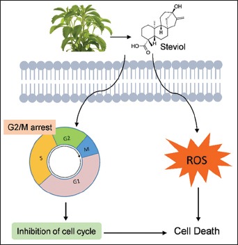
Steviol remarkably inhibited the growth MCF-7 HBCCs in a dose dependent manner
It abolishes cell cycle progression by arresting cells at G2/M phase
Steviol induces the cells to undergo apoptosis
Steviol induces the cells to generate reactive oxygen species (ROS).
Abbreviations used: MCF-7: Michigan Cancer Foundation-7; SRB: Sulforhodamine-B assay; FACS: Fluorescence-activated cell sorter; ROS: Reactive oxygen species; DNA: Deoxyribonucleic acid.
Keywords: Apoptosis, Michigan Cancer Foundation-7, Stevia rebaudiana, Steviol
INTRODUCTION
Worldwide, the majority of cancer deaths in women is due to breast cancer.[1] It also leads to a significant morbidity and mortality among women, and the outcome of disease is mainly affected by metastasis.[2] There are multiple factors involved in the pathogenesis of breast cancer, namely, life style, genetic susceptibility, and hormonal and environmental factors. In general, for in vitro studies, the human breast adenocarcinoma cell line (Michigan Cancer Foundation-7 [MCF-7]) is most widely used. Recent studies suggest that chemo- and radio-therapies are receiving more importance in inducing apoptosis in neoplastic cells by working effectively against most cancer types, but many tumors do not respond to these drug therapies and limit the successful outcomes.
Furthermore, in most cases, these drugs lack the ability to distinguish between normal and cancerous cells and they fail in activating the apoptotic pathways.[3] Therefore, the main emphasis should be laid on searching valuable natural alternative medicinal therapy which may increase the efficiency of complete and safe treatment for breast cancer.
Literatures suggest that approximately 74% of novel anticancer compounds are either natural products or derivatives of natural products.[4,5] Majority of drugs used in the treatment of cancer, whether artificial chemicals or natural products, inhibit the synthesis of new genetic material and cause irreversible damage to deoxyribonucleic acid (DNA) and its precursors. Basic research investigations confirm that, at molecular levels, several dietary chemopreventive compounds are activated.[6,7] A variety of plants and their derived bioactive compounds possess antiproliferative and anticarcinogenic effects toward breast cancer cells.
Stevioside (triglucosylated Steviol), the predominant ent-kaurene diterpene glycoside obtained from the leaves of Stevia rebaudiana Bertoni (Asteraceae family), is an effective noncaloric natural sweetener (around 200–250 times sweeter than sucrose) used in the diet in many countries in Asia and South America.[8] A study by Geuns et al.[9] on biotransformation of stevioside reports that, in the large intestine, bacterial flora of the cecum or colon degrade stevioside into free Steviol which is further transformed in the liver into its glucuronide derivative and excreted from the body through urine. Hutapea et al.[10] studied the in vitro digestibility of stevioside by a variety of digestive enzymes and reported that intestinal microflora hydrolyzed the stevioside to further compounds Steviol and Steviol-16, 17 alpha-epoxide. Finally, Steviol 16, 17 alpha-epoxide was subsequently transformed back into Steviol and excreted as Steviol glucuronide in urine. Studies suggest that stevioside and its related compounds (Steviol and isosteviol) offer therapeutic properties, which include antihypertensive,[11] antitumor,[12] antihyperglycemic,[13] anticancerous,[14] and anti-inflammatory.[15]
It is widely known that apoptosis or a process of programmed cell death is generally indicated by cytosol condensation, marginalized chromatin, protein fragmentation, degradation of DNA, and finally smaller units of apoptotic bodies are formed by the breakdown of cells.[16] A well-regulated process of apoptosis is characterized by one of the two pathways. When a cell senses any stress, it kills itself in intrinsic pathway whereas killing of cells occurs due to signals received from other cells in extrinsic pathway. Both the pathways activate caspases (protein digestive enzymes) which induce cell death and participate in the initiation and execution of apoptosis. Extreme apoptosis leads to atrophy whereas insufficient amount causes cancer (uncontrolled proliferation of cells). Studies report that the essential role of free radicals or reactive oxygen species (ROS) is vice versa in cancer and apoptosis.
Evidences suggest that ROS induces apoptosis and they regulate apoptotic cell death by activating the enzyme caspase which releases mitochondrial cytochrome c. The free radicals inside the cells were created by the ROS which are primarily the by-products of normal cellular metabolism. They initiate carcinogenesis and impose oxidative destruction to proteins, DNA, and lipids,[17] and as a result, they produce highly mutagenic oxidative DNA product (8-oxo-2’-deoxyguanosine). ROS regulates the initiation of apoptotic signaling and produces carcinogenesis by depolarizing mitochondrial membrane and interfering the signal cascade systems such as activated protein-1, nuclear transcription factor c-Jun kinase, kappa B, mitogen-activated protein kinases, and phospholipase A2.[18,19] Although the mechanism involved in ROS is still controversial regarding its role during apoptosis, till now, there is no information reported on the effect of Steviol on human breast cancer. Therefore, the present investigation was carried out to study the role of Steviol on human breast cancer cell line (MCF-7).
Our results showed that Steviol dose dependently inhibits MCF-7 cell growth and arrests cell cycle arrest at G2/M phase. This study indicates the antineoplastic action of Steviol in breast cancer.
MATERIALS AND METHODS
Reagents
Dulbecco's modified Eagle medium (DMEM), streptomycin sulfate, penicillin G, gentamicin sulfate, N-[2-hydroxyethyl] piperazine-N9-2-ethanesulfonic acid (HEPES), sulforhodamine-B (SRB), phosphate-buffered saline (PBS; pH 7.4), and trichloroacetic acid were purchased from Sigma Chemical Co., St. Louis, MO, USA. Dichlorofluorescin diacetate (DCFDA) was purchased from Merck–Calbiochem. Fetal bovine serum (FBS) was procured from GIBCO BRL Laboratories, New York, USA. In addition, all the chemicals used were of analytical grade.
Cell culture
Human breast cancer cell lines (MCF-7) were purchased from the National Centre for Cell Sciences (Pune, India). These cells are regularly cultured according to the procedure described by Shagufta et al. and Gupta et al.[20,21] For culturing the MCF-7 cells, DMEM (pH 7.4) was used which contains streptomycin (100 U/mL), penicillin (200 mg/mL), and gentamicin (50 mg/mL) supplemented with 10 mM HEPES and 10% FBS at 37°C at a humidified atmosphere of 5% CO2 incubator in T-25 tissue culture flask (TPP, Switzerland). Before performing the experiments, the cells were trypsinized which were obtained from a confluent flask and further cultured for 4 days. For the initial 2 days, DMEM phenol red free was used which contains 10% charcoal-stripped FBS to preculture the cells.[22] Subsequently, the cells were exposed to ligand (Steviol) for 48 h. The number of cells required for analyzing cytotoxicity and the exposure of cells with different concentrations of ligands has been described individually below.
Analysis of cell growth and cytotoxicity
To determine toxic, cytostatic, and proliferative effect(s) of Steviol in MCF-7 cells, SRB assay was done according to a procedure given by Shagufta et al.[23] For treatment, the dose of Steviol should be below IC50 value (i.e., sublethal) where the number of live cells should be more than 50% and the viability of cells was analyzed to achieve IC50 value. In brief, under inverted-phase contrast microscope, the MCF-7 cells were counted by a hemocytometer, and in a 96-well plate, 104 cells/well were plated in phenol red-free DMEM (200 μl) and left for 48 h in CO2 incubator at 37°C. The used medium was changed with fresh medium, and a different concentration of Steviol (10–500 μM) was added in triplicate. After completing the incubation with the drug (Steviol), SRB assay was performed.
Cellular morphological alteration studies
For the cellular morphological analysis, 2 × 10⁶ cell was plated in 6-well plates in phenol red-free medium for 48 h. Thereafter, cells were exposed for 48 h with 10–250 μM Steviol. Afterward, microscopic analysis was conducted under inverted phase-contrast microscope.
Analysis of cell cycle kinetics and apoptosis by flow cytometry
For analyzing the cell cycle distribution and apoptosis, 0.2 × 10⁶ MCF-7 cells were seeded in 6-well plates and incubated for 48 h in a phenol red-free DMEM medium supplemented with 10% charcoal-stripped FBS. The adherent cells were exposed to 10–250 μM Steviol and kept for 48 h in a 5% CO2 incubator. Subsequently, the cells were trypsinized and washed with ice-cold PBS. Seventy percent chilled ethanol was used to fix the cells for 1 h at 4°C. Cells were rewashed with chilled PBS twice and resuspended in 500 μl of PBS containing 100 μg/ml RNase and 40 μg/ml of propidium iodide. Becton–Dickinson fluorescence-activated cell sorter (FACS) analysis was used to perform flow cytometric analysis using CellQuest software (Becton Dickinson, San Jose, CA, USA).
Analysis of intracellular reactive oxygen species generation
Generation of ROS was assessed using fluorescent probe 2’,7’-DCFDA staining. MCF-7 cells at a concentration of 0.2 × 10⁶ cells/well were seeded in 6-well plates and exposed to Steviol (10–250 μM) for 24 h. Thereafter, the cells were washed with ice-cold PBS twice and incubated with 10 μM DCFDA at 37°C for 30 min in dark. Rewashing of the cells was performed twice with ice-cold PBS. Subsequently, the cells were trypsinized and fluorescent intensity was measured through flow cytometry on FACS using CellQuest software (DCFDA Ex 480 nm, Em 530 nm).
Statistical analysis
The obtained results were expressed as mean ± standard deviation. To perform statistical analysis, Bonferroni's multiple comparison tests and one-way analysis of variance were used. P < 0.05 was considered statistically significant.
RESULTS
Steviol induces cytotoxicity in human breast cancer cells (MCF-7)
This study was started by investigating the effect of Steviol on viability of cells, SRB assay [Figure 1] was conducted in human breast cancer cell type (MCF-7). The result demonstrates that Steviol induces dose-dependent reduction in viability of MCF-7 cells. 10 μM of Steviol shows insignificant antiproliferative activity; however, an abrupt decline was noticed in MCF-7 cells treated with 25–500 μM. The IC50 value of Steviol in MCF-7 cells was found to be 185 μM. The microscopic analysis confirms the cytotoxicity results [Figure 2] where dose-dependent alteration in morphology of MCF-7 cells as compared to control was observed.
Figure 1.
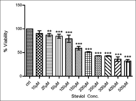
Dose-dependent cytotoxicity evaluation of Steviol in cancerous (MCF-7) cell lines versus controls (untreated MCF-7 cells). Analyzed data represent mean cell viability ± standard error in each concentration of Steviol. Data shown are Mean ± S.D. of three similar experiments, each performed in triplicate. **P<0.01; ***P<0.001 when compared to control
Figure 2.
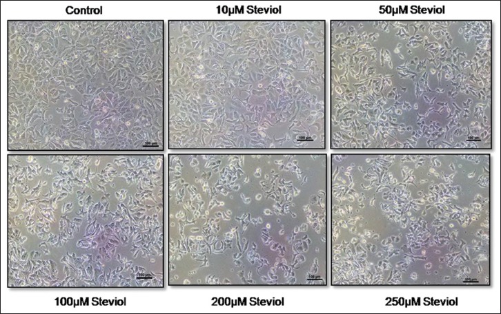
Morphological analysis (using phase-contrast microscope at ×100 magnification) of Steviol-treated MCF-7 cells. Different doses of Steviol decrease the survival rate of MCF-7 cells. MCF-7: Michigan Cancer Foundation-7
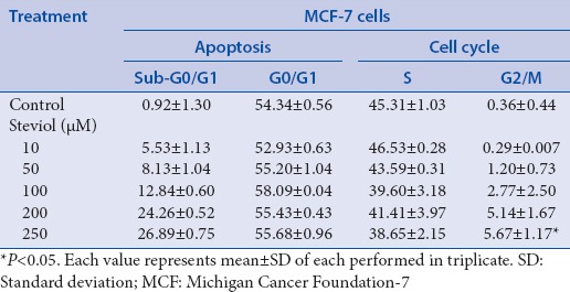
Steviol induces G2/M-phase arrest and apoptosis in MCF-7 cells
It was already specified that cell proliferation was decreased by Steviol and it also induces death of MCF-7 cells; by flow cytometry, its outcome on cell cycle distribution was analyzed. Following treatment with Steviol at concentrations of 10–250 μM for 48 h, accumulation of cells occur significantly in the G2/M phase at a concentration of 250 μM (P < 0.05) which was compared to untreated cells [Figure 3]. The cells gradually increase in G2/M phase indicating that the conditions of cells were neither in replicating nor in resting phase. It was perceived that cells were in a process of death. Moreover, finding of hypodiploid (apoptotic) sub-G0/G1 population of cells confirms its role in inducing apoptosis.
Figure 3.
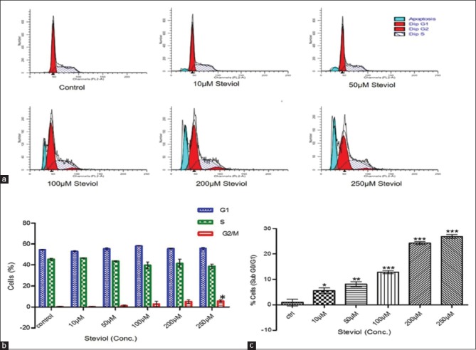
Cell cycle analysis of Steviol-treated MCF-7 cells. Results indicate that Steviol decreased cell proliferation and induced apoptosis in MCF-7 cells. (a) Cell cycle analysis by flow cytometry. (b) Graphical representation of different phases of cell cycle (G1, S, G2/M). (c) Graphical representation of cell population accumulated in sub-G0/G1 phase. MCF-7: Michigan Cancer Foundation-7
In treatment groups, apoptosis was high compared to controls. Significant apoptosis was obtained even at lower dose, i.e., 10 μM Steviol. Interestingly, approximately 26% of cells had undergone apoptosis when treated with 250 μM Steviol for 48 h [Table 1], which was highly significant (P < 0.05) than control cells.
Table 1.
Cell cycle analysis of Steviol-treated MCF-7 cells
Decline in reactive oxygen species generation by Steviol in MCF-7 cells
For measuring the ROS levels within the cells, DCFDA (a ROS-sensitive fluorometric probe) was used. Treatment with Steviol at a concentration of 100 μM onward shows a significant decline in ROS generation after 24 h [Figure 4] when compared to the control. Thus, it was suggested that Steviol has a certain association with the underlying mechanism of apoptosis by impairing mitochondrial function.
Figure 4.
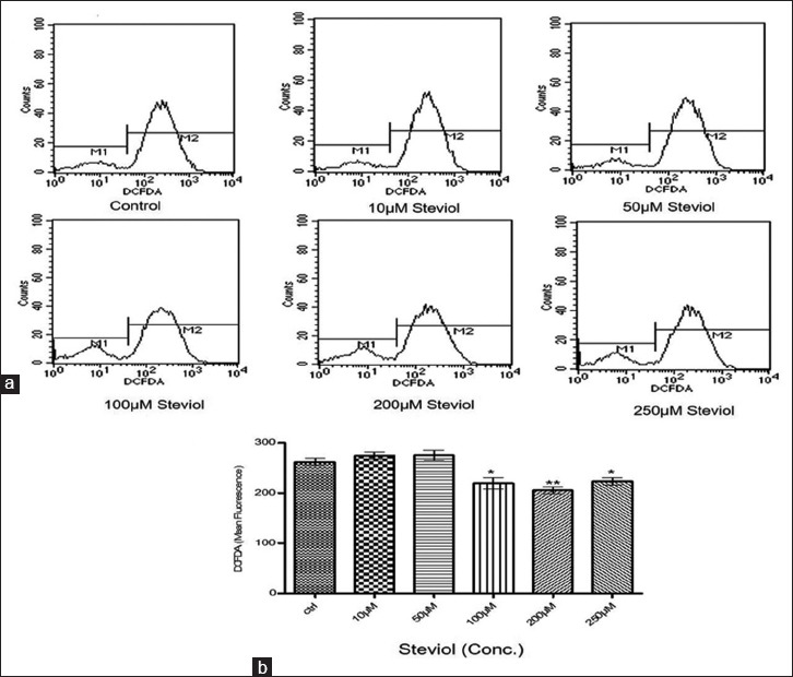
(a) MCF-7 cells (0.2 × 10⁶) were exposed in 6-well plates and exposed with 10–250 μM Steviol for 24 h. Cells were incubated with 10 μM DCFDA for 30 min in dark. Reactive oxygen species generation was detected through flow cytometry. The result signifies the deviation in curve from the control to the treated samples, (b) Graphical representation of Flow data
DISCUSSION
Worldwide, cancer is a well-known health issue with 1 million new cases every year, the most common malignancy in women is breast cancer which comprises around 18% of all female cancers. Medicinal plants and its isolated bioactive compounds are recognized as an alternative source of nontoxic, inexpensive, and efficacious medications to synthetic chemotherapeutic compounds. Currently, researchers are making enormous efforts to synthesize anticancer drugs from plant sources, provided that they kill the cancer cells devoid of unnecessary damage to the normal cells.
The outcome of this study illustrates the cytotoxic or anticancerous effects of Steviol from the plant S. rebaudiana against cultured human breast cancer MCF-7 cells, and its probable mechanisms of action include suppression of cell viability, cell cycle arrest, and induction of apoptosis in cancer cells. Steviol inhibits the proliferation of MCF-7 cells in a dose-dependent manner. Till now, no study has reported on the effect of Steviol on human breast cancer (in vitro or in vivo), so in this aspect, this work has its own originality.
Study on cell growth and cytotoxicity [Figure 1] and cellular morphological analysis [Figure 2] shows that, in MCF-7 cells, a dose-dependent decline was induced by Steviol in the surviving cell percentage when compared to parallel controls. This action of Steviol toward breast cancer cells might be credited to differences in the levels of specific tissue and cytochrome P450 (CYP) isoform expression patterns which mediate cell-specific cytotoxicity.[24] Inducible and constitutive enzymes come under a multigene family of CYPs’ which influences tumors’ response to anticancer drugs. The reason is this enzyme system can either activate or detoxify many anticancer agents.
An essential factor of tumor cell survival is resistance to apoptosis. The advancement of drug resistance (reduced effectiveness of a drug)in cancer cells is the main reason for the unsuccessful cancer treatment. Therefore, new drugs are immediately required which are effective against tumor cells and do not induce toxicity in normal cells.
By studying the fundamental mechanisms, it was observed that, in MCF-7 cells, apoptosis was induced by Steviol through DNA damage which correlates with the fact that, in a sub-G0/G1 phase, a significant amount of cell population was found [Figure 3] and it also causes specific cell-type G2/M growth arrest in MCF-7 cells. Results indicate the genotoxic effect of Steviol, which damages the DNA by inducing some proteins.[25] Moreover, an increase in the number of apoptotic cells was noticed.
According to previously reported studies, leaves of S. rebaudiana contain a significant amount of total phenolic compounds and have a strong antioxidant activity in normal tissues.[26] Mostly, very invasive or metastatic cancer cells retain a balance between proliferation and apoptosis by involving a critical level of oxidative stress. Higher ROS is generally accompanied with the activation of oncogene, which is an early event of malignant transformation, thus the usage of antioxidant supplements is receiving higher attention. Studies have shown that downregulation of ROS is also responsible for the induction of apoptosis.[28,29]
In this study, a sharp decrease in the levels of ROS after 100 μM Steviol doses was observed, which indicates the involvement of ROS in Steviol-mediated induction of apoptosis [Figure 4].
CONCLUSION
From the results, it can be concluded that the naturally occurring pure compound such as Steviol isolated from the leaves of S. rebaudiana induces apoptosis and cell cycle disruption and effectively reduces the number of human breast cancer cells (MCF-7). Moreover, Steviol has antitumor property and involves signaling pathways with ROS and mediated by G2/M arrest. There is a requirement of understanding the regulating pathways or molecular mechanism of action of Steviol to establish its therapeutic applications which is safe for human consumption.
Financial support and sponsorship
This study was financially supported by the Department of Science and Technology, New Delhi, India, under a Women Scientist Project Scheme (WOS-A), vide letter no. SR/WOS-A/LS-668/2012.
Conflicts of interest
There are no conflicts of interest.
Acknowledgements
The authors thank the Director of CDRI for giving permission to carry out the research work at CDRI. The authors also thank Dr. Anil K. Balapure and Dr. Ramesh Sharma for their guidance during the experiments with cell culture techniques. Financial support from the Department of Science and Technology, New Delhi, India, under a Women Scientist Project Scheme (WOS-A), vide letter no. SR/WOS-A/LS-668/2012, is deeply acknowledged.
REFERENCES
- 1.Torre LA, Bray F, Siegel RL, Ferlay J, Lortet-Tieulent J, Jemal A. Global cancer statistics, 2012. CA Cancer J Clin. 2015;65:87–108. doi: 10.3322/caac.21262. [DOI] [PubMed] [Google Scholar]
- 2.Ferlay J, Bray F, Pisani P, Parkin DM. GLOBOCAN 2000: Cancer Incidence, Mortality, and Prevalence Worldwide. Version 1.0, IARC Cancer Base No. 5. Lyon, France: International Agency for Research on Cancer and World Health Organization, IARC Press; 2001. [Google Scholar]
- 3.Benjamin CW, Hiebsch RR, Jones DA. Caspase activation in MCF7 cells responding to etoposide treatment. Mol Pharmacol. 1998;53:446–50. doi: 10.1124/mol.53.3.446. [DOI] [PubMed] [Google Scholar]
- 4.Chen MS, Chen D, Dou QP. Inhibition of proteasome activity by various fruits and vegetables is associated with cancer cell death. In Vivo. 2004;18:73–80. [PubMed] [Google Scholar]
- 5.Cragg GM, Newman DJ. Plants as a source of anti-cancer agents. J Ethnopharmacol. 2005;100:72–9. doi: 10.1016/j.jep.2005.05.011. [DOI] [PubMed] [Google Scholar]
- 6.Russo M, Tedesco I, Iacomino G, Galano G, Russo GL. Dietary phytochemicals in chemoprevention of cancer. Curr Med Chem Immunol Endocr Metab Agents. 2005;5:61–72. [Google Scholar]
- 7.Pan MH, Ho CT. Chemopreventive effects of natural dietary compounds on cancer development. Chem Soc Rev. 2008;37:2558–74. doi: 10.1039/b801558a. [DOI] [PubMed] [Google Scholar]
- 8.Chatsudthipong V, Muanprasat C. Stevioside and related compounds: Therapeutic benefits beyond sweetness. Pharmacol Ther. 2009;121:41–54. doi: 10.1016/j.pharmthera.2008.09.007. [DOI] [PubMed] [Google Scholar]
- 9.Geuns JM, Buyse J, Vankeirsbilck A, Temme EH. Metabolism of stevioside by healthy subjects. Exp Biol Med (Maywood) 2007;232:164–73. [PubMed] [Google Scholar]
- 10.Hutapea AM, Toskulkao CH, Buddhasukh D, Wilairat P, Glinsukon TH. Digestion of stevioside, a natural sweetener, by various digestive enzymes. J Clin Biochem Nutr. 1997;23:177–6. [Google Scholar]
- 11.Lee CN, Wong KL, Liu JC, Chen YJ, Cheng JT, Chan P. Inhibitory effect of stevioside on calcium influx to produce antihypertension. Planta Med. 2001;67:796–9. doi: 10.1055/s-2001-18841. [DOI] [PubMed] [Google Scholar]
- 12.Satishkumar J, Saravanan MM, Seethalakshmi I. In-vitro antimicrobial and antitumor activities of Stevia rebaudiana (Asteraceae) leaf extracts. Trop J Pharm Res. 2008;7:1143–9. [Google Scholar]
- 13.Benford DJ, DiNovi M, Schlatter J. Safety Evaluation of certain food additive: Stevia Glycosided. WHO Food Additives Series. World Health Organization. Vol. 54. Geneva, Switzerland: WHO Press; 2006. pp. 1–649. [Google Scholar]
- 14.Takasaki M, Konoshima T, Kozuka M, Tokuda H, Takayasu J, Nishino H, et al. Cancer preventive agents. Part 8: Chemopreventive effects of stevioside and related compounds. Bioorg Med Chem. 2009;17:600–5. doi: 10.1016/j.bmc.2008.11.077. [DOI] [PubMed] [Google Scholar]
- 15.Das S, Das AK, Murphy RA, Punwani IC, Nasution MP, Kinghorn AD. Evaluation of the cariogenic potential of the intense natural sweeteners stevioside and rebaudioside A. Caries Res. 1992;26:363–6. doi: 10.1159/000261469. [DOI] [PubMed] [Google Scholar]
- 16.Thompson CB. Apoptosis in the pathogenesis and treatment of disease. Science. 1995;267:1456–62. doi: 10.1126/science.7878464. [DOI] [PubMed] [Google Scholar]
- 17.Orrenius S. Mechanisms of oxidative cell damage. In: Pol G, Albano E, Dianzani MU, editors. Free Radicals: From Basic Science to Medicine. Basel, Switzerland: Birkhauser Verlag; 1993. pp. 47–64. [Google Scholar]
- 18.Guyton KZ, Liu Y, Gorospe M, Xu Q, Holbrook NJ. Activation of mitogen-activated protein kinase by H2O2. Role in cell survival following oxidant injury. J Biol Chem. 1996;271:4138–42. doi: 10.1074/jbc.271.8.4138. [DOI] [PubMed] [Google Scholar]
- 19.Bae YS, Kang SW, Seo MS, Baines IC, Tekle E, Chock PB, et al. Epidermal growth factor (EGF)-induced generation of hydrogen peroxide. Role in EGF receptor-mediated tyrosine phosphorylation. J Biol Chem. 1997;272:217–21. [PubMed] [Google Scholar]
- 20.Shrivastava A, Tiwari M, Sinha RA, Kumar A, Balapure AK, Bajpai VK, et al. Molecular iodine induces caspase-independent apoptosis in human breast carcinoma cells involving the mitochondria-mediated pathway. J Biol Chem. 2006;281:19762–71. doi: 10.1074/jbc.M600746200. [DOI] [PubMed] [Google Scholar]
- 21.Gupta A, Dwivedy A, Keshri G, Sharma R, Balapure AK, Singh MM, et al. Rapid synthesis of 4-benzylidene and 4-[bis-(4-methoxyphenyl)-methylene-2-substituted phenyl-benzopyrans as potential selective estrogen receptor modulators (SERMs) using McMurry coupling reaction. Bioorg Med Chem Lett. 2006;16:6006–12. doi: 10.1016/j.bmcl.2006.08.126. [DOI] [PubMed] [Google Scholar]
- 22.Soto AM, Sonnenschein C. The role of estrogens on the proliferation of human breast tumor cells (MCF-7) J Steroid Biochem. 1985;23:87–94. doi: 10.1016/0022-4731(85)90265-1. [DOI] [PubMed] [Google Scholar]
- 23.Shagufta, Srivastava AK, Sharma R, Mishra R, Balapure AK, Murthy PS, et al. Substituted phenanthrenes with basic amino side chains: A new series of anti-breast cancer agents. Bioorg Med Chem. 2006;14:1497–505. doi: 10.1016/j.bmc.2005.10.002. [DOI] [PubMed] [Google Scholar]
- 24.Sridar C, Kent UM, Notley LM, Gillam EM, Hollenberg PF. Effect of tamoxifen on the enzymatic activity of human cytochrome CYP2B6. J Pharmacol Exp Ther. 2002;301:945–52. doi: 10.1124/jpet.301.3.945. [DOI] [PubMed] [Google Scholar]
- 25.Roos WP, Kaina B. DNA damage-induced cell death by apoptosis. Trends Mol Med. 2006;12:440–50. doi: 10.1016/j.molmed.2006.07.007. [DOI] [PubMed] [Google Scholar]
- 26.Shukla S, Mehta A, Mehta P, Bajpai VK. Antioxidant ability and total phenolic content of aqueous leaf extract of Stevia rebaudiana Bert. Exp Toxicol Pathol. 2012;64:807–11. doi: 10.1016/j.etp.2011.02.002. [DOI] [PubMed] [Google Scholar]
- 27.Chen WY, Wu CC, Lan YH, Chang FR, Teng CM, Wu YC. Goniothalamin induces cell cycle-specific apoptosis by modulating the redox status in MDA-MB-231 cells. Eur J Pharmacol. 2005;522:20–9. doi: 10.1016/j.ejphar.2005.08.047. [DOI] [PubMed] [Google Scholar]
- 28.Wang J, Yi J. Cancer cell killing via ROS: To increase or decrease, that is the question. Cancer Biol Ther. 2008;7:1875–84. doi: 10.4161/cbt.7.12.7067. [DOI] [PubMed] [Google Scholar]
- 29.Jeong CH, Joo SH. Downregulation of reactive oxygen species in apoptosis. J Cancer Prev. 2016;21:13–20. doi: 10.15430/JCP.2016.21.1.13. [DOI] [PMC free article] [PubMed] [Google Scholar]