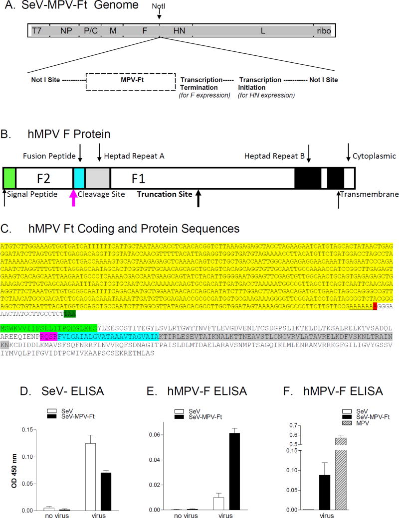
Figure 1. Characterization of recombinant SeV-MPV-Ft.
A. SeV-MPV-Ft rescue: A construct carrying the hMPV F sequence was produced by Invitrogen using GeneArt™Gene Synthesis technology, and confirmed to encompass the correct F sequence. The sequence was then recombined between F and HN genes at a unique NotI site in a vector carrying the complete SeV genome. Virus was rescued and amplified. B. hMPV F Protein: A cartoon of the hMPV F protein is shown indicating functional segments. Shown are positions of the signal peptide and F2 segment. Within the F1 segment are the fusion peptide, heptad repeats A and B, and transmembrane and cytoplasmic domains. The site of predicted F protein truncation is shown. C. hMPV-Ft coding and protein sequences: The N-terminal coding sequence of the hMPV-Ft gene is shown. The bases highlighted in yellow were unaltered from the original clone. The red highlight indicates the position of the A deletion, followed by a stop codon (highlighted in dark green) resulting from a frame-shift. The predicted protein sequence is shown below the gene sequence. The signal peptide, cleavage site, fusion peptide and heptad repeat A are highlighted in light green, pink, blue, and grey, respectively, corresponding to the similar color scheme in panel B. D, E and F. ELISAs with SeV-specific or hMPV-F-specific monoclonal antibodies for the detection of hMPV F expression: Uninfected and infected cell cultures were tested for viral protein expression in assays with SeV-specific (Panel D) or hMPV-specific (Panels E and F) monoclonal antibodies.

