Abstract
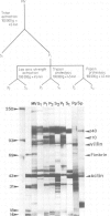
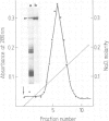
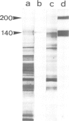
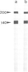
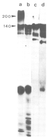
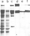
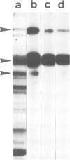
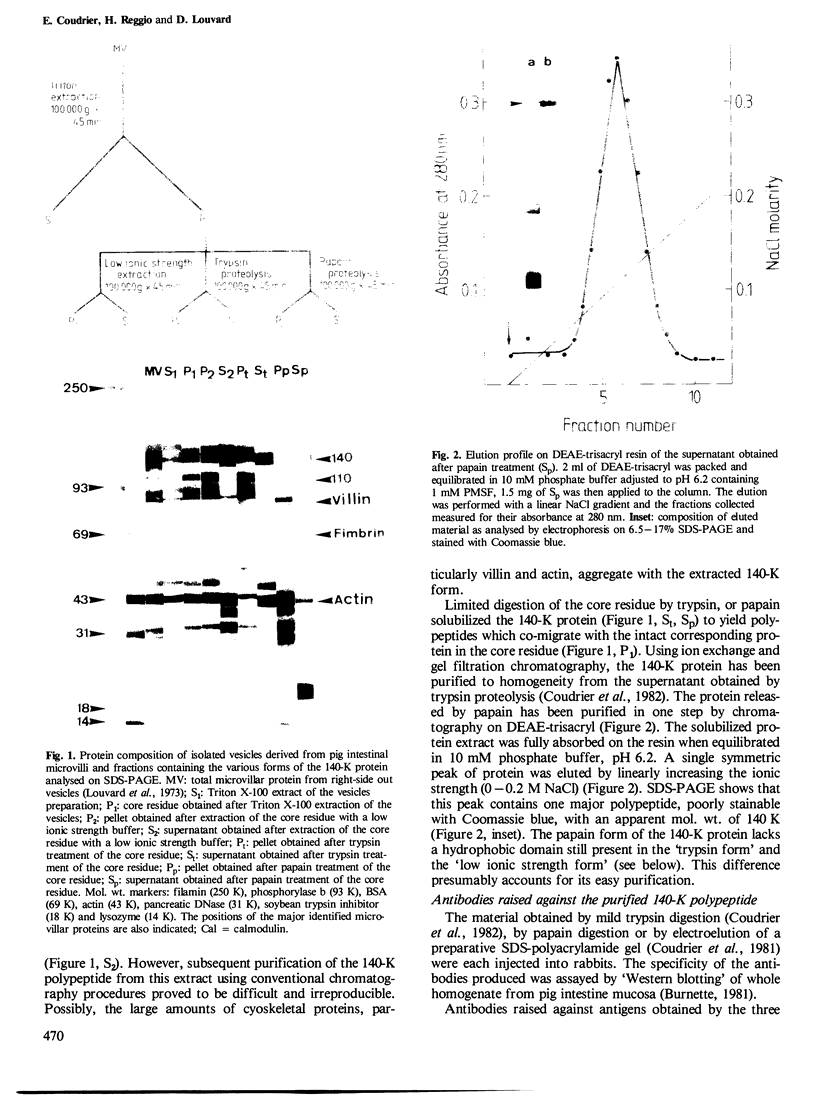
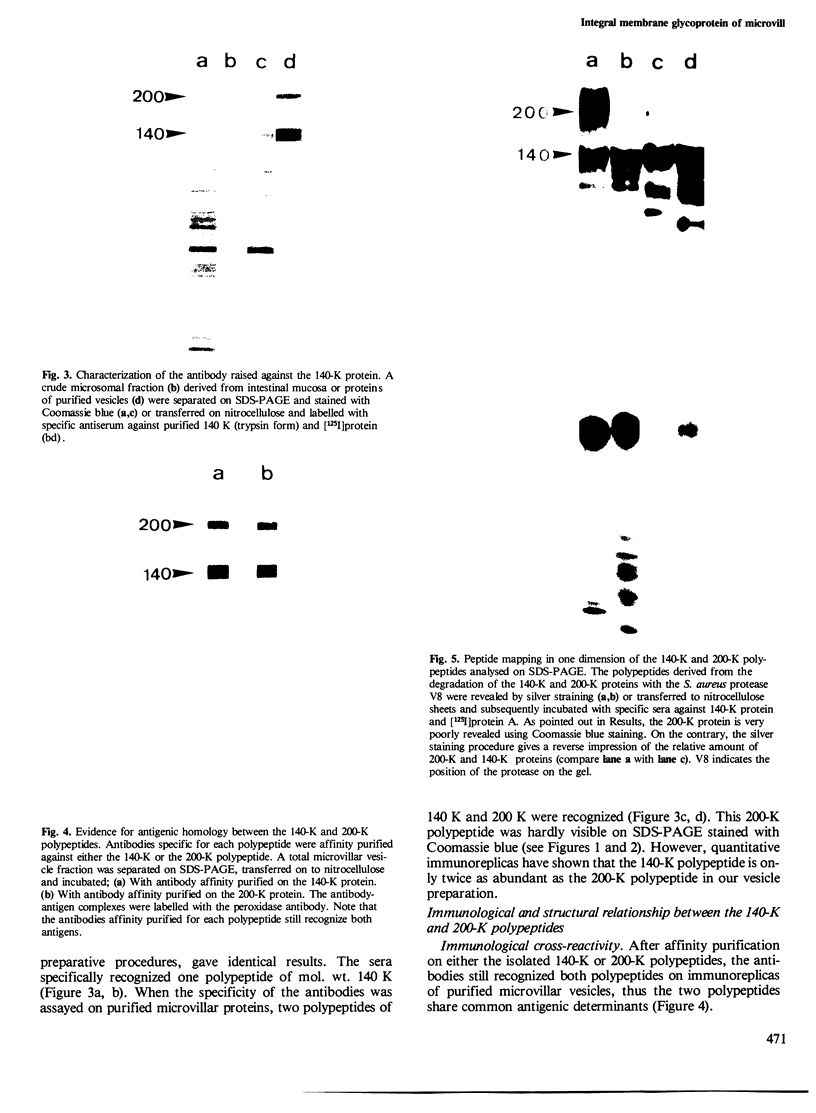
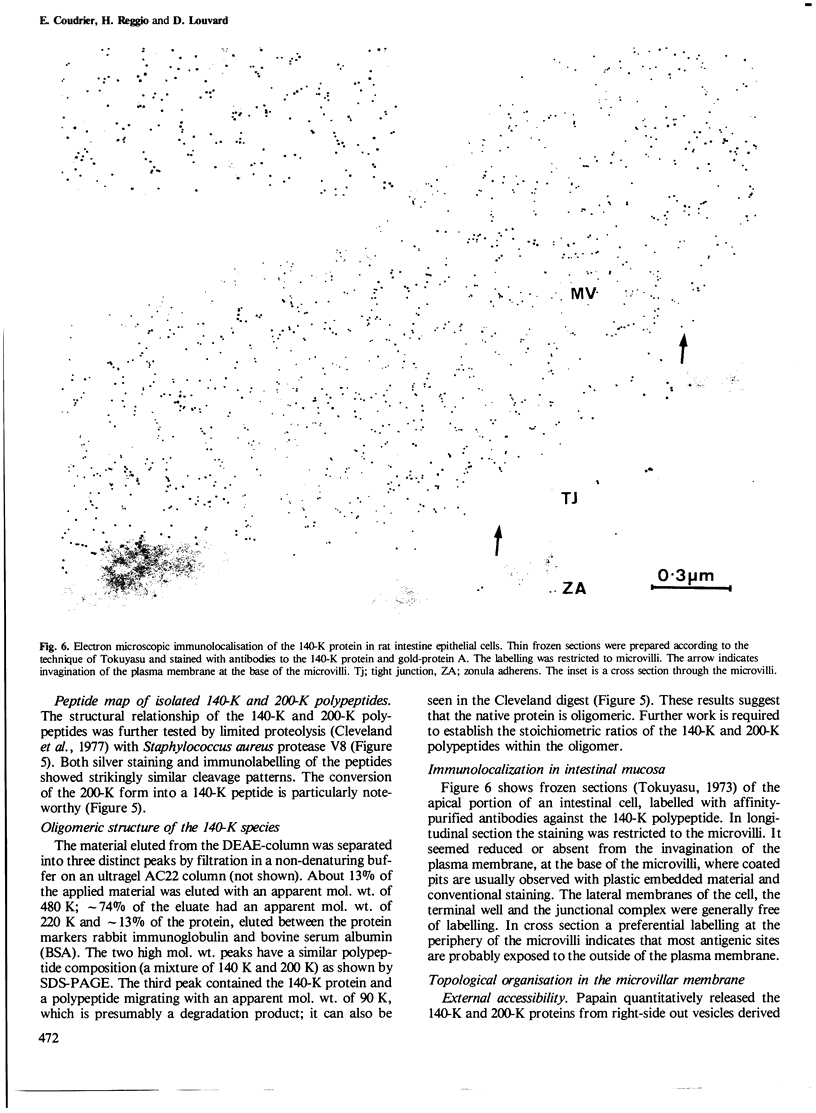
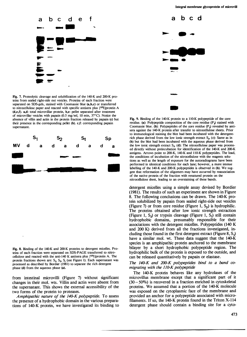
An integral membrane glycoprotein of pig intestinal microvilli which exists in two polypeptide forms [mol. wt. 140 K and 200 K as measured by SDS-polyacrylamide gel electrophoresis (SDS-PAGE)] was purified to homogeneity and characterized. The 200-K form is probably a precursor of the 140-K species. We have localized the glycoprotein by electron microscope immunochemistry using specific antibodies and determined its topological organization with respect to the membrane bilayer. Triton X-100 treatments which solubilize most other microvillar membrane glycoproteins from purified, closed, right-side out vesicles do not efficiently extract this protein. The protein can be partially solubilized from the detergent-insoluble residue, either by treatment with proteases (trypsin or papain) or by exposure to low ionic strength buffer in the presence of chelating agents and detergents. Once solubilized by papain or trypsin, the protein co-migrates on SDS-PAGE with the protein obtained by low ionic strength extraction. However, the form of the protein released by papain does not bind detergents and exhibits hydrophilic properties. Our observations are consistent with the 140-K protein having a small hydrophobic domain that anchors it to the microvillar membrane. The 140-K glycoprotein binds in vitro to a 110-K protein of the core cytoskeleton residue. These observations suggest that the 140-K glycoprotein may be a transmembrane protein which may in vivo provide attachment sites for direct or indirect association with polypeptides of the microvillus cytoskeleton.
Full text
PDF
Images in this article
Selected References
These references are in PubMed. This may not be the complete list of references from this article.
- Bordier C. Phase separation of integral membrane proteins in Triton X-114 solution. J Biol Chem. 1981 Feb 25;256(4):1604–1607. [PubMed] [Google Scholar]
- Bretscher A., Osborn M., Wehland J., Weber K. Villin associates with specific microfilamentous structures as seen by immunofluorescence microscopy on tissue sections and cells microinjected with villin. Exp Cell Res. 1981 Sep;135(1):213–219. doi: 10.1016/0014-4827(81)90313-x. [DOI] [PubMed] [Google Scholar]
- Bretscher A., Weber K. Fimbrin, a new microfilament-associated protein present in microvilli and other cell surface structures. J Cell Biol. 1980 Jul;86(1):335–340. doi: 10.1083/jcb.86.1.335. [DOI] [PMC free article] [PubMed] [Google Scholar]
- Bretscher A., Weber K. Localization of actin and microfilament-associated proteins in the microvilli and terminal web of the intestinal brush border by immunofluorescence microscopy. J Cell Biol. 1978 Dec;79(3):839–845. doi: 10.1083/jcb.79.3.839. [DOI] [PMC free article] [PubMed] [Google Scholar]
- Bretscher A., Weber K. Purification of microvilli and an analysis of the protein components of the microfilament core bundle. Exp Cell Res. 1978 Oct 15;116(2):397–407. doi: 10.1016/0014-4827(78)90463-9. [DOI] [PubMed] [Google Scholar]
- Bretscher A., Weber K. Villin is a major protein of the microvillus cytoskeleton which binds both G and F actin in a calcium-dependent manner. Cell. 1980 Jul;20(3):839–847. doi: 10.1016/0092-8674(80)90330-x. [DOI] [PubMed] [Google Scholar]
- Burnette W. N. "Western blotting": electrophoretic transfer of proteins from sodium dodecyl sulfate--polyacrylamide gels to unmodified nitrocellulose and radiographic detection with antibody and radioiodinated protein A. Anal Biochem. 1981 Apr;112(2):195–203. doi: 10.1016/0003-2697(81)90281-5. [DOI] [PubMed] [Google Scholar]
- Cleveland D. W., Fischer S. G., Kirschner M. W., Laemmli U. K. Peptide mapping by limited proteolysis in sodium dodecyl sulfate and analysis by gel electrophoresis. J Biol Chem. 1977 Feb 10;252(3):1102–1106. [PubMed] [Google Scholar]
- Coudrier E., Reggio H., Louvard D. Immunolocalization of the 110,000 molecular weight cytoskeletal protein of intestinal microvilli. J Mol Biol. 1981 Oct 15;152(1):49–66. doi: 10.1016/0022-2836(81)90095-4. [DOI] [PubMed] [Google Scholar]
- Coudrier E., Reggio H., Louvard D. The cytoskeleton of intestinal microvilli contains two polypeptides immunologically related to proteins of striated muscle. Cold Spring Harb Symp Quant Biol. 1982;46(Pt 2):881–892. doi: 10.1101/sqb.1982.046.01.082. [DOI] [PubMed] [Google Scholar]
- Craig S. W., Lancashire C. L. Comparison of intestinal brush-border 95-Kdalton polypeptide and alpha-actinins. J Cell Biol. 1980 Mar;84(3):655–667. doi: 10.1083/jcb.84.3.655. [DOI] [PMC free article] [PubMed] [Google Scholar]
- Geiger B., Tokuyasu K. T., Singer S. J. Immunocytochemical localization of alpha-actinin in intestinal epithelial cells. Proc Natl Acad Sci U S A. 1979 Jun;76(6):2833–2837. doi: 10.1073/pnas.76.6.2833. [DOI] [PMC free article] [PubMed] [Google Scholar]
- Glenney J. R., Jr, Weber K. Calmodulin-binding proteins of the microfilaments present in isolated brush borders and microvilli of intestinal epithelial cells. J Biol Chem. 1980 Nov 25;255(22):10551–10554. [PubMed] [Google Scholar]
- Graham R. C., Jr, Karnovsky M. J. The early stages of absorption of injected horseradish peroxidase in the proximal tubules of mouse kidney: ultrastructural cytochemistry by a new technique. J Histochem Cytochem. 1966 Apr;14(4):291–302. doi: 10.1177/14.4.291. [DOI] [PubMed] [Google Scholar]
- Hauri H. P., Quaroni A., Isselbacher K. J. Biogenesis of intestinal plasma membrane: posttranslational route and cleavage of sucrase-isomaltase. Proc Natl Acad Sci U S A. 1979 Oct;76(10):5183–5186. doi: 10.1073/pnas.76.10.5183. [DOI] [PMC free article] [PubMed] [Google Scholar]
- Howe C. L., Mooseker M. S., Graves T. A. Brush-border calmodulin. A major component of the isolated microvillus core. J Cell Biol. 1980 Jun;85(3):916–923. doi: 10.1083/jcb.85.3.916. [DOI] [PMC free article] [PubMed] [Google Scholar]
- Howe J. G., Hershey J. W. A sensitive immunoblotting method for measuring protein synthesis initiation factor levels in lysates of Escherichia coli. J Biol Chem. 1981 Dec 25;256(24):12836–12839. [PubMed] [Google Scholar]
- Laemmli U. K. Cleavage of structural proteins during the assembly of the head of bacteriophage T4. Nature. 1970 Aug 15;227(5259):680–685. doi: 10.1038/227680a0. [DOI] [PubMed] [Google Scholar]
- Louvard D., Maroux S., Baratti J., Desnuelle P., Mutaftschiev S. On the preparation and some properties of closed membrane vesicles from hog duodenal and jejunal brush border. Biochim Biophys Acta. 1973 Feb 16;291(3):747–763. doi: 10.1016/0005-2736(73)90478-1. [DOI] [PubMed] [Google Scholar]
- Louvard D., Maroux S., Vannier C., Desnuelle P. Topological studies on the hydrolases bound to the intestinal brush border membrane. I. Solubilization by papain and Triton X-100. Biochim Biophys Acta. 1975 Jan 28;375(2):235–248. [PubMed] [Google Scholar]
- Louvard D., Semeriva M., Maroux S. The brush-border intestinal aminopeptidase, a transmembrane protein as probed by macromolecular photolabelling. J Mol Biol. 1976 Oct 5;106(4):1023–1035. doi: 10.1016/0022-2836(76)90350-8. [DOI] [PubMed] [Google Scholar]
- Maroux S., Louvard D. On the hydrophobic part of aminopeptidase and maltases which bind the enzyme to the intestinal brush border membrane. Biochim Biophys Acta. 1976 Jan 21;419(2):189–195. doi: 10.1016/0005-2736(76)90345-x. [DOI] [PubMed] [Google Scholar]
- Matsudaira P. T., Burgess D. R. Identification and organization of the components in the isolated microvillus cytoskeleton. J Cell Biol. 1979 Dec;83(3):667–673. doi: 10.1083/jcb.83.3.667. [DOI] [PMC free article] [PubMed] [Google Scholar]
- Mooseker M. S. Brush border motility. Microvillar contraction in triton-treated brush borders isolated from intestinal epithelium. J Cell Biol. 1976 Nov;71(2):417–433. doi: 10.1083/jcb.71.2.417. [DOI] [PMC free article] [PubMed] [Google Scholar]
- Mooseker M. S., Tilney L. G. Organization of an actin filament-membrane complex. Filament polarity and membrane attachment in the microvilli of intestinal epithelial cells. J Cell Biol. 1975 Dec;67(3):725–743. doi: 10.1083/jcb.67.3.725. [DOI] [PMC free article] [PubMed] [Google Scholar]
- Olmsted J. B. Affinity purification of antibodies from diazotized paper blots of heterogeneous protein samples. J Biol Chem. 1981 Dec 10;256(23):11955–11957. [PubMed] [Google Scholar]
- Tokuyasu K. T. A study of positive staining of ultrathin frozen sections. J Ultrastruct Res. 1978 Jun;63(3):287–307. doi: 10.1016/s0022-5320(78)80053-7. [DOI] [PubMed] [Google Scholar]
- Tokuyasu K. T. A technique for ultracryotomy of cell suspensions and tissues. J Cell Biol. 1973 May;57(2):551–565. doi: 10.1083/jcb.57.2.551. [DOI] [PMC free article] [PubMed] [Google Scholar]
- Tokuyasu K. T., Singer S. J. Improved procedures for immunoferritin labeling of ultrathin frozen sections. J Cell Biol. 1976 Dec;71(3):894–906. doi: 10.1083/jcb.71.3.894. [DOI] [PMC free article] [PubMed] [Google Scholar]


