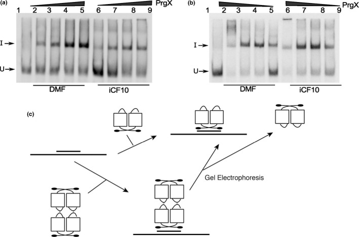
Figure 8.

Binding of Apo‐PrgX and PrgX/I to DNA only containing one operator sequence. EMSA assays were performed using 8 fmol of digoxigenin‐labeled LT DNA probes with increasing amounts of protein. PrgX was preincubated with DMF or I for 5 min before addition of probes. (a) XBS1 DNA template has only the XBS1 binding site. PrgX concentration used: lanes 2–5 and lanes 6–9: 10, 25, 100, and 200 nmol L−1, respectively. (b) XBS2 DNA template has only the XBS2 binding site. PrgX concentration used: lanes 2–5 and lanes 6–9: 200, 100, 25, and 10 nmol L−1, respectively. (c) Binding PrgX to DNA contains a single‐binding site illustrated by cartoon. Gel electrophoresis dissociated PrgX‐C or PrgX‐I tetramers, resulting in band shifts similar to those produced by PrgX dimers
