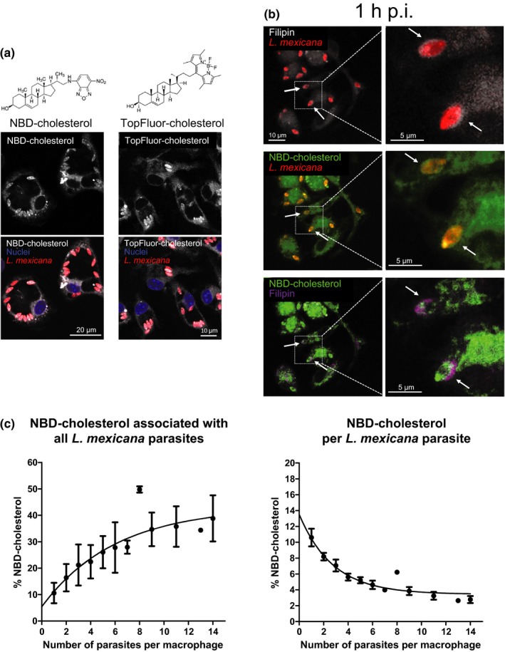
Figure 5.

Fluorophore‐labeled cholesterol does not accumulate around the parasite. (a) Macrophages were preincubated with NBD‐ or TopFluor‐cholesterol for 16 hr. Afterward, macrophages were infected with Leishmania mexicana amastigotes (MOI of 5) for 2 hr. Seventy‐two hours after infection, cells were fixed and subsequently stained with DAPI (nuclei, excited at 405 nm). Both NBD and TopFluor groups were excited at 488 nm. (b) Distribution of filipin‐stainable and fluorophore‐labeled cholesterol in the early phases of infection with L. mexicana. Mouse bone marrow‐derived macrophages were preincubated with NBD‐cholesterol for 16 hr and then infected with L. mexicana amastigotes (MOI of 5) for 2 hr. An hour after infection, cells were fixed and subsequently stained with filipin (excited at 405 nm). For a better identification, filipin‐stained cholesterol in the presence of NBD‐cholesterol is visualized in purple. Arrows indicate Leishmania parasites and/or filipin around the parasites. All images are confocal and were acquired using a Zeiss LSM 780 microscope, 63× oil‐immersion objective, and processed using identical conditions. (c) Relative fluorescence corresponding to NBD‐cholesterol incorporated by all the parasites (left panel) or a single parasite (right panel) in an infected macrophage is represented as a function of the number of parasites internalized by the macrophages over a period of 48 hr postinfection. The exponential curve regressions were calculated using GraphPad Prism
