

Abstract
Capillaria spp infection was diagnosed in a 3 year old dog with history of pollakiuria, dysuria and haematuria. Confirmatory diagnosis was done by demonstration of the parasitic ova in the urinary sediment, cystitis by ultrasonography, presence of proteins and leukocytes in the urine. Dog was successfully treated with fenbendazole and ivermectin along with supportive therapy. Recovery was noticed after 2 weeks of treatment with clear and transparent urine and absence of parasitic ova in urinary sediment.
Keywords: Capillaria spp, Cystitis, Dog, Urinalysis
Introduction
Capillaria plica is a worldwide distributed nematode parasite of the urinary tract in carnivores. Capillaria spp eggs passed through the urine and contaminate the environments which become source of infection to other dogs (Inforzato and Santos 2009). Usually, dogs affected with urinary capillariosis do not show any signs but due to heavy infection, dogs may exhibit pollakiuria, cystitis, dysuria and inappropriate micturition (Studzinska et al. 2015). Capillaria spp infection in the bladder of dogs, rarely reported and it is self limiting in nature (Sarma et al. 2011; Basso et al. 2014). The purpose of this report is diagnosis of cystitis caused by Capillaria spp and it’s therapeutic management in a dog.
Case history and observations
A three year old male dog presented to the Teaching Veterinary Clinical Complex, College of Veterinary Science, Proddatur with history of pollakiuria, occasional haematuria. Upon examination dog showed dysuria and restlessness. Temperature, pulse and respiration recorded were 101.4 °F, 96/min and 28/min respectively. Whole blood, urine, peripheral blood smears were collected for laboratory examination (Reddy et al. 2014a). Abdominal ultrasonography was also done.
Peripheral blood smear examination did not reveal any haemoprotozoans. The haematological values were haemoglobin 12.8 gm/dL, total erythrocyte count 5.68 × 10⁶/cumm, total leucocyte count 13.6 × 10³/cumm, Neutrophil (8704/cumm), Lymphocyte (3808/cumm), Eosinophil (952/cumm) and Monocyte (136/cumm). The ultrasound evaluation of urinary bladder showed thickened hyperechogenicity bladder wall (Fig. 1). The urine appeared to be cloudy, alkaline in nature (pH 7.5) and uristic urine test strip was positive for blood, leukocytes, protein and nitrite. Dog was treated for cystitis with cefpodoxime proxetil (@5 mg/kg body weight, PO, SID) and prednisolone (@1 mg/kg Body weight, PO) for 2 weeks. After treatment, cloudiness of the urine was reduced but presence of pollakiuria was noticed.
Fig. 1.
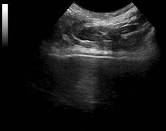
Thickened urinary bladder wall indicative of cystitis
Again urine was collected and microscopic examination of the urinary sediments revealed the presence of Capillaria plica ova along with few erythrocytes, leucocytes and epithelial cells. Ova were in size of 55–65 × 25–30 μm, barrel-shaped, with two opercules with polar plugs (Fig. 2).
Fig. 2.

Presence of Capillaria spp. ova in urinary sediment examination
Treatment and discussion
After confirmation of Capillaria spp infection, dog was treated with oral fenbendazole @ 50 mg/kg body weight/day for 10 days and two doses of ivermectin @ 0.2 mg/kg body weight subcutaneously at weekly interval. Two weeks after treatment, complete clinical recovery was noticed with absence of pollakiuria and dysuria. Repeated urine examination did not reveal any abnormalities. Based on the ultrasonography and urine analysis it was treated for cystitis with cefpodoxime which acts on both gram positive and negative bacteria (Reddy et al. 2014b). On the first day of presentation urine sediment was not examined because it is not a routine activity and case was diagnosed to be cystitis. We thought cystitis might be due to a bacterial infection alone.
After incomplete response to the antibiotic therapy, examination of urine sediment was carriedout to detect the further abnormalities. Diagnosis of infection was according by demonstration of parasitic ova in urinary sediment (Studzinska et al. 2015). Ultrasonographically inflammatory contents were noticed as hyperechoic foci or debris in bladder along with thickening of bladder wall which confirms the cystitis in this case. Adult Capillaria spp worms affect mainly urinary bladder but may be occasionally localized in the ureters and renal pelvis. Presence of the adult worms in the bladder damage the mucus membranes causes inflammation which leads to secondary bacterial infection (Deplazes et al. 2013). Similar condition was observed and treated by different authors with different drugs with fenbendazole, albendazole, levamisole and ivermectin (Basso et al. 2014, Studzinska et al. 2015). We preferred fenbendazole for this case because of easy availability. Dogs with refractory cystitis should be checked for occurrence of Capillaria spp.
Conclusion
Present study reports about the diagnosis of Capillaria spp infection through urine sediment examination, cystitis by ultrasonography and their successful therapeutic approach in a dog.
Acknowledgments
Authors are thankful to the authorities of the Sri Venkateswara Veterinary University, Tirupati for providing necessary facilities.
References
- Basso W, Spänhauer Z, Arnold S, Deplazes P. Capillaria plica (syn. Pearsonema plica) infection in a dog with chronic pollakiuria: challenges in the diagnosis and treatment. Parasitol Int. 2014;63(1):140–142. doi: 10.1016/j.parint.2013.09.002. [DOI] [PubMed] [Google Scholar]
- Deplazes P, Eckert J, von Samson-Himmelstjerna G, Zahner H. Lehrbuch der Parasitologie für die Tiermedizin. 3. Stuttgart: Enke; 2013. [DOI] [PubMed] [Google Scholar]
- Inforzato GR, Santos WRM. Capilariose em gatos. Revista Eletronica de Medicina Veterinaria. 2009;7(12):75–78. [Google Scholar]
- Reddy BS, Sivajothi S, Reddy LSSV, Raju KGS. Clinical and laboratory findings of Babesia infection in dogs. J Parasit Dis. 2014 doi: 10.1007/s12639-014-0491-x. [DOI] [PMC free article] [PubMed] [Google Scholar]
- Reddy BS, Kumari KN, Rao VV. Rayulu VC (2014b) Efficacy of cefpodoxime with clavulanic acid in the treatment of recurrent pyoderma in dogs. ISRN Vet Sci. 2014;467010:5. doi: 10.1155/2014/467010. [DOI] [PMC free article] [PubMed] [Google Scholar]
- Sarma DK, Goswami S, Talukdar SK, Kalita D. Urinary bladder worm in a dog and its successful treatment. Intas Polivet. 2011;12(I):107. [Google Scholar]
- Studzinska MB, Gałek JO, Kutrzepa MD, Tomczuk K. Diagnosis and therapy of Capillaria plica infection: report and literature review. Acta Parasitol. 2015;60(3):563–566. doi: 10.1515/ap-2015-0081. [DOI] [PubMed] [Google Scholar]