Abstract
Rift Valley fever virus (RVFV) is an arthropod-borne bunyavirus that can cause serious and fatal disease in humans and animals. RVFV is a negative-sense RNA virus of the Phlebovirus genus in the Bunyaviridae family. The main envelope RVFV glycoproteins, Gn and Gc, are encoded on the M segment of RVFV and known inducers of protective immunity. In an attempt to develop a safe and efficacious RVF vaccine, we constructed and tested a vectored equine herpesvirus type 1 (EHV-1) vaccine that expresses RVFV Gn and Gc. The Gn and Gc genes were custom-synthesized after codon optimization and inserted into EHV-1 strain RacH genome. The rH_Gn-Gc recombinant virus grew in cultured cells with kinetics that were comparable to those of the parental virus and stably expressed Gn and Gc. Upon immunization of sheep, the natural host, neutralizing antibodies against RVFV were elicited by rH_Gn-Gc and protective titers reached to 1:320 at day 49 post immunization but not by parental EHV-1, indicating that EHV-1 is a promising vector alternative in the development of a safe marker RVFV vaccine.
Main text
Rift Valley fever virus (RVFV) is an arthropod-borne virus that can cause serious health problems in both animals and humans [1, 2]. The disease caused by RVFV in ruminants is characterized by an acute hepatitis, abortion in pregnant animals and high mortality rates, especially in newborns [3, 4]. In humans, the virus usually leads to a mild flu-like febrile illness but in some cases, it can cause severe symptoms, such as hemorrhagic fever, hepatitis, encephalitis, and retinal degeneration [5–7]. RVFV can be transmitted from infected animals to humans, especially when humans are in contact with infected animals. Of particularly high risk are blood and aborted fetuses including the amniotic fluid and secundina [6, 8]. RVFV was first isolated from sheep during an epizootic in the Rift Valley of Kenya in 1931. RVFV is an enveloped RNA virus and belongs to the Phlebovirus genus in the Bunyaviridae family. The genome of the Bunyaviridae is comprised of three segments of negative-sense, single-stranded RNA that are referred to as S (small), M (medium), and L (large) with a total genome size of approximately 11.9 kb [9–11]. The M segment encodes the two major envelope surface glycoproteins Gn and Gc and two non-structural proteins NSm1 and NSm2. The Gn and Gc with molecular masses of 57- and 55-KDa, respectively [12, 13], form a heterodimer processed from a polyprotein by host proteases in the endoplasmic reticulum (ER). The glycoproteins are the main target of protective immunity against RVFV infection [14, 15]. Antibodies against surface Gn and Gc can effectively neutralize RVFV by blocking virus-receptor interactions and virus-cell entry [15]. In addition, it may also play a role in complement-mediated clearance of RVFV [13, 16]. Hence, Gn and Gc are the main targets for vaccine development [12, 13, 16–23].
Although the live attenuated [24] and inactivated vaccines [25–27] have been licensed for veterinary use, they still have some drawbacks. The ideal RVFV vaccine would be the one that (i) is safe, (ii) elicits rapid humoral immune responses that neutralize RVFV, and (iii) induces long-term protective immunity. Therefore, this study presents a different approach, using an EHV-1 strain RacH as the delivery vector. Equine herpesvirus type 1 (EHV-1) is a member of the genus Varicellovirus in the subfamily Alphaherpesvirinae. It possesses a double-stranded DNA genome of 150 Kbp in length. EHV-1 is capable of entering a wide variety of cell types of different origins and its attenuation could be attributed to deletion of both copies of gene 67 [28–30]. The EHV-1 vaccine strain RacH has been cloned as an infectious bacterial artificial chromosome (BAC) [31] and developed as a universal live virus vector against various viruses. RacH has a proven safety record and can induce both humoral and cellular immune responses to transgenes introduced in the vector and provide protection in vaccinated animals, including mice, dogs, cattle and swine [32–38]. In the present study, we describe the construction and evaluation of a RacH-vectored vaccine expressing Gn and Gc of RVFV (rH_Gn-Gc). We show that recombinant EHV-1 stably expresses Gn-Gc and induces a Gn-Gc-specific neutralizing antibody response in a natural host of RVFV, sheep.
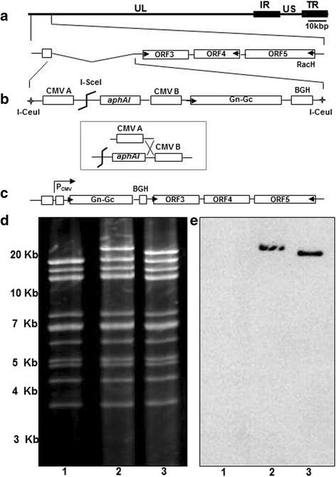
The Gn-Gc sequence of an Egyptian isolate of RVFV (ZH-501 strain; GenBank accession number DQ380200.1) was commercially synthesized after codon optimization (Genscript). Gn-Gc sequences were PCR-amplified from the commercial plasmid using Phusion high-fidelity DNA polymerase (New England Biolabs) with oligonucleotides primers P1 (TATGGATCCATGGCTGGAATTGCTATGACT) and P2 (TATGCGGCCGCTTAATTAATCTAGATTATCT) and cloned into the BamHI/NotI site of pEP-CMV-in [39] to generate pEP_Gn-Gc. The expression cassette containing RVF Gn-Gc under the control of HCMV IE promoter was released from pEP_Gn-Gc by digestion with SpeI and SphI, and subcloned into the SpeI/SphI sites of pUC19_ORF1/2, resulting in the transfer plasmid pUC19_ORF1/2-Gn-Gc. By digestion of pUC19_ORF1/2-Gn-Gc with I-CeuI, a 6.7 kbp fragment containing the Gn-Gc gene expression cassette, a kanamycin resistance gene (aphAI) and two flanking sequences was released and inserted in lieu of ORF1/2 of pRacH1-EF1 using two-step Red-mediated recombination (Fig. 1) as previously described [39]. The EHV-1 RacH BAC clone, pRacH1-EF1 (termed pH 1-EF1 in this study) was generated previously by replacing the HCMV IE promoter upstream of egfp gene in the mini-F with human elongation factor promoter 1α (EF-1α) [36, 37]. In the first recombination, insertion of Gn-Gc sequences and the aphA1 gene resulted in kanamycin-resistant intermediates that differed from parental pH 1-EF1 BAC in the EcoRV restriction pattern. As predicted in silico, the insertion of the cassette resulted in an EcoRV fragment of 21,535 bp in size compared to the 16,411 bp in the parental pH 1-EF1 (Fig. 1d). In the second recombination step, the aphA1 gene was removed, which led to the reduction in size of the 21,535 bp EcoRV fragment to 20,557 bp (Fig. 1d). The results of the RFLP analysis were confirmed by Southern blotting, which revealed that only the 21,535 and 20,557 bp EcoRV bands in the intermediate and resolved recombinant, respectively, were reactive with Gn-Gc-specific probes P3 (GCCCGATTCTTTTGTGTGCT) and P4 (AATCCGTGAAGAGGCCTGGA) (Fig. 1e). Nucleotide sequencing using oligonucleotides primers P5 (GCCGAGCGAGTTCGGCATCCT), P6 (GCCATCCTGGACCAGAACAA), P7 (GCAGGAGATCAGGAAGGCCT), P8 (CCAGCGCCATCATCGAGACC), P9 (GAGAAGCAGAAGCCCTACTT), P10 (GTGCGTGGAGAGCGAGCTGC), P11 (AGATGGAGGGCAGCCTGGCC), P12 (TCGGTCTTGGCCAGCAGCTT), P13 (GGAGCCACTGGCTCAGCTCT), P14 (GGGTGGAAGTCGGTGAAGGT), P15 (GTTCATGTCCAGCACCTCGT), P16 (CGTTGCTGCCCTTCTTGAAG), P17 (CTTGCGGTGTCGTCCTCTCC), and P18 (CTTCCGCTTGCTCTCCTCCT) further confirmed the correct insertion of the gene at the left genomic terminus of the pH 1-EF1 clone that otherwise appeared unaltered (data not shown). From the above results, we concluded that the generated recombinant pH1_Gn-Gc BAC harbored the RVFV Gn-Gc sequences in the targeted locus.
Fig. 1.

Generation of recombinant EHV-1 expressing Gn-Gc protein of RVFV (rH_Gn-Gc). Schematic illustration of the construction of rH_Gn-Gc vaccine vector based on pRacH1. a Depiction of the left terminus of the unique-long segment of EHV-1 strain RacH infectious BAC clone pH 1-EF1, in which ORF1 and ORF2 are naturally deleted. b A fragment released from transfer plasmid pUC19-ORF1/2-Gn-Gc by I-CeuI digestion was used to recombine with RacH genome, result in incorporation of Gn-Gc gene of RVFV, HCMV promoter and kanamycin resistance gene in the ORF1/ORF2 locus of the RacH genome. c After I-SceI digestion, kanamycin was removed in the following step of en passent mutagenesis to generate the final arrangement of rH_Gn-Gc genome. d and e Restriction fragment length polymorphisms and southern blot of pH1_EF1, the cloning intermediate and the final pH_Gn-Gc construct. An ethidium bromide-stained agarose gel is shown in the left panel with EcoRV restriction patterns of pH_EF1 (lanes 1), the kanamycin-resistant intermediate (lanes 2) and pH_Gn-Gc (lanes 3). GeneRuler 1 kb Plus DNA Ladder (Thermo Scientific) was used for determination of DNA fragment sizes. In the right panel, a southern blot of the same gel is shown after hybridization with a digoxigenin-labeled Gn-Gc RVFV probe
Parental RacH virus (rH) and the recombinant rH expressing Gn and Gc of RVFV (rH_Gn-Gc) were propagated in rabbit kidney (RK13) cells. Cultures were maintained in modified Eagle’s medium (MEM) (Biochrom) supplemented with 5% fetal bovine serum (FBS, Biochrom), 100 U/ml penicillin, and 100 μg/ml streptomycin (1% penicillin–streptomycin). Reconstitution of recombinant and parental viruses was achieved by transfection of pH1_Gn-Gc or pH 1-EF1 DNA into RK13 cells using polyethylenimine (PEI) (Polysciences). Reconstitution of EHV-1 gp2-encoding sequences with subsequent removal of mini-F sequences was achieved by co-transfection of 1 μg BAC DNA and 10 μg plasmid DNA p71H containing the full-length ORF71, which encodes gp2, in RK13 cells [40]. Three days after co-transfection, nonfluorescing-virus plaques were picked and purified to homogeneity by two rounds of plaque purification, and virus stocks were prepared and stored at −80 °C for further use.
To compare the in vitro growth properties of rH_Gn-Gc with those of parental rH, plaque diameters and single-step growth kinetics were determined. Plaque areas of rH_Gn-Gc were compared to those of parental rH virus, which was set as 100%. Mean percentages and standard deviations were calculated from three independent experiments. The Shapiro-Wilks test was used to assess for normality and Student’s t-test was employed to compare the mean areas of the plaques of the examined viruses. Our results shown that the average diameter of rH_Gn-Gc plaques was reduced in size by approximately 17% compared to parental virus (Fig. 2a); however, this reduction did not reach statistical significance (p = 0.31). To determine single-step growth kinetics, RK13 cells seeded in 12-well plates were infected at a multiplicity of infection (moi). of 3. Viruses were allowed to attach for 1 h at 4 °C, followed by a penetration step of 1.5 h at 37 °C. After washing twice with PBS, infected cells were treated with ice-cold citrate buffered saline for 3 min to remove residual virus. At different time points (0, 4, 8, 12, 24 and 36 h p.i.), supernatants and cells were harvested separately, and intracellular and extracellular viral titers were determined using plaque assay. Single-step growth curves were determined in three independent experiments and means and standard deviations were computed and plotted. Student’s t-test was used to test the differences of viral growth kinetics of examined viruses. Both viruses exhibited comparable virus titers during the 36 h observation period, with respect to both extracellular and intracellular titers (Fig. 2b). Virus titers at the end of the observation period were virtually identical between the analyzed viruses. From these results, we concluded that the insertion of transgene did not have a marked effect on viral growth in vitro.
Fig. 2.

Comparison of in vitro growth properties of rH_Gn-Gc with those of parental virus. a RK13 cells were infected by viruses at moi of 0.001 and overlaid. Fifty plaques per virus were photographed and the areas were measured. b The single-step growth kinetics of those viruses was analyzed and revealed no significant differences in growth properties of parental and recombinant viruses. Error bars represent standard deviations. These results are representative of three independent experiments
To evaluate expression of the Gn or Gc by rH_Gn-Gc, indirect immunofluorescence (IF) was used as described before [30, 36]. RK13 cells were infected either with rH_Gn-Gc or rH for 24 h and then incubated with rabbit anti-RVFV(CT) (ProSci catalog no. 4521) or rabbit anti-RVFV(IN) (ProSci catalog no. 4519), that recognize the RVFV Gn or Gc, respectively, for 1 h at RT. After extensive washing with PBS, the secondary antibody, anti-rabbit IgG conjugated with Alexa 488 (Invitrogen), was added at a 1:500 dilution and incubated for 30 min at RT. After thorough washing, plaques were inspected by using an inverted fluorescence microscope Zeiss Axiovert 100 and plaques recorded with the Axiocam (Zeiss). In the case of rH_Gn-Gc, virus plaques were reactive with both RVFV(IN) and RVFV(CT) pAb, whereas those induced by parental virus were not (Fig. 3a). Gn and Gc expression were also assessed by western blot analysis as described before [30, 36]. Expression of Gn and Gc was detected with the same antibodies and horseradish peroxidase-conjugated goat anti-rabbit pAb that was obtained from Southern Biotech. Expression of β-actin was assessed as a loading control using rabbit anti-β-actin polyclonal antibody (pAb) purchased from Cell Signaling Technologies. Reactive bands were visualized by enhanced chemoluminescence (ECL plus, Amersham). Proteins of approximately 57- and 55-kDa in size were reactive with the anti-RVFV(IN) and anti-RVFV(CT) antibody, respectively, in lysates of cells infected with the Gn-Gc expressing rH_Gn-Gc, but was absent in cells that were mock-infected or infected with parental virus (Fig. 3b). Our findings are in agreement with previous reports indicating that the Gn (57-KDa) and Gc (55-KDa) are produced from a single protein precursor [12, 13]. We concluded from our results that the recombinant rH_Gn-Gc efficiently expressed the RVFV Gn and Gc proteins in vitro.
Fig. 3.

Expression of Gn-Gc protein after infection with parental rH and rH_Gn-Gc virus. a Immunofluorescence staining of RK13 cells infected with either parental rH or rH_Gn-Gc virus. Plaques were stained with anti-RVFV(IN) (i and ii) or anti-RVFV(CT) (iii and iv) pAb, that was reactive with RVFV Gn or Gc, respectively, followed by Alexa Flour488-conjugated goat anti-rabbit IgG. Plaques in infected cells with rH_Gn-Gc visualized by fluorescence microscopy (i and iii), whereas those induced by rH virus were not (ii and iv). b Cell lysates either mock infected, infected by rH or rH_Gn-Gc were separated by 10% SDS-PAGE and analysed by Western blot. Expression of Gn and Gc was detected using pAb-RVFV(IN) (left panel) and -RVFV(CT) (right panel), respectively. Expression of β-actin was determined as a loading control. The PageRulerTM Prestained protein ladder (Thermo Scientific) was used for determination of protein sizes
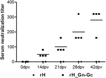
To test whether the rH_Gn-Gc virus could induce an RVFV specific antibody response in vivo, serological studies were done in sheep to determine whether rH_Gn-Gc was capable of inducing neutralizing antibody responses against RVFV in the natural host. All animals were screened with an enzyme linked immunosorbent assay to test for the presence of antibodies against RVFV Gn and Gc before immunization (data not shown). All sheep used in these studies were housed in isolation rooms at the Veterinary Serum Vaccine Research Institute, Cairo, Egypt. Animal care procedures were in accordance with state animal welfare guidelines under the supervision of an ethics committee. One- to five-year-old sheep were allocated randomly to two groups, with 4 sheep in group 1 and 2 animals in group 2. In group 1, sheep were immunized twice in a 3-week interval with rH_Gn-Gc (1 × 10⁵ PFU/ml) by intramuscular (IM) inoculation. In group 2 (control group), sheep were inoculated by the same route and virus amount with parental rH virus. Serum neutralizing antibodies to RVFV were determined in serum samples, collected from both group at the indicated day post vaccination (0, 14, 21, 35 and 42) by standard serum neutralization test (SNT) as described previously [41]. For SNT, RVFV strain ZH501 isolated from a human patient during the outbreak of 1977 in Egypt and kindly provided by the Naval Medical Research Unit 3 (NAMRU-3) Cairo, Egypt, was used. The virus was propagated on baby hamster kidney 21 (BHK-21) cells at the Veterinary Serum Vaccine Research Institute, Cairo, Egypt. Serum samples were examined by SNT, which revealed that all animals immunized with rH_Gn-Gc mounted high antibody titers against RVFV (Fig. 4). As expected, all animals in the rH-immunized group did not induce any RVFV-specific antibody (Fig. 4). Reduction in plaques size of RVFV by 50% (PRNT50) compared with control was used to quantify titer of neutralizing antibody. Our results showed that the endpoint protection titer (50% protective titer) ranged between 1:40 to 1:80 in immunized animals after first immunization dose, while the protective titers reached to 1:320 by day 49 of immunization (Fig. 4). These titers are within the range associated with protection against RVF challenge in sheep in a previous study [42]. We concluded from our findings that the engineered EHV-1 vector expressing RVFV Gn-Gc is able to induce robust neutralizing antibody responses in immunized natural hosts for RVFV.
Fig. 4.

Neutralizing antibody response induced by rH_Gn_Gc. Sheep were primed and boosted with either rH or rH_Gn-Gc. Blood samples were collected from immunized sheep at indicated days 0, 14, 21, 28 and 42. Serum from immunized sheep was titrated by a standard serum neutralization test (SNT). Each dot represents an individual sheep
Currently, no RVFV vaccines for humans are commercially available, but live attenuated [24] and inactivated vaccines [25–27] have been licensed for veterinary use in endemic countries. While the live attenuated vaccine is able to induce long-lasting immunity and satisfactory protection if administrated properly, its safety has been questionable. Abortions in pregnant ewes and illness in European cattle [24] were reported, as was the potential recombination with field strains and reversion to virulence during the vaccine manufacturing process. Therefore, new approaches are necessary to develop safe and effective RVFV vaccines. Several viral recombinant vectored vaccines have been developed. Those are based on vaccinia virus [43], Newcastle disease virus [12], adenovirus [13], Venezuelan equine encephalitis virus [44] or capripoxvirus [18, 45].
In this study, we explored the feasibility of using EHV-1 as a vehicle to deliver Gn-Gc of RVFV. The potential of EHV-1 as a universal vector for immunization has been previously demonstrated, including its high packaging capacity, broad cell tropism, and the lack of pre-existing anti-vector immunity in non-equine animals [46]. As a live vector, EHV-1 strain RacH has been developed and proved useful in inducing both humoral and cellular immune responses and providing protection in a number of experimental systems and of different animals, including mice, dogs, swine and cattle [32–38]. RVFV Gn-Gc are essential and sufficient for immune protection, as reported in the previous studies using baculovirus and sheeppox expression of these same two RVFV proteins [23]. Gn-Gc sequences under the control of the HCMV IE promoter was inserted in the ORF1 locus, which encodes a protein mediating evasion of T-cell immunity [47, 48]. In line with previous studies [32–37], insertion of Gn-Gc into the EHV-1 genome did not affect in vitro growth characteristics and the recombinant virus was able to replicate in cell culture as efficiently as the parental virus and stably expressed Gn-Gc. Importantly, when we inoculated the recombinant virus into sheep, a RVFV-specific neutralizing antibody response was induced following IM administration in sheep. Antibody titers were maintained at high levels up to the time point when the experiments were terminated.
In summary, we developed a recombinant EHV-1 vaccine encoding RVFV Gn-Gc and evaluated its potential as a vaccine by measurement of RVFV-specific neutralizing antibody in sheep. Our results show that EHV-1 could be used as an alternative live vector for RVFV immunization in sheep. Future study will be designed to determine whether the recombinant EHV-1-vectored Gn-Gc vaccine is capable to protect sheep against challenge infection.
Acknowledgments
The authors would like to thank Veterinary Serum Vaccine Research Institute, Cairo, Egypt for providing animal facilities to achieve this study.
Funding
This work was supported by a restricted grant from the Freie Universität Berlin to N.O. and in part by grant from the Egyptian Ministry of Education of A.S.
Availability of data and materials
The datasets supporting the results of this article are included within the article.
Abbreviations
- aphI
Kanamycin resistance gene
- BAC
Bacterial artificial chromosome
- DNA
Deoxyribonucleic acid
- egfp
Enhanced green fluorescence protein
- EHV-1
Equine herpesvirus type1
- ER
Endoplasmic reticulum
- Gc
Glycoprotein C
- Gn
Glycoprotein N
- HCMV
Human cytomegalovirus
- IM
Intramuscular
- L
Large
- M
Medium
- moi
Multiplicity of infection
- Ns
Non-structural protein
- ORF
Open reading frame
- PEI
Polyethylenimine
- PFU
Plaques forming unit
- rH
Recombinant equine herpesvirus RacH strain
- RK13
Rabbit kidney cells
- RNA
Ribonucleic acid
- RVFV
Rift Valley Fever Virus
- S
Small
- SNT
Serum Neutralization test
Authors’ contributions
AS contributed to this work by designing, performing the experiments, analyzing the data and drafting of the manuscript. ME, AMD, and GM contributed to analyzing the data and drafting of the manuscript. NO contributed to the designing, analyzing the data, drafting of the manuscript, and giving final approval of the version to be published. All authors read and approved the final manuscript.
Ethics approval
All sheep used in these studies were housed in isolation rooms at the Veterinary Serum Vaccine Research Institute, Cairo, Egypt and all animals were not killed for this scientific research. Animal care procedures were in accordance with state animal welfare guidelines under the supervision of an ethics committee.
Consent for publication
Not applicable.
Competing interests
The authors declare that they have no competing interests.
Publisher’s Note
Springer Nature remains neutral with regard to jurisdictional claims in published maps and institutional affiliations.
Contributor Information
Abdelrahman Said, Phone: +49-30-838-51822, Email: abdosaid79@gmail.com.
Nikolaus Osterrieder, Phone: +49-30-838-51822, Email: no.34@fu-berlin.de.
References
- 1.Flick R, Bouloy M. Rift Valley fever virus. Curr Mol Med. 2005;5(8):827–834. doi: 10.2174/156652405774962263. [DOI] [PubMed] [Google Scholar]
- 2.Gerdes GH. Rift Valley fever. Rev Sci Tech. 2004;23(2):613–623. doi: 10.20506/rst.23.2.1500. [DOI] [PubMed] [Google Scholar]
- 3.Peters CJ, Liu CT, Anderson GW, Jr, Morrill JC, Jahrling PB. Pathogenesis of viral hemorrhagic fevers: Rift Valley fever and Lassa fever contrasted. Rev Infect Dis. 1989;11(Suppl 4):S743–S749. doi: 10.1093/clinids/11.Supplement_4.S743. [DOI] [PubMed] [Google Scholar]
- 4.Balkhy HH, Memish ZA. Rift Valley fever: an uninvited zoonosis in the Arabian peninsula. Int J Antimicrob Agents. 2003;21(2):153–157. doi: 10.1016/S0924-8579(02)00295-9. [DOI] [PubMed] [Google Scholar]
- 5.Easterday BC. Rift valley fever. Adv Vet Sci. 1965;10:65–127. [PubMed] [Google Scholar]
- 6.Madani TA, Al-Mazrou YY, Al-Jeffri MH, Mishkhas AA, Al-Rabeah AM, Turkistani AM, et al. Rift Valley fever epidemic in Saudi Arabia: epidemiological, clinical, and laboratory characteristics. Clin infect Dis. 2003;37(8):1084–1092. doi: 10.1086/378747. [DOI] [PubMed] [Google Scholar]
- 7.Laughlin LW, Meegan JM, Strausbaugh LJ, Morens DM, Watten RH. Epidemic Rift Valley fever in Egypt: observations of the spectrum of human illness. Trans R Soc Trop Med Hyg. 1979;73(6):630–633. doi: 10.1016/0035-9203(79)90006-3. [DOI] [PubMed] [Google Scholar]
- 8.Mohamed M, Mosha F, Mghamba J, Zaki SR, Shieh WJ, Paweska J, et al. Epidemiologic and clinical aspects of a Rift Valley fever outbreak in humans in Tanzania, 2007. Am J Trop Med Hyg. 2010;83(2 Suppl):22–27. doi: 10.4269/ajtmh.2010.09-0318. [DOI] [PMC free article] [PubMed] [Google Scholar]
- 9.Muller R, Poch O, Delarue M, Bishop DH, Bouloy M. Rift Valley fever virus L segment: correction of the sequence and possible functional role of newly identified regions conserved in RNA-dependent polymerases. J Gen Virol. 1994;75(Pt 6):1345–1352. doi: 10.1099/0022-1317-75-6-1345. [DOI] [PubMed] [Google Scholar]
- 10.Giorgi C, Accardi L, Nicoletti L, Gro MC, Takehara K, Hilditch C, et al. Sequences and coding strategies of the S RNAs of Toscana and Rift Valley fever viruses compared to those of Punta Toro, Sicilian Sandfly fever, and Uukuniemi viruses. Virology. 1991;180(2):738–753. doi: 10.1016/0042-6822(91)90087-R. [DOI] [PubMed] [Google Scholar]
- 11.Collett MS. Messenger RNA of the M segment RNA of Rift Valley fever virus. Virology. 1986;151(1):151–156. doi: 10.1016/0042-6822(86)90114-5. [DOI] [PubMed] [Google Scholar]
- 12.Kortekaas J, de Boer SM, Kant J, Vloet RP, Antonis AF, Moormann RJ. Rift Valley fever virus immunity provided by a paramyxovirus vaccine vector. Vaccine. 2010;28(27):4394–4401. doi: 10.1016/j.vaccine.2010.04.048. [DOI] [PubMed] [Google Scholar]
- 13.Holman DH, Penn-Nicholson A, Wang D, Woraratanadharm J, Harr MK, Luo M, et al. A complex adenovirus-vectored vaccine against Rift Valley fever virus protects mice against lethal infection in the presence of preexisting vector immunity. Clin Vaccine Immunol. 2009;16(11):1624–1632. doi: 10.1128/CVI.00182-09. [DOI] [PMC free article] [PubMed] [Google Scholar]
- 14.Gerrard SR, Nichol ST. Synthesis, proteolytic processing and complex formation of N-terminally nested precursor proteins of the Rift Valley fever virus glycoproteins. Virology. 2007;357(2):124–133. doi: 10.1016/j.virol.2006.08.002. [DOI] [PMC free article] [PubMed] [Google Scholar]
- 15.Huiskonen JT, Overby AK, Weber F, Grunewald K. Electron cryo-microscopy and single-particle averaging of Rift Valley fever virus: evidence for GN-GC glycoprotein heterodimers. J Virol. 2009;83(8):3762–3769. doi: 10.1128/JVI.02483-08. [DOI] [PMC free article] [PubMed] [Google Scholar]
- 16.Lopez-Gil E, Lorenzo G, Hevia E, Borrego B, Eiden M, Groschup M, et al. A single immunization with MVA expressing GnGc glycoproteins promotes epitope-specific CD8+−T cell activation and protects immune-competent mice against a lethal RVFV infection. PLoS Negl Trop Dis. 2013;7(7):e2309. doi: 10.1371/journal.pntd.0002309. [DOI] [PMC free article] [PubMed] [Google Scholar]
- 17.Faburay B, Lebedev M, McVey DS, Wilson W, Morozov I, Young A, et al. A glycoprotein subunit vaccine elicits a strong Rift Valley fever virus neutralizing antibody response in sheep. Vector Borne Zoonotic Dis. 2014;14(10):746–756. doi: 10.1089/vbz.2014.1650. [DOI] [PMC free article] [PubMed] [Google Scholar]
- 18.Soi RK, Rurangirwa FR, McGuire TC, Rwambo PM, DeMartini JC, Crawford TB. Protection of sheep against Rift Valley fever virus and sheep poxvirus with a recombinant capripoxvirus vaccine. Clin Vaccine Immunol. 2010;17(12):1842–1849. doi: 10.1128/CVI.00220-10. [DOI] [PMC free article] [PubMed] [Google Scholar]
- 19.Mandell RB, Koukuntla R, Mogler LJ, Carzoli AK, Freiberg AN, Holbrook MR, et al. A replication-incompetent Rift Valley fever vaccine: chimeric virus-like particles protect mice and rats against lethal challenge. Virology. 2010;397(1):187–198. doi: 10.1016/j.virol.2009.11.001. [DOI] [PMC free article] [PubMed] [Google Scholar]
- 20.de Boer SM, Kortekaas J, Antonis AF, Kant J, van Oploo JL, Rottier PJ, et al. Rift Valley fever virus subunit vaccines confer complete protection against a lethal virus challenge. Vaccine. 2010;28(11):2330–2339. doi: 10.1016/j.vaccine.2009.12.062. [DOI] [PubMed] [Google Scholar]
- 21.Ikegami T, Makino S. Rift valley fever vaccines. Vaccine. 2009;27(Suppl 4):D69–D72. doi: 10.1016/j.vaccine.2009.07.046. [DOI] [PMC free article] [PubMed] [Google Scholar]
- 22.Papin JF, Verardi PH, Jones LA, Monge-Navarro F, Brault AC, Holbrook MR, et al. Recombinant Rift Valley fever vaccines induce protective levels of antibody in baboons and resistance to lethal challenge in mice. Proc Natl Acad Sci U S A. 2011;108(36):14926–14931. doi: 10.1073/pnas.1112149108. [DOI] [PMC free article] [PubMed] [Google Scholar]
- 23.Schmaljohn CS, Parker MD, Ennis WH, Dalrymple JM, Collett MS, Suzich JA, et al. Baculovirus expression of the M genome segment of Rift Valley fever virus and examination of antigenic and immunogenic properties of the expressed proteins. Virology. 1989;170(1):184–192. doi: 10.1016/0042-6822(89)90365-6. [DOI] [PubMed] [Google Scholar]
- 24.Botros B, Omar A, Elian K, Mohamed G, Soliman A, Salib A, et al. Adverse response of non-indigenous cattle of European breeds to live attenuated Smithburn Rift Valley fever vaccine. J Med Virol. 2006;78(6):787–791. doi: 10.1002/jmv.20624. [DOI] [PubMed] [Google Scholar]
- 25.Kark JD, Aynor Y, Peters CJ. A Rift Valley fever vaccine trial: 2. Serological response to booster doses with a comparison of intradermal versus subcutaneous injection. Vaccine. 1985;3(2):117–122. doi: 10.1016/0264-410X(85)90060-X. [DOI] [PubMed] [Google Scholar]
- 26.Kark JD, Aynor Y, Peters CJ. A rift valley fever vaccine trial. I. Side effects and serologic response over a six-month follow-up. Am J Epidemiol. 1982;116(5):808–820. doi: 10.1093/oxfordjournals.aje.a113471. [DOI] [PubMed] [Google Scholar]
- 27.Meadors GF, Gibbs PH, Peters CJ. Evaluation of a new Rift Valley fever vaccine: safety and immunogenicity trials. Vaccine. 1986;4(3):179–184. doi: 10.1016/0264-410X(86)90007-1. [DOI] [PubMed] [Google Scholar]
- 28.Hubert PH, Birkenmaier S, Rziha HJ, Osterrieder N. Alterations in the equine herpesvirus type-1 (EHV-1) strain RacH during attenuation. Zentralblatt fur Veterinarmedizin Reihe B Journal of veterinary medicine Series B. 1996;43(1):1–14. doi: 10.1111/j.1439-0450.1996.tb00282.x. [DOI] [PubMed] [Google Scholar]
- 29.Neubauer A, Meindl A, Osterrieder N. Mutations in the US2 and glycoprotein B genes of the equine herpesvirus 1 vaccine strain RacH have no effects on its attenuation. Berliner und Munchener tierarztliche Wochenschrift. 1999;112(9):351–354. [PubMed] [Google Scholar]
- 30.Osterrieder N, Neubauer A, Brandmuller C, Kaaden OR, O'Callaghan DJ. The equine herpesvirus 1 IR6 protein influences virus growth at elevated temperature and is a major determinant of virulence. Virology. 1996;226(2):243–251. doi: 10.1006/viro.1996.0652. [DOI] [PubMed] [Google Scholar]
- 31.Rudolph J, Osterrieder N. Equine herpesvirus type 1 devoid of gM and gp2 is severely impaired in virus egress but not direct cell-to-cell spread. Virology. 2002;293(2):356–367. doi: 10.1006/viro.2001.1277. [DOI] [PubMed] [Google Scholar]
- 32.Rosas C, Van de Walle GR, Metzger SM, Hoelzer K, Dubovi EJ, Kim SG, et al. Evaluation of a vectored equine herpesvirus type 1 (EHV-1) vaccine expressing H3 haemagglutinin in the protection of dogs against canine influenza. Vaccine. 2008;26(19):2335–2343. doi: 10.1016/j.vaccine.2008.02.064. [DOI] [PMC free article] [PubMed] [Google Scholar]
- 33.Rosas CT, Tischer BK, Perkins GA, Wagner B, Goodman LB, Osterrieder N. Live-attenuated recombinant equine herpesvirus type 1 (EHV-1) induces a neutralizing antibody response against West Nile virus (WNV) Virus Res. 2007;125:69–78. doi: 10.1016/j.virusres.2006.12.009. [DOI] [PubMed] [Google Scholar]
- 34.Rosas CT, König P, Beer M, Dubovi EJ, Tischer BK, Osterrieder N. Evaluation of the vaccine potential of an equine herpesvirus type 1 vector expressing bovine viral diarrhea virus structural proteins. J Gen Virol. 2007;88:748–757. doi: 10.1099/vir.0.82528-0. [DOI] [PubMed] [Google Scholar]
- 35.Rosas CT, Paessler S, Ni H, Osterrieder N. Protection of mice by equine herpesvirus type 1 based experimental vaccine against lethal Venezuelan equine encephalitis virus infection in the absence of neutralizing antibodies. Am J Trop Med Hyg. 2008;78(1):83–92. [PubMed] [Google Scholar]
- 36.Said A, Damiani A, Ma G, Kalthoff D, Beer M, Osterrieder N. An equine herpesvirus 1 (EHV-1) vectored H1 vaccine protects against challenge with swine-origin influenza virus H1N1. Vet Microbiol 2011;154(1–2):113-123. doi: 10.1016/j.vetmic.2011.07.003. S0378–1135(11)00385–3. [DOI] [PubMed]
- 37.Ma G, Eschbaumer M, Said A, Hoffmann B, Beer M, Osterrieder N. An equine herpesvirus type 1 (EHV-1) expressing VP2 and VP5 of serotype 8 bluetongue virus (BTV-8) induces protection in a murine infection model. PLoS One. 2012;7(4):e34425. doi: 10.1371/journal.pone.0034425. [DOI] [PMC free article] [PubMed] [Google Scholar]
- 38.Said A, Lange E, Beer M, Damiani A, Osterrieder N. Recombinant equine herpesvirus 1 (EHV-1) vaccine protects pigs against challenge with influenza a(H1N1)pmd09. Virus Res. 2013;173(2):371–376. doi: 10.1016/j.virusres.2013.01.004. [DOI] [PubMed] [Google Scholar]
- 39.Tischer BK, von Einem J, Kaufer B, Osterrieder N. Two-step red-mediated recombination for versatile high-efficiency markerless DNA manipulation in Escherichia Coli. BioTechniques. 2006;40(2):191–197. doi: 10.2144/000112096. [DOI] [PubMed] [Google Scholar]
- 40.von Einem J, Wellington J, Whalley JM, Osterrieder K, O'Callaghan DJ, Osterrieder N. The truncated form of glycoprotein gp2 of equine herpesvirus 1 (EHV-1) vaccine strain KyA is not functionally equivalent to full-length gp2 encoded by EHV-1 wild-type strain RacL11. J Virol. 2004;78(6):3003–3013. doi: 10.1128/JVI.78.6.3003-3013.2004. [DOI] [PMC free article] [PubMed] [Google Scholar]
- 41.Fagbo S, Coetzer JA, Venter EH. Seroprevalence of Rift Valley fever and lumpy skin disease in African buffalo (Syncerus Caffer) in the Kruger National Park and Hluhluwe-iMfolozi park, South Africa. J S Afr Vet Assoc. 2014;85(1):1075. doi: 10.4102/jsava.v85i1.1075. [DOI] [PubMed] [Google Scholar]
- 42.Faburay B, Wilson WC, Gaudreault NN, Davis AS, Shivanna V, Bawa B, et al. A recombinant Rift Valley fever virus glycoprotein subunit vaccine confers full protection against Rift Valley fever challenge in sheep. Sci Rep. 2016;6:27719. doi: 10.1038/srep27719. [DOI] [PMC free article] [PubMed] [Google Scholar]
- 43.Kakach LT, Suzich JA, Collett MS. Rift Valley fever virus M segment: phlebovirus expression strategy and protein glycosylation. Virology. 1989;170(2):505–510. doi: 10.1016/0042-6822(89)90442-X. [DOI] [PubMed] [Google Scholar]
- 44.Gorchakov R, Volkova E, Yun N, Petrakova O, Linde NS, Paessler S, et al. Comparative analysis of the alphavirus-based vectors expressing Rift Valley fever virus glycoproteins. Virology. 2007;366(1):212–225. doi: 10.1016/j.virol.2007.04.014. [DOI] [PMC free article] [PubMed] [Google Scholar]
- 45.Ayari-Fakhfakh E, do Valle TZ, Guillemot L, Panthier JJ, Bouloy M, Ghram A, et al. MBT/pas mouse: a relevant model for the evaluation of Rift Valley fever vaccines. J Gen Virol. 2012;93(Pt 7):1456–1464. doi: 10.1099/vir.0.042754-0. [DOI] [PubMed] [Google Scholar]
- 46.Trapp S, von Einem J, Hofmann H, Kostler J, Wild J, Wagner R, et al. Potential of equine herpesvirus 1 as a vector for immunization. J Virol. 2005;79(9):5445–5454. doi: 10.1128/JVI.79.9.5445-5454.2005. [DOI] [PMC free article] [PubMed] [Google Scholar]
- 47.Ma G, Feineis S, Osterrieder N, Van de Walle GR. Identification and characterization of equine herpesvirus type 1 pUL56 and its role in virus-induced downregulation of major histocompatibility complex class I. J Virol. 2012;86(7):3554–3563. doi: 10.1128/JVI.06994-11. [DOI] [PMC free article] [PubMed] [Google Scholar]
- 48.Said A, Azab W, Damiani A, Osterrieder N. Equine Herpesvirus type 4 UL56 and UL49.5 proteins Downregulate cell surface major Histocompatibility complex class I expression independently of each other. J Virol. 2012;86(15):8059–8071. doi: 10.1128/JVI.00891-12. [DOI] [PMC free article] [PubMed] [Google Scholar]
Associated Data
This section collects any data citations, data availability statements, or supplementary materials included in this article.
Data Availability Statement
The datasets supporting the results of this article are included within the article.


