Abstract
It is generally accepted that myopic defocus is a more potent signal to the emmetropization system than hyperopic defocus: one hour per day of myopic defocus cancels out 11 hours of hyperopic defocus. However, we have recently shown that the potency of brief episodes of myopic defocus at inhibiting eye growth depends on the time of day of exposure. We here ask if this will also be true of the responses to brief periods of hyperopic defocus: may integration of the signal depend on time of day? If so, are the rhythms in axial length and choroidal thickness altered? Hyperopic defocus: Birds had one eye exposed to hyperopic defocus by the wearing of −10D lenses for 2 or 6 hrs at one of 3 times of day for 5 days: Morning (7 am; n=13; n=6), Mid-day (12pm: n=20; 10am–4pm: n=8), or Evening (7pm: n=12; 2–8pm: n=11). A separate group wore monocular lenses continually as a control (n=12). Form deprivation: Birds wore a diffuser over one eye for 2 hrs at one of 3 times of day for 5 days: Morning (n=12); Noon (n=19) or Evening (n=6). For all groups, ocular dimensions were measured using high-frequency A-scan ultrasonography at noon on the first day, under inhalation anesthesia. On day 5, eye dimensions were re-measured at 12 pm, and refractive errors were measured using a Hartinger’s refractometer. A subset of birds in the 2-hr lens group (morning, n=8; mid-day, n=8; evening, n=6), and the deprivation group (n=6 per time point), were measured at 6 pm, 12 am, 6 am and 12 pm on the last day of exposure, to obtain the parameters of the diurnal rhythms in axial length and choroidal thickness. The effects of 2 hrs of defocus depended on time of day of exposure: it stimulated eye growth when exposure was in the morning and inhibited it at noon (change in vitreous chamber, X-C; ANOVA p<0.0005; 120 µm vs −77 µm/5d, respectively; t-tests: p=0.001; p=0.01; post-hoc tests: p=0.002). For noon, experimental eyes were more hyperopic (1.4 D; p<0.0001). Similar to 2 hrs defocus, longer exposures at mid-day inhibited growth and produced hyperopia (X-C: −167 µm; t-test p=0.005; RE: 1.8 D; p=0.03). The effects of 2 hrs of FD were similar to those of hyperopic defocus in inhibiting growth for noon exposures, but FD inhibited growth in the morning as well (X-C: Morning: −122 µm; noon: −92 µm; ttests p=0.006 and p=0.016 respectively). Experimental eyes were more hyperopic (1.8 D; 1.0 D; p<0.05). The rhythms in axial length are altered for the morning exposures in both conditions. Form deprivation at noon that caused inhibition caused the phases of the two rhythms to shift toward one another (6:00 and 10:45 for choroid and axial length). Our findings imply that the retinal “integrator”, and/or scleral growth mechanisms exhibit diurnal rhythms. Furthermore, they suggest that reading activities early in the day may be contraindicated in school children at risk of becoming myopic.
Keywords: Hyperopic, myopia, defocus, form-deprivation, circadian, rhythms, choroid, axial length
Introduction
Myopia, or near-sightedness, is becoming epidemic in parts of Asia (Goh and Lam 1994; Lin and al. 1996; Fan et al. 2004; He et al. 2004; Lin et al. 2004; Qian et al. 2009), and is steadily increasing in the developed parts of the world (review: (Pan et al. 2012). There is a strong association between level of education and myopia incidence (Curtin 1985; Morgan and Rose 2013), which is generally believed to be due to the intensive near-work associated with reading activities. While genetic factors are also undoubtedly involved in the etiology of school myopia, research focusing on the influence of environmental factors is most likely to lead to successful interventions. For instance, reading could expose the eyes to hyperopic defocus due to the nearness of the page when there is accommodative lag or insufficiency (Gwiazda et al. 1995), so it has been suggested that taking frequent breaks from reading to expose eyes to myopic defocus might be a means of preventing myopia development (Zhu et al. 2003; Wallman and Winawer 2004). Other potential environmental influences on the emmetropization system include the amount of time spent out-of-doors (Rose et al. 2008) (Ngo et al. 2013). The various factors involved in any of these visual or environmental influences need to be studied to optimize potential treatment options and efficacies.
In the field of animal models of emmetropization, a long held tenet has been that the temporal integration of hyperopic blur (Schmid and Wildsoet 1996), like that of form deprivation (Napper et al. 1997), takes much longer than that for myopic defocus (review: (Winawer et al. 2001; Wallman and Winawer 2004; Zhu and Wallman 2009). Specifically, the hyperopic blur or deprivation conditions must prevail for nearly an entire day length in order for these signals to be integrated and responded to with stimulation of eye growth and the development of myopia. In fact, even very brief exposures to myopic defocus can override much longer periods of either form deprivation or hyperopic blur and result in growth inhibition and hyperopia. However, we have recently found that there are subtle differences in the responses that can be uncovered when the time-of-day of the defocus is varied: specifically, myopic defocus in the afternoon or evening is more effective at inhibiting eye growth than that given in the early morning (Nickla et al. 2017). This is true both for brief myopic defocus imposed on an emmetropic eye, and for brief myopic defocus alternating with prolonged hyperopic defocus, evincing a diurnal susceptibility of the eye’s response mechanism to the defocus signal. This led us to ask whether the same could be true of the responses to brief hyperopic defocus, or even to brief form deprivation: Could response differences be uncovered by varying time of day? Is it possible that the eye is more sensitive at a specific time, such that growth stimulation could be elicited when given at certain times, but not others? We tested this hypothesis, and found that, indeed, the responses, both axial and choroidal, were dependent on time of day for both conditions. Specifically, brief hyperopic defocus resulted in significant growth stimulation when exposures were in the early morning, and unexpectedly caused growth inhibition when given mid-day. Similarly, brief form deprivation also resulted in growth inhibition at midday. Finally, in the group receiving hyperopic defocus, the rhythms in axial length were altered for both time points under which ocular growth was altered.
Methods
Subjects
White Leghorn chicks (Gallus gallus domesticus) were hatched in an incubator and raised from day one in temperature – controlled brooders. The light cycle was 14L/10D; the light level in the brooders at chick height was about 500 lux. Food and water were supplied ad libitum. Care and use of the animals conformed to the ARVO Resolution for the Care and Use of Animals in Research.
Paradigms
Hyperopic defocus
Starting at 12 days of age, birds received either 2 daily hours or 6 daily hours of hyperopic defocus induced by the wearing of a monocular −10 D lens at one of the following times of day for 5 days: “morning”: 7:00–9:00 (n=13) or 7:00–13:00 (n=6); “mid-day”: 12:00–14:00 (n=20) or 10:00–16:00 (n=8) or “evening”: 19:00–21:00 (n=12) or 14:00–20:00 (n=11). All groups had a total of 5 exposures. A separate group wore the monocular lenses continually, as a “constant lens-wear” control group (n=12). The L/D cycle was 14/10 in all groups (7 am–9 pm). Lenses were attached to Velcro rings the matching one of which was glued to the feathers around the eyes. The fellow eyes were untreated.
Form deprivation
Birds wore monocular diffusers starting at 12 days, for 2 hours daily, at one of the following times of day (same times as above) for 5 days: morning (n=12), mid-day (n=19) or evening (n=6). For all groups, ocular dimensions were measured using high-frequency A-scan ultrasonography at noon on the first day of the experiment, under inhalation anesthesia (for details see: (Nickla et al. 1998)). On day 5 of the experiment, eyes were re-measured with ultrasound at 12 pm, and refractive errors were measured using a Hartinger’s refractometer. Furthermore, a subset of birds in both the 2 hour lens-wearing group (morning, n=8; mid-day, n=8; evening, n=6), and the form deprivation group (n=6 in all 3 groups), were also measured at 6 pm, 12 am, 6 am and 12 pm on the last day (day 5–6) of exposure, to obtain the parameters of the diurnal rhythms in axial length and choroidal thickness (see below for details). Data on the rhythms for continuous lens-wearing controls have been published (Nickla 2005), and so were not measured. Measurements at night were done under a photographic safe light; measurements typically took about 5 minutes per eye, after which the birds were returned to the dark cage.
Data Analysis
For the ocular dimensions and refractive errors, the data are expressed as experimental minus control eyes, in figures 1 and 4, and paired t-tests were used to assess significant inter-ocular differences. To assess between group differences, one-way ANOVAs were used on the experimental groups (morning, noon and evening), with post-hoc Bonferroni tests.
Figure 1.
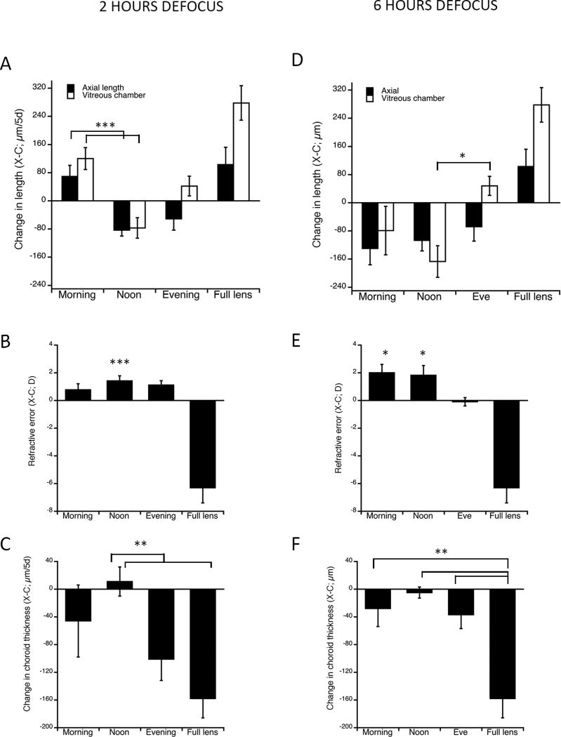
Effects on ocular dimensions of 2 hours (left column) and 6 hours of −10 D lens-induced hyperopic defocus given at different times of day. All data are interocular (X-C) differences. A and D. Effects on axial length (black bars) and vitreous chamber depth (white bars). B and E. End refractive error. C and F. Effects on choroidal thickness. Error bars in all graphs are sems. *p<0.05, **p<0.005, ***p<0.0005.
Ocular growth rate was defined as the change in axial length (from the front of the cornea to the front of the sclera) and change in vitreous chamber depth (from back of the lens to the front of the retina) over the experimental interval, from noon to noon. Choroidal thickness was also measured. The diurnal rhythms in both eye length and choroidal thickness, as determined by the 6-hour interval measurements on the last day of the experiment, were assessed in two ways: first, we calculated the change over each of the four 6-hour intervals, which includes the “steady state” eye growth occurring over the 24 hour period (i.e the slope of the longitudinal data). These data are plotted as bar graphs in the figures showing the rhythm data for lens-wearing eyes (Figures 2 and 3). One-way ANOVAs (StatPlus; AnaystSoft) were used to assess between group differences for each of the 4 intervals. Second, the “steady state” growth rate (or change in choroidal thickness) was subtracted from the longitudinal data, as described in Nickla et al. (Nickla et al. 1998), in order to determine the fit to a sine function. To control for the large between-animal variability, these data were normalized to their mean, and the means of these longitudinal data are plotted as line graphs. 2-way ANOVAs were used on these data to determine between-group interactions as a function of time of day. Sine waves with a fixed 24-hour period were fit to both all individual data for both axial length and choroid thickness (“individual sine peak” and “individual sine amp” in Table 1), to assess phase, as well as to the mean normalized data, as one way to determine phase and amplitude (“mean sine peak” and “mean sine amp” in Table 1). Only those eyes whose data could be fit to a sine function (numbers indicated in parenthesis in Table 1) were used in the determination of “individual sine peak”, and phase differences between groups were analyzed using circular statistics. “Goodness of fit” required that the mean standard deviation of the residuals of the sine wave fit was less than 85% of the standard deviation of the raw data. The other two ways of assessing “amplitude” are described below. One- and two-way ANOVAs were used to analyze the effect of “time of day”, and to determine the interaction as indicated for both axial length and choroid thickness. Where appropriate, post-hoc t-tests were used to test between-group differences.
Figure 2.
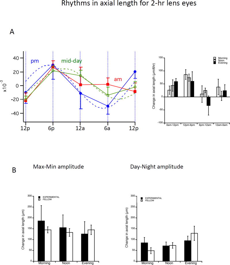
Effects on the rhythms in axial length for the 2-hour lens-wearing group. A. Left: Longitudinal data (length as a function of time) normalized to the mean for all three groups, and 24-hour sine wave fits to data (dotted lines). Note that the morning group (red line) cannot be well-fit to a sine function. Right: Change in length per 6-hour interval for all 3 groups. There are no significant between-group differences at any interval, by ANOVA. B. Amplitude of the rhythms derived by two analyses (described in Methods): Maximum vs minimum (left) and Day vs night (right) for experimental (black bars) and fellow (white bars) eyes. Error bars in all graphs are sems.
Figure 3.
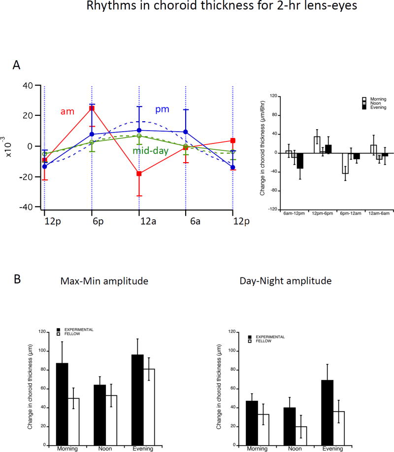
Effects on the rhythms in choroidal thickness for the 2-hour lens-wearing group. A. Left: Longitudinal data (length as a function of time) normalized to the mean for all three groups, and 24-hour sine wave fits to data (dotted lines). Note that the morning group (red line) cannot be well-fit to a sine function. Right: Change in length per 6-hour interval for all 3 groups. There are no significant between-group differences at any interval, by ANOVA. B. Amplitude of the rhythms derived by two analyses (described in Methods): Maximum vs minimum (left) and Day vs night (right) for experimental (black bars) and fellow (white bars) eyes. Error bars in all graphs are sems.
TABLE 1.
Choroid and axial rhythm parameters of amplitudes (“amp” in µm) with sems (parentheses), and acrophases for all groups.
| Negative lenses | Form deprivation | |||||
|---|---|---|---|---|---|---|
| MORN (n=8) |
NOON (n=8) |
EVE (n=6) |
MORN (n=6) |
NOON (n=6) |
EVE (n=6) |
|
| CHOROID | ||||||
| Max-Min amp | 87 (23) | 64 (9) | 96 (17) | 96 (28) | 128 (19) | 107 (15) |
| Night-Day amp | 47 (8) | 40 (11) | 69 (17) | 74 (20) | 71 (14) | 78 (23) |
| Mean sine amp | ----- | 11 | 27 | 54 | ----- | 55 |
| Mean sine peak | ----- | 22:45 | 0:20 | 6:00 | ----- | 23:30 |
| Individual sine peak | ----- 6/8 | ----- 6/8 | 22:30 (#5) | 5:45 (#4) | ----- 4/6 | 23:20 (#4) |
| AXIAL | ||||||
| Max-Min amp | 186 (71) | 155 (59) | 126 (57) | 144 (21) | 108 (15) | 90 (5) |
| Day-Night amp | 85 (25) | 71 (18) | 95 (21) | 113 (13) | 95 (21) | 66 (14) |
| Mean sine amp | ----- | 44 | 62 | 66 | 71 | 33 |
| Mean sine peak | ----- | 19:10 | 17:00 | 10:45 | 15:15 | 16:45 |
| Individual sine peak | ----- 5/8 | 20:45 (#5) | 17:20 (#6) | 10:00 (#4) | 14:35 (#5) | 15:35 (#5) |
| Individual sine amp | 94 (33) | 67 (15) | 91 (20) | 71 (7) | 103 (16) | 75 (30) |
Shaded data: Max-min and Night-day amplitude; analyses described in methods.
Un-shaded data derived from longitudinal data fit to a 24-hour sine function. “Mean sine amp” and “mean sine peak” are from the fit to the mean normalized data shown in figures. “Individual sine peak” and “individual sine amp” are from individual eyes fit to sine waves with a 24-hr period (# is n for both parameters); circular statistics were used to analyze phase differences.
---- Data are not sinusoidal. Ratios for these indicate numbers of “oscillators”.
We used two other methods of assessing “amplitude” apart from the mean of the sine wave fit mentioned above: In one, we took the minimum vs maximum differences from the 5 diurnal measurements, regardless of the time of day; this is referred to as “max-min amp” in Table 1. In the second, we restricted the maximum to either the day, for axial length, or night, for choroid thickness, and the minimum to either night, for axial length, or day, for the choroid thickness; this is referred to as “night-day amp” in Table 1 (both in shaded cells). This second one is a more accurate means of assessing if there is a diurnal excursion (i.e. if there is a day vs night change). The degree of similarity between the two methods of assessing “amplitude” also serves as a measure of whether the excursion is a diurnal one (that is, has its peak and trough about 12 hours apart).
Results
HYPEROPIC DEFOCUS: 2 OR 6 HOURS
Effects on ocular dimensions
2 hours
2 hours of hyperopic defocus stimulated growth of the vitreous chamber when given in the morning (Figure 1A, white bars: ANOVA: p<0.0005; X-C: 120 µm/5d; paired ttest p=0.001) but unexpectedly, it inhibited growth when given mid-day (X-C: −77 µm/5d; p=0.01). The change in axial length (black bars) showed a similar trend (ANOVA: p=0.003; morning: p=0.1; mid-day: p=0.0002). These interocular changes between morning vs midday groups were significantly different for both axial length and vitreous chamber depth (p<0.0001 and p=0.00006 respectively). Evening exposures had no significant effects on either axial length or vitreous chamber depth. All 2-hr defocus groups grew significantly slower than full-lens controls for vitreous chamber elongation (ANOVA p<0.00001; control vs experimental: 278 vs 120, −77 and 42 µm/5d respectively; p<0.01 for all). For axial length, the 2-hour morning group did not differ from full-lens controls.
The growth inhibition for the mid-day group resulted in significant hyperopia (Figure 1B: X-C: 1.4 D; p<0.00001), and all 2-hr defocus groups were significantly more hyperopic than full-lens controls, however, there were no significant between-group differences for the experimental groups by a one-way ANOVA (p>0.1). For choroidal thickness changes, the growth inhibition in the mid-day group is associated with a lack of choroidal thinning, but the only significant between-group difference for experimental groups was between the mid-day and evening (Figure 1C: X-C: 8 vs −100 µm/5d; p<0.005). The choroidal thinning in the morning and evening groups did not differ from that in control eyes with full time lens-wear (−46 and −100 vs −158; p>0.1 for both).
6 hours
The effects of longer, 6 hour periods of hyperopic defocus on vitreous chamber growth were generally similar to those of 2 hours, in that mid-day exposure resulted in growth inhibition (Figure 1D: vitreous chamber: ANOVA: p=0.079; X-C: −167/5d; t-test p=0.005; axial length: X-C: −107 µm; t-test p=0.007). By contrast, however, morning exposure did not show growth stimulation, and in fact tended toward growth inhibition (axial length X-C: −130; p=0.069; vitreous chamber: −79 µm; p>0.05). Evening exposures had no significant effects on either axial length (−68 µm; p>0.1) or vitreous chamber depth (61 µm; p>0.1). The only significant between-group difference for the experimental groups was for vitreous chamber growth in the noon vs evening groups (p<0.05). The refractive errors for both morning and mid-day groups were significantly hyperopic (Figure 1E: 2.0 D and 1.8 D, respectively; t-test p<0.05 for both). There were no significant between-group differences in the choroidal responses by a one-way ANOVA (p>0.1; Figure 1F). Choroids in all 3 experimental groups thinned significantly less than those in the full-lens controls (p<0.05 for all comparisons).
Effects on ocular rhythms
Axial length
We measured ocular dimensions at 6-hour intervals over 24 hours on the last day for the 2-hour lens-wearing eyes in order to study the rhythms (Figure 2). For eyes given defocus at both mid-day (growth inhibited) and evening (no effect on growth), the mean rhythms in axial length were well-fit to a sinusoid with a period of 24 hours, similar to that of normal eyes ((Nickla et al. 1998); Figure 2A, left; green and blue lines). The mean phase of these rhythms differed, with that for the mid-day group being phase-delayed relative to the evening group (8:45 pm vs 5:20 pm; p<0.05 by circular statistics). However, the mean of the rhythms in eyes given defocus in the morning, and which showed growth stimulation, could not be fit to a sine function (red line). This was because in this group, only 4/8 eyes could be fit to a 24 hour sine function; of these 4, one had a very small (19 µm) amplitude, and the other 3 had disparate acrophases. The data for the changes over the 6 hour intervals showed no between-group differences at any time interval (Figure 2A, right).
The amplitudes of the rhythms as derived from the individual sine wave fits did not differ (mid-day vs eve: 67 vs 91 µm; p=0.3; Table 1). Nor did they differ when amplitude was derived from the maximum vs minimum (morning, mid-day and evening: 186 µm, 155 µm, 126 µm respectively; ANOVA p=0.2; figure 2B, left; Table 1) or the day vs night (85 µm, 71 µm, 95 µm respectively; ANOVA p=0.6; figure 2B, right; Table 1). There were also no significant differences between experimental and fellow eye amplitudes (compare black and white bars in Fig. 2B).
Choroidal thickness
The rhythm in choroidal thickness was essentially normal for only the evening group (although the data were not as sinusoidal as usual; Figure 3A; blue line): there was a mean peak at 12:20 am (10:30 PM from individual waves; Table 1), and a mean amplitude of 27 µm (Table 1, mean sine amp). Six out of 8 choroids in both the morning and mid-day groups showed oscillations around the 6-hour intervals (Table 1); for mid-day, while the mean can be fit to a 24-hour sinusoid, the mean amplitude is only 11 µm (green line; Table 1, mean sine amp). There were no significant effects on rhythm “amplitude” by either means of analysis (Figure 3B: morning, mid-day, evening: max vs min (left): 87 µm, 64 µm, 96 µm; day vs night (right): 47 µm, 40 µm, 69 µm; ANOVA p>0.1 for both). Neither were there any significant differences between amplitudes for experimental versus fellow control eyes (compare black and white bars).
FORM DEPRIVATION: 2 HOURS
Effects on ocular dimensions
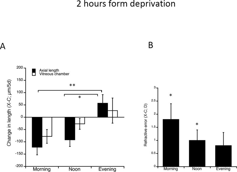
2 hours of form deprivation resulted in growth inhibition for axial length (black bars, Figure 4A), both when given in the morning and when given at mid-day (X-C morning: −122 µm; mid-day: −92 µm; t-tests p=0.006; p=0.016 respectively). There was no effect of deprivation in the evening. Both morning and mid-day groups grew significantly slower than eyes given defocus in the evening (ANOVA p=0.006; post-hoc Bonferroni tests: morning vs evening: p=0.006; noon vs evening: p=0.014). The data for vitreous chamber depth tended to show the same effects, but were not statistically significant. In accord with the growth inhibition for the morning and mid-day exposures, both groups were significantly hyperopic (Figure 4B: X-C: 1.8 D; 1.0 D respectively; t-test p<0.05 for both). There were no between-group differences in the choroidal responses (data not shown).
Figure 4.
Effects on ocular dimensions of 2 hours form-deprivation given at different times of day. All data are interocular (X-C) differences. A. Effects on axial length (black bars) and vitreous chamber depth (white bars). B. End refractive error. Error bars in all graphs are sems. *p<0.05, **p<0.01
Effects on ocular rhythms
Axial length
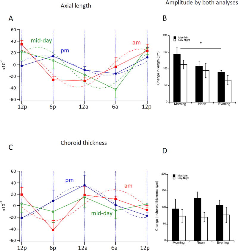
The mean rhythms in axial length in eyes receiving form deprivation at mid-day (growth inhibited; Figure 5A: green line) or in the evening (no effect on growth; blue line) were well-fit to a sinusoidal function with a period of 24 hours, similar to groups given hyperopic defocus at those times (compare to Figure 2A). There were no significant differences in phase when analyzed by circular statistics (Table 1, “individual sine peak”: noon and eve: 2:35 pm vs 3:35 pm; p>0.1). The phase data for both these groups were very consistent: 9/10 of the sinusoids had acrophases between 1:45 pm and 5:45 pm. The results for the eyes receiving deprivation in the morning (red line), however, differed: While the mean rhythm was also well-fit to a sine function (4/6 eyes, with acrophases between 9:30 am and 11 am), the mean phase was significantly advanced relative to that of the other groups (Table 1, “individual sine peak”: 10:00 vs 2:35 pm and 3:35 pm; p<0.05). This result also differed from the data for the axial rhythm for the “morning” group for lens-wearing eyes (compare to Figure 2A). A 2-way ANOVA showed that time of day accounted for the differences between groups (p=0.0002; interaction p<0.00001). There were no significant between-group differences in the 6-hour interval data (data not shown).
Figure 5.
Effects on the rhythms in axial length and choroid thickness for the 2 hour form deprivation group. A. Left: Longitudinal data (length as a function of time) for axial length normalized to the mean for all three groups, and 24-hour sine wave fits to data (dotted lines). B. Amplitude of the rhythms derived by two analyses (described in Methods): Maximum vs minimum (black bars) and Day vs night (stippled bars) for experimental eyes. C. Longitudinal data (length as a function of time) for choroidal thickness normalized to the mean for all three groups, and 24-hour sine wave fits to data (dotted lines). Note that the noon group cannot be well-fit to a sine function. D. Amplitude of the rhythms derived by two analyses (described in Methods): Maximum vs minimum (black bars) and Day vs night (stippled bars) for experimental eyes. *p<0.05
For the min-max amplitude analysis (Figure 5B, black bars), the morning exposure group was significantly larger than that of the evening group (144 µm vs 90 µm; ANOVA p=0.04; morning vs evening, p=0.04). This did not hold true for either the day-night analysis (white bars, morning, noon, evening: 113 µm, 95 µm, 66 µm respectively, ANOVA p=0.1) or the individual sine wave fits (71 µm, 103 µm and 75 µm; ANOVA p=0.4; Table 1).
Choroidal thickness
The rhythms in choroid thickness for eyes given deprivation in the evening were normal in phase (Figure 5C; blue lines), similar to the evening lens-exposure group (compare to Figure 3A): the mean peak was at 11:30 pm (11:20 pm from individual waves; Table 1), and the mean amplitude was 55 µm. The mean for the morning group was also (somewhat) sinusoidal, with a mean peak at 6 am (Figure 5C, red lines: 5:45 am from individual waves), with a mean amplitude of 54 µm. The mid-day group mean, however, showed oscillations around 6 hour intervals; 4/6 choroids were oscillators. There was no significant interaction between group and time of day by a 2-way ANOVA (p>0.1). There were no significant effects on rhythm “amplitude”, by either means of analysis (Figure 5D; Table 1) (morning, mid-day, evening: max vs min, black bars: 96 µm, 128 µm, 107 µm; ANOVA p=0.5; day vs night, white bars: 74 µm, 71 µm, 78 µm; ANOVA p=0.9).
Discussion
Our main findings are that brief periods of hyperopic defocus affected eye growth differentially depending on the time of day of exposure: 2 hours of defocus resulted in growth stimulation compared to fellow eyes when given in the morning, but caused inhibition when given at mid-day. Longer 6-hour periods also caused growth inhibition when given at mid-day, but tended towards growth inhibition when given in the morning. 2 hours of form deprivation inhibited growth when given both in the morning and at midday. For the lens-wearing eyes only, the growth stimulation in the morning exposure group was associated with abnormal rhythms in both axial length and choroidal thickness.
Temporal integration of hyperopic blur and form deprivation
It has been generally accepted that nearly constant exposure to either hyperopic defocus or form deprivation is required for the integration of the signal to effect eye growth stimulation (Schmid and Wildsoet 1996; Napper et al. 1997). However, we here show that different effects can be unmasked when looking at brief exposures to either condition, at different times of day: morning caused growth stimulation while mid-day caused inhibition. In the seminal paper on the integration characteristics of hyperopic defocus, Schmid and Wildsoet tested various daily periods of −10 D lens-wear over 5 days, the same magnitude of defocus and the same experiment duration as did we, but their shortest period of lens-wear was 1 hour, while ours was 2 hours. The graph of their growth data for the 1- hour −10 D group shows that, while not significant, there was, in fact, a 60 µm decrease in experimental eyes vs fellow controls, for both vitreous chamber depth and axial length, which is similar to our results of decreases of 77 and 83 µm, respectively. This suggests that our study corroborates their results, despite their lack of statistical significance. While there are several possible explanations for the statistical discrepancies between the two studies (they used younger chicks, 1 vs 2 hours of exposure, and a different breed), it is most likely to be simply a difference in measurement precision due to their lower-frequency ultrasound system. It remains a mystery however, why we found that only mid-day exposure caused a growth inhibition (morning caused stimulation and evening had no effect) while their data for their two combined exposure times of 7am and 6pm tended to show the inhibition. It is also unclear how to explain our finding of growth stimulation resulting from the 2-daily hours of morning exposures in light of their lack of effect, but it is plausible that their 1-hour exposure duration was insufficient to elicit the stimulation.
Two hours of form deprivation also caused growth inhibition, both when given in the morning and when given at mid-day. It has long been accepted that the temporal integration characteristics of form deprivation are similar to those of hyperopic defocus: a mere 2 daily hours of unobstructed vision largely prevents the development of myopia, and even 15 minutes of “vision” causes a significant reduction in myopia (Napper et al. 1997). However, the minimal duration of occlusion that was tested in the cited study was 8 hours (with 4 hours of “vision”), because the purpose was to determine the minimum amount of normal vision to prevent growth stimulation, so there was no need to look at longer than 4-hour durations of vision, or shorter periods of deprivation, hence there is no discrepancy.
Our anomalous finding that both brief hyperopic defocus and brief deprivation can cause the opposite of the expected effect, that of growth inhibition rather than stimulation at certain times, was unexpected. We suspect that the small degree of myopic defocus presumably experienced upon removal of the lens or diffuser is the cause. It is known that the temporal integration of myopic defocus is rapid (Schmid and Wildsoet 1996; Zhu et al. 2005), and also that the myopic defocus signal is more effective later in the day, evincing a diurnal sensitivity in the “integrator” (Nickla et al. 2017). These facts would explain the finding that removing the lens or diffuser after 2 hours of defocus in the mid-day, or after 6 hours of defocus in the morning (removal would occur at 1 pm), would cause a growth inhibition, whereas the 2-hours of morning defocus after removal would not. By the same token, that the evening removal of either lens or diffusers would be followed by darkness would explain the lack of inhibition in this condition. What is still questionable, however, is the growth inhibition found in the 2-hour morning diffuser group: we speculate that the magnitude of the myopic defocus experienced after diffuser-wear as opposed to lens-wear was greater, and hence elicited an inhibition rather than a stimulation.
There is evidence supporting a causal association between thicker choroids and growth inhibition. First, all brief daily visual manipulations that inhibit the myopia development in form deprived or lens-wearing eyes are associated with transient choroidal thickening responses (Nickla 2007). Second, there is a significant negative correlation between choroidal thickness and ocular growth rate in young normal eyes (Nickla 2005; Nickla and Totonelly 2015). If it is true that choroidal thickness, or thickening, is mechanistically linked with scleral growth inhibition, we hypothesized that the anomalous finding of growth inhibition for both of the lens groups and for form deprivation at mid-day, was a result of either a more robust choroidal thickening response upon removal of the lens or diffuser at this time versus other times, or that choroids are thicker in the noon versus the morning group at the time of placement of the device on the eye. We did a pilot experiment that looked at choroidal thickness, starting from immediately prior to 2 hours of lens wear, and at 0, 1, 2 and 3 hours after lens-removal, for both morning and noon exposures, and found that the choroids in the noon group were indeed significantly thicker compared to fellow eyes than those in the morning group immediately prior to lens-placement (X-C: 87; p<0.05 vs 43 µm; p>0.05), but there was no difference in the thickening response, supporting the second of the above hypotheses.
Implications of the effects on rhythm parameters
There are three main findings regarding the alterations in the rhythms in axial length. First, hyperopic blur at mid-day, which resulted in growth inhibition, caused a phase-delay relative to the rhythm in eyes given blur in the evening (which has no effect on growth, and whose rhythms are normal). Second, eyes given defocus in the morning, which resulted in growth stimulation, showed abnormally-shaped patterns that could not be fit to a 24-hour sine wave. Third, eyes given form-deprivation in the morning, which tended to inhibit growth, showed a phase advance relative to the other times of deprivation.
Are any of these alterations in the axial length rhythm relevant to emmetropization? The growth inhibition caused by defocus or deprivation at mid-day (or deprivation in the morning) was not associated with any consistent effect on the rhythms in axial length, either for phase or for amplitude. However, if we compare the data for the 2-hour hyperopic defocus group to that of eyes receiving 2 hours of myopic defocus at the same times (Nickla et al. 2017); Table 2), some interesting similarities are revealed: first, both types of defocus at mid-day (growth inhibition for both) caused significant phase-delays relative to the rhythms in eyes exposed to evening defocus. Second, both types of defocus in the morning (myopic defocus at this time prevents the compensatory inhibition; hyperopic defocus stimulates growth) caused abnormal 6-hour oscillations. These oscillations are similar to what was seen in the rhythms in eyes receiving 2 hours of light (vision) at mid-night, which caused eventual growth stimulation (Nickla and Totonelly 2016). To summarize, when defocus, either myopic or hyperopic, was given at mid-day, the growth inhibition was associated with a phase-delay in the axial rhythm relative to that for evening defocus. When defocus was given in the morning, or when light is given in the middle of the night, all of which either disinhibited (myopic defocus) or stimulated (hyperopic defocus and night light) growth, the axial length rhythms showed abnormal oscillations around 6 hours.
Table 2.
Summary of effects on ocular growth and axial and choroidal rhythms for 4 different “brief” visual treatments.
| 2 hr myopic defocus* | 2 hr hyperopic defocus | 2 hr form deprivation | 2 hr night light** |
|||||||
|---|---|---|---|---|---|---|---|---|---|---|
| AM | NOON | PM | AM | NOON | PM | AM | NOON | PM | 12 AM | |
| Growth | Prevents inhibition | ↓ | ↓ | ↑ | ↓ | No effect | ↓ | ↓ | No effect | ↑ |
| Axial | Oscillate | Delayed# | ##No effect | Oscillate | Delayed# | ##No effect | Advanced# | ##No effect | ##No effect | Oscillate |
| Choroid | No effect | No effect | No effect | Oscillate | Oscillate | No effect | Delayed | Oscillate | No effect | Oscillate |
Direction of phase shift relative to ## group
↓ Inhibits growth; ↑ Stimulates growth
Data from Nickla et al., 2017.
Data from Nickla & Totonelly, 2016.
Regarding the rhythm in choroidal thickness, the only ones that appeared normal (24-hour sine wave and acrophase at mid-night) were those in eyes given defocus or deprivation in the evening, in which eye growth was unaltered. In the morning and mid-day groups, when there were effects on eye growth, choroids either oscillated around 6 hours, or were phase- delayed (morning deprivation). Similar abnormalities were also seen in eyes given light at mid-night, when growth was stimulated. They were not found in eyes given myopic defocus at any time of day (Nickla et al. 2017), therefore hyperopic defocus appears to be more detrimental than myopic defocus on this rhythm.
To conclude, out of the 4 visual manipulations discussed: myopic and hyperopic defocus, form deprivation and light at mid-night, the only times of day at which there were not alterations in either rhythm are for exposures in the evening, when there was no effect on eye growth. Exposures at morning or mid-day, which resulted in eye growth changes were all associated with abnormalities in at least one of the rhythms, if not both. The only consistent effect on phase was the phase-delay in the axial rhythms found for the mid-day exposures for both myopic and hyperopic defocus, which are both associated with growth inhibition. This shift is in a similar direction (delay), and has similar peaks (6:30 pm and 7:10 pm respectively, for mean sine peaks) as that found in eyes exposed to full-time myopic defocus from positive lenses (6:30 pm; (Nickla 2005)), or in eyes recovering from deprivation myopia (8:00 pm; (Nickla et al. 1998)), both conditions that result in extreme growth inhibition. However, this phase shift is not a consistent characteristic of growth inhibition, because it was not seen in the eyes given myopic defocus in the evening (Nickla et al. 2017), or in the morning or mid-day form-deprived eyes whose growth was inhibited (this manuscript).
Altered rhythms in axial length and choroidal thickness are presumably a consequence of changes in other rhythms (dopamine? IOP?), that themselves are driven by ocular clock(s). The complex alterations found in response to brief visual stimuli in our current results do not conform to any simple association, such as consistent changes in phase for growth inhibition, and vice versa. The different visual signals (plus or minus defocus, form deprivation) could impinge on the same oscillating component (dopamine?) or on separate ones (one increases dopamine, another increases acetylcholine?). Our best chance of addressing the underlying mechanism(s) for these alterations is to look at candidate molecular signals, such as dopamine, and clock genes, under various conditions.
Practical clinical implications
We have previously reported that brief myopic defocus in the morning largely prevents the compensatory inhibitory growth response to the defocus (Nickla et al. 2017), and brief hyperopic defocus in the morning permits the compensatory stimulatory response to the defocus. This suggests that, in chicks, the signal integrator is more sensitive to hyperopic blur than to myopic blur in the morning, and/or that the sclera is more susceptible to increase growth than to slow growth early in the day, both of which may have practical implications for school children in terms of myopia control therapies, if our results are translatable: First, the greater sensitivity to hyperopic blur suggests that scheduling extensive reading activities first thing in the morning, which may impose hyperopic defocus, would be contraindicated. Second, if imposing brief periods of myopic blur might someday be used as a therapy, our results would suggest that timing these for later in the day rather than earlier would be best. Third, if increasing the amount of time spent outdoors is used as a preventative treatment for myopia control in children (Morgan et al. 2012), our results showing a differential time-of-day sensitivity to visual stimuli of various kinds increases the plausibility that this putative inhibitory effect may depend on the timing of the outdoor exposure, due to the spectral differences experienced over the course of a day. Because the rhythms in axial length and choroidal thickness in humans are similar in phase to those of chicks (Stone et al. 2004; Brown et al. 2009; Chakraborty et al. 2011), we believe that the above clinical implications will prove to be applicable.
In conclusion, brief periods of both myopic and hyperopic defocus have effects on ocular growth that are dependent on time of day, indicating that the temporal integration of the visual signal, and/or the scleral growth mechanisms, are diurnally-regulated. Furthermore, the times of exposure that lead to ocular growth changes (inhibition or stimulation) are associated with various alterations in the diurnal rhythms in axial length and/or choroidal thickness. These results provide strong evidence that circadian rhythms are an essential element of emmetropization. They might also have important implications for the timing of drug therapies, and for myopia-preventative activities for school-age children.
Highlights.
Brief hyperopic defocus in the morning elicits eye growth stimulation.
Brief hyperopic defocus mid-day elicits eye growth inhibition.
Morning defocus is associated with alterations in rhythms in axial length.
The results have implications regarding timing of reading activities in school.
Acknowledgments
The authors thank Karl Englehart, Jessica Hahm and Tiana Camarillo for their help in collecting some of these data. This work was funded by NIH-NEI-013636 and NIH-NEI-025307.
Footnotes
Publisher's Disclaimer: This is a PDF file of an unedited manuscript that has been accepted for publication. As a service to our customers we are providing this early version of the manuscript. The manuscript will undergo copyediting, typesetting, and review of the resulting proof before it is published in its final citable form. Please note that during the production process errors may be discovered which could affect the content, and all legal disclaimers that apply to the journal pertain.
References
- Brown JS, Flitcroft DI, Ying G, Francis EL, Schmid GF, Quinn GE, Stone RD. In vivo human choroidal thickness measurements: Evidence for diurnal fluctuations. Invest Ophthalmol Vis Sci. 2009;50:5–12. doi: 10.1167/iovs.08-1779. [DOI] [PMC free article] [PubMed] [Google Scholar]
- Chakraborty R, Read SA, Collins MJ. Diurnal variations in axial length, choroidal thickness, intraocular pressure, and ocular biometrics. Invest. Ophthalmol. Vis. Sci. 2011;52:5121–5129. doi: 10.1167/iovs.11-7364. [DOI] [PubMed] [Google Scholar]
- Curtin BJ. The Myopias. Philadephia: Harper & Row, Publ; 1985. [Google Scholar]
- Fan DS, Lam DSC, Lam R. Prevalence, incidence and progression of myopia of school children in Hong Kong. Invest. Ophthalmol. Vis. Sci. 2004;45:1071–1075. doi: 10.1167/iovs.03-1151. [DOI] [PubMed] [Google Scholar]
- Goh WS, Lam CS. Changes in refractive trends and optical components of Hong Kong Chinese aged 19–39 years. Ophthal Physiol Opt. 1994;14:378–382. [PubMed] [Google Scholar]
- Gwiazda J, Bauer J, Thorn F, Held R. A dynamic relationship between myopia and blur-driven accommodation in school-aged children. Vision Res. 1995;35:1299–1304. doi: 10.1016/0042-6989(94)00238-h. [DOI] [PubMed] [Google Scholar]
- He M, Zeng J, Liu Y, Xu J, Pokharel G, Ellwein L. Refractive error and visual impairment in urban children in southern China. Invest. Ophthalmol. Vis. Sci. 2004;45:793–799. doi: 10.1167/iovs.03-1051. [DOI] [PubMed] [Google Scholar]
- Lin LL. Epidemiological study of ocular refractions among school children (aged 6 through 18) in Taiwan. Invest. Ophthalmol. Vis. Sci. 1996;37:S1002. [Google Scholar]
- Lin LL, Shih YF, Hsiao CK, J CC. Prevalence of myopia in Taiwanese schoolchildren: 1983–2000. Ann. Acad. Med. Singapore. 2004;33:27–33. [PubMed] [Google Scholar]
- Morgan I, Rose KA. Myopia and international educational performance. Ophthal Physiol Opt. 2013;33:329–338. doi: 10.1111/opo.12040. [DOI] [PubMed] [Google Scholar]
- Morgan I, Xiang F, Rose KA, Chen Q, He M. Two year results from the Guangzhou outdoor activity longitudinal study. 2012 ARVO E-Abstract #2735. [Google Scholar]
- Napper G, Brennan N, Barrington M, Squires M, Vessey G, Vingrys A. The effect of an interrupted daily period of normal visual stimulation on form deprivation myopia in chicks. Vision Res. 1997;37(12):1557–1564. doi: 10.1016/s0042-6989(96)00269-6. [DOI] [PubMed] [Google Scholar]
- Ngo C, Saw S-M, Dharani R, Flitcroft DI. Does sunlight (bright lights) explain the protective effects of outdoor activity against myopia? Ophthalmic Physiol. Optics. 2013;33:368–372. doi: 10.1111/opo.12051. [DOI] [PubMed] [Google Scholar]
- Nickla D. The phase relationships between the diurnal rhythm in axial length and choroidal thickness and the association with ocular growth rate in chicks. J. Comp. Physiol. A. 2005;192:399–407. doi: 10.1007/s00359-005-0077-2. [DOI] [PubMed] [Google Scholar]
- Nickla D. Transient increases in choroidal thickness are consistently associated with brief daily visual stimuli that inhibit ocular growth in chicks. Exp. Eye Res. 2007;84:951–959. doi: 10.1016/j.exer.2007.01.017. [DOI] [PubMed] [Google Scholar]
- Nickla D, Thai P, Zanerkia-Trahan R, Totonelly K. Myopic defocus in the evening is more effective at inhibiting eye growth than defocus in the morning: Effects on rhythms in axial length and choroidal thickness in chicks. Exp. Eye Res. 2017;154:104–115. doi: 10.1016/j.exer.2016.11.012. [DOI] [PMC free article] [PubMed] [Google Scholar]
- Nickla D, Totonelly K. Choroidal thickness predicts ocular growith in normal chicks but not in eyes with experimentally altered growth. Clin. Exp. Optom. 2015;98:564–570. doi: 10.1111/cxo.12317. [DOI] [PMC free article] [PubMed] [Google Scholar]
- Nickla D, Totonelly K. Brief light exposure at night disrupts the circadian rhythms in eye growth and choroidal thickness in chicks. Exp. Eye Res. 2016;146:189–195. doi: 10.1016/j.exer.2016.03.003. [DOI] [PMC free article] [PubMed] [Google Scholar]
- Nickla DL, Wildsoet C, Wallman J. Visual influences on diurnal rhythms in ocular length and choroidal thickness in chick eyes. Exp. Eye Res. 1998;66:163–181. doi: 10.1006/exer.1997.0420. [DOI] [PubMed] [Google Scholar]
- Pan CW, Ramamurthy D, Saw S-M. World-wide prevalence and risk factors for myopia. Ophthal Physiol Opt. 2012;32:3–16. doi: 10.1111/j.1475-1313.2011.00884.x. [DOI] [PubMed] [Google Scholar]
- Qian YS, Chu RY, He JC. Incidence of myopia in high school students with and without red-green vision deficiency. Invest. Ophthalmol. Vis. Sci. 2009;50:1598–1605. doi: 10.1167/iovs.07-1362. [DOI] [PubMed] [Google Scholar]
- Rose K, Morgan I, Ip J, Kifley A, Huynh S, Smith W, Mitchell P. Outdoor activity reduces the prevalence of myopia in children. Ophthalmology. 2008;116:1229–1230. doi: 10.1016/j.ophtha.2007.12.019. [DOI] [PubMed] [Google Scholar]
- Schmid K, Wildsoet CF. Effects of the compensatory responses to positive and negative lenses of intermittent lens wear and ciliary nerve section in chicks. Vision Res. 1996;36:1023–1036. doi: 10.1016/0042-6989(95)00191-3. [DOI] [PubMed] [Google Scholar]
- Stone RD, Quinn GE, Francis EL, Ying G, Flitcroft DI, Parekh P, Brown J, Orlow J, Schmid G. Diurnal axial length fluctuations in human eyes. Invest Ophthalmol Vis Sci. 2004;45:63–70. doi: 10.1167/iovs.03-0294. [DOI] [PubMed] [Google Scholar]
- Wallman J, Winawer J. Homeostasis of eye growth and the question of myopia. Neuron. 2004;43:447–468. doi: 10.1016/j.neuron.2004.08.008. [DOI] [PubMed] [Google Scholar]
- Winawer J, Khan S, Wallman J. Enduring responses to spectacle lens wear in chicks. Invest. Ophthalmol. Vis. Sci. 2001;42(4):S1609. [Google Scholar]
- Zhu X, Park T, Winawer J, Wallman J. In a matter of minutes, the eye can know which way to grow. Invest Ophthalmol Vis Sci. 2005;46:2238–2241. doi: 10.1167/iovs.04-0956. [DOI] [PubMed] [Google Scholar]
- Zhu X, Wallman J. Temporal properties of compensation for positive and negative spectacle lenses in chicks. Invest Ophthalmol Vis Sci. 2009;50:37–46. doi: 10.1167/iovs.08-2102. [DOI] [PMC free article] [PubMed] [Google Scholar]
- Zhu X, Winawer J, Wallman J. Potency of myopic defocus in spectacle lens compensation. Invest. Ophthalmol. Vis. Sci. 2003;44:2818–2827. doi: 10.1167/iovs.02-0606. [DOI] [PubMed] [Google Scholar]


