FIG 5 .
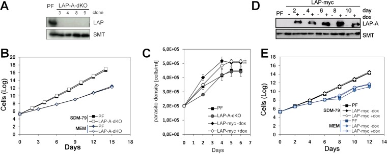
Growth phenotype of LAP-A-dKO and LAP-A-OE cells in different media and effect of leucine depletion. dKO mutants were obtained with PFs (LAP-A-dKO), and cell proliferation was assessed. (A) Western blot analysis indicating successful elimination of LAP-A expression in different cell clones. (B) Cumulative growth curves of PF T. brucei cultured in SDM-79 containing FBS and MEM containing dialyzed FBS. (C) Cumulative growth curves of parental, TbLAP-A-OE (LAP-myc), and LAP-A-dKO cells grown in MEM devoid of leucine. (D) Western blot analysis indicating the level of expression of LAP-A at different time points. Cells were diluted every 48 h. For Western blot analyses, anti-TbLAP-A antibody was used at a 1:15,000 dilution; the loading control was rabbit anti-TbSMT antibody at a 1:10,000 dilution. Each lane contained 2.5× 10⁶ parasites. (E) Cumulative growth curves of LAP-A-OE PF parasites grown in SDM-79 and glucose-free MEM. dox, doxycycline.

