

Abstract
A 2-year-old girl presented to the emergency department with a 3-day history of a painful stiff neck after getting a kick to her head from her older brother. Her general practitioner had recently started her on oral antibiotics for otitis media. Plain film imaging of her cervical spine on admission revealed anterior subluxation of C2 on C3 suggestive of bifacetal dislocation. Subsequent CT imaging confirmed malalignment of the upper cervical spine. The patient was admitted and worked up with MRI of the cervical spine which unexpectedly revealed a large 4×2 cm retropharyngeal abscess extending from C1 to C4. No associated structural abnormality of the spine was detected. This case report highlights the life-threatening causes of torticollis (retropharyngeal abscess and cervical spine injury), and summarises the anatomy and normal variants that one should expect on interpretation of cervical spine imagery.
Background
This case report highlights the importance of a detailed and focused history and examination. Clues in the history can guide the clinician to the true cause of the presenting symptom. Furthermore, the common causes of torticollis in a child are briefly described with a broad overview of what to expect when imaging the paediatric cervical spine. This case aims to highlight the difficulty in interpretation of cervical spine imagery in a paediatric population and provides some tips to help differentiate the ‘normal’ from the ‘abnormal’ cervical spine.
Case presentation
A 2-year-old girl was admitted urgently to the emergency department reporting of ongoing neck stiffness and pain that had begun ∼3 days prior.
Her mother had witnessed her daughter accidentally receive a ‘kick to the head’ by her son, when playing outside. She observed that her daughter was holding her head ‘in a straight and awkward position since the kick’.
Of note in her history, her general practitioner had recently started her on oral antibiotics (co-amoxiclav) for a diagnosis of otitis media. She had initially presented at that time with upper respiratory symptoms and was irritable and restless.
Her medical history and family histories were unremarkable. The patient had reached all her developmental milestones and her vaccinations were up to date.
On examination, the patient was holding her head in extension and side flexion (to the right) with loss of the normal cervical lordosis. She was moving all limbs independently. She was markedly tender and irritable to palpation of the upper cervical spinous processes. Movement arcs of the neck were not examined further on arrival to the emergency department due to the questionable presence of a traumatic injury.
On admission to the emergency department, the patient had no fever with a mildly increased heart rate. All other vital signs were normal.
Plain film imaging and blood investigations including full blood count and inflammatory markers were performed (figure 1) which revealed a C reactive protein (CRP) of 41 mg/L. She had a normal white cell count (WCC) and neutrophil count.
Figure 1.
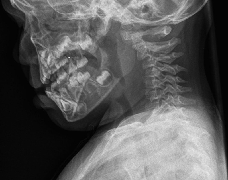
Cervical spine radiograph demonstrating subluxation of C2 vertebral body on C3.
Plain radiography was suggestive of acute anterior subluxation of C2 on C3 representing bilateral facet joint dislocation. Full spinal precautions were undertaken.
CT was then performed for further evaluation (figure 2).
Figure 2.
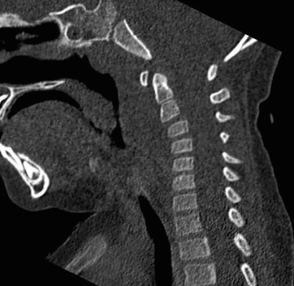
CT cervical spine demonstrating subluxation of C2 vertebral body on C3.
The CT confirmed the findings of the radiograph suggesting acute C2 on C3 facet joint subluxation. The patient was admitted overnight for observation and analgesia.
Over the next 12 hours, the patient's influenza-like symptoms had not subsided and the patient remained quite irritable and restless. The patient then spiked a temperature of 38.5°C overnight.
As she failed to settle over the subsequent 12 hours, MRI was performed (figure 3).
Figure 3.
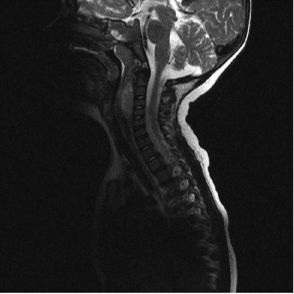
MRI neck demonstrating 4×2 cm retropharyngeal abscess.
In contrast to the CT, MRI did not reveal a C2 on C3 facet joint subluxation but instead revealed a 4×2 cm retropharyngeal abscess. No boney or ligamentous pathology was noted on MRI.
The patient underwent an urgent incision and drainage (I&D) of the abscess via a transoral approach under care of the ear, nose and throat (ENT) surgeons. Intraoperative specimens sent for culture sensitivity and microscopy grew a non-typeable Haemophilus influenza, which was sensitive to erythromycin. Antibiotic therapy was started thereafter.
Outcome and follow-up
Following I&D and initiation on appropriate antibiotic treatment, the patient responded quickly. She was discharged on oral antibiotics and was well on review in the outpatient department 6 months postoperatively.
Discussion
Torticollis is a deformity characterised by lateral inclination of the head, torsion of the neck and deviation of the face.1 It is not a diagnosis but a symptom of diverse conditions and is associated with a broad range of childhood illnesses.2 Kiwak3 and Mc Daniel et al4 summarise the aetiology of torticollis into congenital and acquired conditions (see box 1). Congenital torticollis manifests at 1–4 weeks of age and accounts for 0.3–2.0% of the total incidence.5
Box 1. Aetiology of torticollis4.
Congenital
Congenital muscular torticollis,
Klippel-fiel syndrome, Sprengel's deformity,
Congenital articular and ligamentous lesion,
Arnold-Chiari malformation, spina bifida.
Acquired
Traumatic
Subluxations, dislocations, fractures.
Infection/inflammatory
Upper respiratory tract illness, cervical adenitis;
Retropharyngeal abscess, osteomyelitis, tuberculosis;
Rheumatoid arthritis.
Neoplasm
Neurogenic: posterior fossa, spinal cord, vestibular system;
Vertebral column: osteoid osteoma.
Neurogenic
Syringomyelia, ocular dysfunction, bulbar palsies.
Idiopathic
Atlantoaxial rotatory instability.
Joint dysfunction.
Of acquired causes, injury involving the sternocleidomastoid or trapezius muscles is the most common cause identified.6 Acute infection follows muscular injury as the second most common cause of acquired torticollis accounting for up to 20% of the total incidence.6
Acute infections causing torticollis include viral or streptococcal pharyngitis, parapharyngeal abscess, retropharyngeal abscess and upper respiratory tract infections. Of these conditions, retropharyngeal abscess is considered life threatening.7 Retropharyngeal abscess occurs most commonly in the 2–4 years old population8 and is associated with severe inflammation of the retropharyngeal lymph nodes (located in the retropharyngeal space).9 Clinical findings suggestive of retropharyngeal abscess include fever, irritability, dysphagia, drooling, odynophagia and respiratory distress.8
The management of children presenting with retropharyngeal abscess depends on (1) presence of airway compromise (2) the size of the abscess. With airway compromise, immediate I&D is recommended.10 The management of a small abscess (cross-sectional area of <2 to 3 cm2) without airway compromise is a topic of debate currently with some authors offering a conservative approach of intravenous antibiotics,10 with others offering immediate surgery.11
Several modes of imaging were used in this study to help delineate a true diagnosis. Both plain radiographs and CT result in exposure of ionising radiation to the patient in question. As the clinician's main concern initially was of trauma-induced subluxation, CT was deemed the most appropriate imaging modality to delineate any true subluxation or dislocation.
CT scanning is also quite useful for detecting soft tissue anomalies. Boucher et al12 demonstrated a 100% sensitivity and 45% specificity of detecting retropharyngeal abscess on CT. Hurley and Heran13 identified CT as the imaging modality of choice in identifying deep neck infections. MRI is useful for assessing the extent of soft tissue involvement and for delineating vascular complications. In this case report the retropharyngeal swelling should have been identified on the initial CT, preventing the need for further imaging in the form of MRI.
Other life-threatening causes of torticollis include cervical spine injury. Overall, cervical spine injury is rare in children with the majority of cases occurring as a result of motor vehicle accidents.14 Symptoms suggestive of cervical spine injury include local pain, muscle spasm and decreased range of motion of the neck.15 Initial management of suspected cases of cervical spine injury involves immobilisation in a neutral position.16
This clinical case highlights the importance of focused history taking and paying attention to the details in history and physical examination in traumatic patients. As depicted above, blunt head and neck trauma may cause different signs and symptoms in patients such as swelling, tenderness, tracheal deviation, stridor, cervical pain in comparison to the infective symptoms displayed with a retropharyngeal abscess.
Management of blunt neck trauma in paediatric population follows the basic principles of ATLS. One must immediately immobilise the cervical spine in cases of suspected cervical spine injury. The desired position is with the entire spine immobilised in a neutral position on a firm surface. This may be achieved via manual inline immobilisation initially followed by application of a semirigid cervical collar, side head supports and strapping. The routine ‘ABCs’ must be undertaken prior to performing a secondary survey. Immobilisation must continue until the cervical spine is clinically and radiologically cleared, if cervical spine injury suspected.
The appropriateness of performing cervical spine imaging may be aided by the National X-Ray Utilisation Study (NEXUS) algorithm. The NEXUS guidelines state that in the history of blunt trauma, if the patient possesses midline tenderness, imaging is recommended.17 In our case, the young patient was restless and irritable on palpation in the midline, hence imaging was performed.
Radiological evaluation of cervical spine injury must consist of three views if possible (lateral, anteroposterior, and open-mouth odontoid views). A wide range of normal anatomical variants and synchondroses can make radiological evaluation very difficult in the paediatric patient.
Particular variants to familiarise oneself with on plain film interpretation include pseudosubluxation of C2 on C3, prevertebral soft-tissue widening and the ‘pseudo-Jefferson fracture’.
As illustrated in this case, pseudosubluxation of C2 on C3 can be difficult to differentiate from true subluxation (see figure 4). The posterior cervical line may be implemented here to help delineate ‘normal’ from ‘abnormal’ anatomy. The posterior cervical line involves a line that is drawn from the anterior aspect of the spinous process of C1 to the anterior aspect of the spinous process of C3.18 The anterior edges of the spinous processes of C1, C2 and C3 should line up within 1 mm of each other on flexion and extension radiographs.18 If this line does not overlap the anterior aspect of the spinous process of C2 by 2 mm or more, this can represent the presence of a bilateral pars interarticularis (hangman fracture) of C2.19
Figure 4.
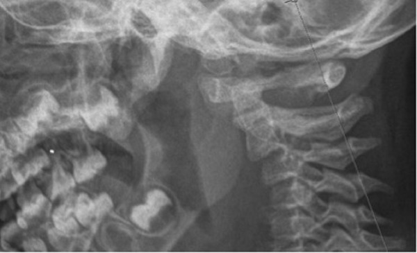
Cervical spine radiograph demonstrating correct interspinous alignment.
The prevertebral space is situated between the prevertebral fascia anteriorly and the vertebral bodies posteriorly.20 A space <6 mm at the level of C3 is considered normal in children.21
Widening of the prevertebral space is an important finding to identify as it may represent an abscess, haematoma or bony injury. Evaluation of this space in children is complicated as widening can be mimicked in exhalation such as with a crying child or cervical spine flexion such as with an uncooperative child. Owing to this anomaly, Lustrin et al19 has advised repeating lateral plain films in mild extension and in inspiration in cases of suspected widening.
The Jefferson burst fracture consists of fractures of the arches of C1 and subsequent lateral displacement of C1 with respect to C2.22 This fracture type can be appreciated on open-mouth odontoid views. In children, pseudospread of the atlas on the axis can occur. This is termed as ‘pseudo-Jefferson fracture’. Up to 6 mm of displacement of the lateral masses relative to the dens is common in patients up to 4 years old and may be seen in patients up to 7 years old.23
Overall this clinical scenario helps to differentiate between two of the life-threatening causes of torticollis namely cervical spine injury and retropharyngeal abscess. Interestingly, there have been several case reports in the literature linking trauma as a potential causative factor for development of a neck abscess.24 25
Ursic et al24 describe an almost identical picture to our case albeit in an adult. Here, the patient in question received blunt trauma to the neck. He was initially worked up in the emergency department (with normal haematological and biochemical profiles) and subsequently discharged. Three days later the patient re-presented to the emergency department with infective symptoms and neck pain and elevated CRP and WCC. Imaging further reveals a neck abscess in the anterior triangle of the neck which required urgent drainage. This case highlighted by Ursic et al24 again demonstrates the importance of a focused clinical history and examination. Clinicians should have a low threshold for suspicion of a neck abscess in adult and paediatric patients alike given the presence of infective symptomatology and neck pain. One could postulate from both cases that the presence of blunt trauma could indeed be the responsible pathogenesis for the formation of a neck abscess when no predisposing factors are present.
Furthermore, Selbst et al25 presented another similar case involving blunt trauma to the cervical spine of an adolescent. In this case, the adolescent reported of neck stiffness and fever on a background of receiving blunt trauma from a motor vehicle. Similar to our case, the NEXUS guidelines would promote the usage of cervical spine imagery in said case potentially pulling the clinician down a wrong or inappropriate avenue of diagnosis. The clinical impression here again was cervical spine injury with the authors negating the clinical findings of fever, neck stiffness and a sore throat.25 Work up imagery did go on to reveal a neck abscess and I&D was performed.
The two above cases demonstrate the formation of neck abscess postblunt trauma in an adolescent and an adult. The case we present demonstrates the formation of retropharyngeal abscess postblunt trauma in a paediatric patient. This may or may not be related to the blunt trauma itself. In any regard, the clinical symptomatology provided by the patient should be enough for the clinician to consider a diagnosis of neck abscess.
Pertinent questions to ask in this situation include assessment of infective symptomatology. Although the presence of trauma may sidetrack the clinician, concomitant pathology may always be present. Be careful to exclude infective symptoms including chills, fevers, night sweats and system questioning, for example, cough (LRTI), ear pain, runny nose (ENT), presence of insidious neck pain prior to alleged trauma (deep neck infection), and dysphagia and odonophagia (retropharyngeal abscess).
In summary, torticollis is a symptom associated with a broad range of childhood illnesses. Clinicians should take the time to take a focused history and examination to help out rule any life-threatening causes. Blunt trauma may indeed predispose patients to abscess formation. Retropharyngeal abscess is a life-threatening emergency and can be treated either conservatively or operatively depending on abscess size and presence of airway compromise. When cervical spine injury is suspected, immobilisation is vital. Baseline knowledge of suspected anatomical variants is paramount in interpretation of paediatric cervical spine imagery.
Learning points.
Retropharyngeal abscess is a recognised cause of torticollis.
Never underestimate a full clinical history and examination. Subtle hints were available in the story to point the clinician towards the correct diagnosis.
Developmental anatomical knowledge is paramount in interpreting cervical spine imagery in children.
Footnotes
Contributors: CKMD is the lead author responsible for data acquisition and design of case. NMG was responsible for analysis and acquisition of imagery. CNF was responsible for planning of study. FS was responsible for conception of study.
Competing interests: None declared.
Patient consent: Obtained.
Provenance and peer review: Not commissioned; externally peer reviewed.
References
- 1.Snyder EM, Coley BD. Limited value of plain radiographs in infant torticollis. Pediatric 2006;118:e1779–84. 10.1542/peds.2006-1624 [DOI] [PubMed] [Google Scholar]
- 2.McAloon J. Pediatric management problems (torticolis). Pediatr Nurs 1986:371–80. [PubMed] [Google Scholar]
- 3.Kiwak KJ. Establishing an etiology for torticolis. Postgrad Med 1984:126–34. 10.1080/00325481.1984.11698622 [DOI] [PubMed] [Google Scholar]
- 4.McDaniel A, Hirsch BE, Kornblut AD, et al. Torticollis in infancy and adolescence. Ear Nose Throat J 1984:478–87. [PubMed] [Google Scholar]
- 5.Cheng JC, Wong MW, Tang SP, et al. Clinical determinants of the outcome of manual stretching in the treatment of congenital muscular torticollis in infants. A prospective study of eight hundred and twenty-one cases. J B J Surg Am Vol 2001;83:679–87. 10.2106/00004623-200105000-00006 [DOI] [PubMed] [Google Scholar]
- 6.Pharisa C, Lutz N, Roback MG, et al. Neck complaints in the pediatric emergency department: a consecutive case series of 170 children. Pediatr Emerg Care 2009;25:823–6. 10.1097/PEC.0b013e3181c06062 [DOI] [PubMed] [Google Scholar]
- 7.Bredenkamp JK, Maceri DR. Inflammatory torticollis in children. Arch Otolaryngol Head Neck Surg 1990;116:310–13.. 10.1001/archotol.1990.01870030074012 [DOI] [PubMed] [Google Scholar]
- 8.Craig FW, Schunk JE. Retropharyngeal abscess in children: clinical presentation, utility of imaging, and current management. Pediatric 2003;111:1394 10.1542/peds.111.6.1394 [DOI] [PubMed] [Google Scholar]
- 9.Hasegawa J, Tateda M, Hidaka H, et al. Retropharyngeal abscess complicated with torticollis: case report and review of the literature. Tohoku J Exp Med 2007;213:99–104. 10.1620/tjem.213.99 [DOI] [PubMed] [Google Scholar]
- 10.McClay JE, Murray AD, Booth T. Intravenous antibiotic therapy for deep neck abscesses defined by computed tomography. Arch Otolaryngol Head Neck Surg 2003;129:1207 10.1001/archotol.129.11.1207 [DOI] [PubMed] [Google Scholar]
- 11.Saluja S, Brietzke SE, Egan KK, et al. A prospective study of 113 deep neck infections managed using a clinical practice guideline. Laryngology 2013;123:3211 10.1002/lary.24168 [DOI] [PubMed] [Google Scholar]
- 12.Boucher C, Dorion D, Fisch C. Retropharyngeal abscesses: a clinical and radiologic correlation. J Otolaryngol 1999;28:134–7. [PubMed] [Google Scholar]
- 13.Hurley MC, Heran MK. Imaging studies for head and neck infections. Infect Dis Clin North Am 2007;21:305 10.1016/j.idc.2007.04.001 [DOI] [PubMed] [Google Scholar]
- 14.Dietrich AM, Ginn-Pease ME, Bartkowski HM, et al. Pediatric cervical spine fractures: predominantly subtle presentation. J Pediatr Surg 1991;26:995–1000. 10.1016/0022-3468(91)90850-S [DOI] [PubMed] [Google Scholar]
- 15.Baker C, Kadish H, Schunk JE. Evaluation of pediatric cervical spine injuries. Am J Emerg Med 1999;17:230 10.1016/S0735-6757(99)90111-0 [DOI] [PubMed] [Google Scholar]
- 16.Curran C, Dietrich AM, Bowman MJ, et al. Pediatric cervical-spine immobilization: achieving neutral position? J Trauma 1995;39:729–32. 10.1097/00005373-199510000-00022 [DOI] [PubMed] [Google Scholar]
- 17.Hoffman JR, Mower WR, Wolfson AB. Validity of a set of clinical criteria to rule out injury to the cervical spine in patients with blunt trauma. National Emergency X-Radiography Utilization Study Group. N Engl J Med 2000;343:94–9. 10.1056/NEJM200007133430203 [DOI] [PubMed] [Google Scholar]
- 18.Swischuk LE. Anterior displacement of C2 in children: physiologic or pathologic. Radiology 1977;122:759–63. 10.1148/122.3.759 [DOI] [PubMed] [Google Scholar]
- 19.Lustrin ES, Karakas SP, Ortiz AO, et al. Pediatric cervical spine: normal anatomy, variants, and trauma. Radiographics 2003;23:539–60. 10.1148/rg.233025121 [DOI] [PubMed] [Google Scholar]
- 20.Debnam JM, Guha-Thakurta N. Retropharyngeal and prevertebral spaces: anatomic imaging and diagnosis. Otolaryngol Clin North Am 2012;45:1293–310. 10.1016/j.otc.2012.08.004 [DOI] [PMC free article] [PubMed] [Google Scholar]
- 21.Warner WC. Rockwood and Wilkins’ fractures in children. In: Beaty JH, Kasser JR eds. Cervical spine injuries in children. Lippincott Williams & Wilkins, 2001;809–46. [Google Scholar]
- 22.Haus BM, Harris MB. Case report: nonoperative treatment of an unstable Jefferson fracture using a cervical collar. Clin Orthop Relat Res 2008;466:1257–61. 10.1007/s11999-008-0143-5 [DOI] [PMC free article] [PubMed] [Google Scholar]
- 23.Roche C, Carty H. Spinal trauma in children. Pediatr Radiol 2001;31:677–700. 10.1007/s002470100532 [DOI] [PubMed] [Google Scholar]
- 24.Ursic CM, Shah SV, Kaviani A. Neck abscess after blunt cervical trauma. J Trauma 2001;51:146–8. 10.1097/00005373-200107000-00024 [DOI] [PubMed] [Google Scholar]
- 25.Selbst SM, Fein JA, Friday J, et al. Retropharyngeal abscess after blunt trauma in an adolescent. Pediatr Emerg Care 1998;14:406–8. 10.1097/00006565-199812000-00007 [DOI] [PubMed] [Google Scholar]