Insect vectors play a key role in dissemination of viruses that cause important diseases in humans, animals, and plants. Specific understanding of insect–virus interactions leading to successful transmission is a central problem in vector biology and critical to developing effective control strategies. In this issue of PNAS, Sin et al. (1) provide evidence that the genetic determinants of insect transmissibility for tomato spotted wilt virus (TSWV) reside on the middle RNA (M RNA) segment encoding the viral membrane glycoproteins (GPs) [N-terminal GP (GN) and C-terminal GP(GC)]. This virus is transmitted between plants by insects called thrips (Thripidae, Thysanoptera) and is the type member of the genus Tospovirus. The tospoviruses are the only plant-infecting members in the family Bunyaviridae, which consists of many viruses that cause diseases in animals and humans (2). Thus, tospoviruses and their thrips vectors are ideal model systems for elucidating processes of virus infection in disparate hosts that can be extended to viruses of importance to human health.
The complex nature of the interplay between thrips, tospoviruses, and their shared plant hosts was first recognized with the discovery that TSWV multiplies in its insect vectors (3, 4). This discovery opened rich and exciting avenues of exploration, including understanding biological and molecular interactions underlying TSWV pathogenesis in plant and insect hosts and the role these processes play in virus evolution. Findings of the last decade show that insect inoculation of tospoviruses into a plant host cannot occur without viral passage across at least three insect organs (the midgut, visceral muscle cells, and salivary glands) that include six membrane barriers (2). Previous hypotheses that tospovirus GPs are essential determinants of thrips acquisition were based on several pieces of experimental evidence: (i) assembly-deficient TSWV isolates could be passed mechanically between plants but were not insect transmissible (5, 6); (ii) TSWV GPs were detected binding the insect midgut during acquisition (7); (iii) TSWV GPs and not other viral proteins were shown to bind thrips proteins in overlay assays (8, 9); and (iv) GN binds the insect vector midgut and inhibits TSWV acquisition (10).
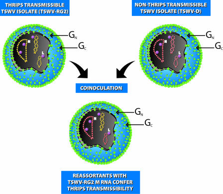
Sin et al. (1) created reassortants by coinoculating plants with a thrips-transmissible TSWV isolate (TSWV-RG2) and a thrips-nontransmissible TSWV isolate (TSWV-D). Their observation that only reassortants with the M RNA from TSWV-RG2 could confer thrips transmissibility (Fig. 1) provided a clear link between this genome segment and determinants of insect transmission. In an exciting extension of this experiment, Sin et al. showed that a single nonsynonymous nucleotide substitution in the ORF encoding GN/GC had no apparent effect on virion assembly, but could eliminate insect transmissibility. For viruses in the genus Tospovirus, the unequivocal association of a phenotype not only to an individual viral RNA segment but to a specific nucleotide without using a reverse genetics approach is without precedent. This result is particularly satisfying because it rules out the possibility that insect nontransmissibility was caused by failure to form virions or other undocumented changes in the large (L) and small (S) RNA segments. Furthermore, the observation that the TSWV M RNA segment was required for insect transmission, but not for plant infection, supports the important role insect vectors play in the evolution of TSWV populations. It seems likely that coevolution between insect vectors and tospoviruses, the potential for TSWV reassortment, and the role of M RNA in determining transmissibility all play critical roles in the emergence of new vector–virus relationships.
Fig. 1.
The observations of Sin et al. (1) show that the genetic determinants of insect transmission of TSWV lie on the viral M RNA in the ORF encoding GN and GC. Two parental TSWV isolates were used to coinoculate plants and generate isolates in which the RNA segments reassorted. One parental isolate, TSWV-D, was not transmitted by insects, whereas isolate TSWV-RG2 was transmissible by two insect vector species, Frankliniella occidentalis and F. fusca. Among the viral reassortants only those containing the M RNA segment from TSWV-RG2 conferred thrips transmissibility. This evidence showing that reassortant viral populations arise from mixed infections with altered traits for insect transmission has important implications for understanding TSWV–thrips coevolution and the emergence of new virus–vector relationships. N, nucleocapsid; L, virion-associated RNA-dependent RNA polymerase. Illustration by Eileen J. Rendahl (Rendahl Graphics and Illustration, Davis, CA).
Viral membrane GPs are important in virus entry to host cells for all members of the bunyaviridae that have been studied (11–14). Reassortment studies with hantaviruses and orthobunyaviruses show that virulence (15–17) and insect transmission (18) maps to the M RNA segment; furthermore, mutations in the GN and GC proteins of other bunyaviruses attenuate disease (15, 19) and insect infection (14). Mutation at amino acid 515 of GN from Ile to Thr in the GN transmembrane domain of Hantaan virus (genus Hantavirus) resulted in attenuation of disease in newborn mice (15). Interestingly, this mutation is remarkably similar to that observed by Sin et al. (1) in that both occur near the C terminus of GN (in the transmembrane domain or cytoplasmic tail) and the observed mutants both changed to Thr. Sin et al. (1) broadly define the viral determinants of thrips transmission and illuminate the importance of knowing the exact location of the GN/GC maturation cleavage site. The TSWV GPs are encoded as a polyprotein that is cleaved to generate GN and GC (20). Although the cleavage site has not been empirically determined, sequence analysis indicates that a canonical signal peptidase motif immediately downstream from a hydrophobic signal peptide may indicate its location. Other bunyaviruses are cleaved by signal peptidases (21), and if this site functions in cleavage, then the Pro at position 459 in SLI 81 [described by Sin et al. (1)] resides near the C terminus of GN and is possibly in a transmembrane domain. Loss of insect transmissibility in the presence of the Pro-459–Thr mutation raises several interesting possibilities. The mutation may change the structure of the GPs or alter posttranslational modifications important in protein function. Examination of other viral GPs revealed that multiple domains interact in protein function and mutations in one domain can impact functions of another. For example, transition of paramyxovirus F protein to a fusogenic conformation depends on interaction of residues in the ectodomain, transmembrane domain, and cytoplasmic tail (22). Mutations in all regions altered fusogenic capabilities of the protein (22). Likewise, mutation in the transmembrane domain of TSWV SLI 81 GN could alter the function of GN and/or GC. If the TSWV GPs function in attachment and entry as heterooligomeric complexes, the mutation reported by Sin et al. (1), which is likely in the C terminus of GN, could also disrupt GC function.
The future holds many possibilities for elucidating the function of the TSWV GPs in virus transmission by thrips. Sin et al. (1) validate the hypothesis that, like the animal–infecting bunyaviruses, the GPs encoded by plant-infecting members of the family are necessary for infection of animal host cells. Modeling the structure of a bunyavirus GC protein revealed that the GPs may be class II fusion proteins, and sequence comparisons showed TSWV GN protein shares sequence similarity with another viral attachment protein, sindbis virus E2 (23). These data are supported by findings that TSWV GN likely plays a role in virus attachment to thrips midgut epithelial cells (10), and that GC is cleaved at acidic pH, consistent with it serving as a fusion protein activated at low pH (24). Determining the sequence of mature GPs and solving their crystal structure will be critical to understanding their function and importance of the amino acid at position 459. Interesting questions to be addressed will include examining the steps at which virus transmission is blocked, including virus stability in the insect gut, attachment, fusion with host cells, replication in insect cells, or cell-to-cell spread in the insect. Experiments with SLI 81 combined with the use of soluble forms of GPs to manipulate insect transmissibility will create a new understanding of the roles of GN and GC. Ideally, examining GN and GC function in the context of an easily manipulated virion would enable researchers to characterize GP function. One way to achieve this objective would be to generate pseudotyped virions that express GN and GC. Mutational analysis of soluble GPs and pseudotyped virions would extend our current understanding of virus binding and entry and enable the generation of specific mutations that would complement the reassortant system described by Sin et al. (1).
An important lesson to be learned from Sin et al. (1) is that clever deployment of new biological tools can temporarily overcome the lack of a reverse genetics system in the Bunyaviridae and advance our understanding of tospovirus–thrips relationships. Researchers working with positive-sense RNA viruses have had the option of using reverse genetics for making functional assignments to viral proteins for >20 years (25). It is impossible to overstate the positive impact of this technology on the advancement of plant virology. In contrast, reverse genetics systems have only recently been established for relatively few animal-infecting negative-sense RNA viruses (26), and there are no example of such systems for plant-infecting viruses. Combining genetic reassortment and mutational analysis with functional assays involving recombinant GPs will allow us to delve deeper into the specific processes governing virus entry. Clearly, this work signals an era in which understanding of TSWV–thrips vector interactions will come of age. Thrips vectors of TSWV provide a model system in which much can be learned about steps involved in virus fusion, interaction with receptors, binding, and entry to cells. This knowledge, coupled with the similarities between tospoviruses and their animal-infecting counterparts in the Bunyarvidae, will lead to an overarching understanding of pathogenesis and vector–virus relationships in this large virus family. Translation of these fundamental advances into creative new control strategies that extend beyond tospoviruses to animal-infecting members of the Bunyaviridae promises to make this an exciting area of research to follow.
See companion article on page5168.
References
- 1.Sin, S.-H., McNulty, B. C., Kennedy, G. G. & Moyer, J. W. (2005) Proc. Natl. Acad. Sci. USA 102, 5168–5173. [DOI] [PMC free article] [PubMed] [Google Scholar]
- 2.Whitfield, A. E., Ullman, D. E. & German, T. L. (2005) Annu. Rev. Phytopathol., in press. [DOI] [PubMed]
- 3.Ullman, D. E., German, T. L., Sherwood, J. L., Westcot, D. M. & Cantone, F. A. (1993) Phytopathology 83, 456–463. [Google Scholar]
- 4.Wijkamp, I., van Lent, J., Kormelink, R., Goldbach, R. & Peters, D. (1993) J. Gen. Virol. 74, 341–349. [DOI] [PubMed] [Google Scholar]
- 5.Nagata, T., Inoue-Nagata, A. K., van Lent, J., Goldbach, R. & Peters, D. (2002) J. Gen. Virol. 83, 663–671. [DOI] [PubMed] [Google Scholar]
- 6.Resende, R. d. O., de Haan, P., de Avila, A. C., Kitajima, E. W., Kormelink, R., Goldbach, R. & Peters, D. (1991) J. Gen. Virol. 72, 2375–2385. [DOI] [PubMed] [Google Scholar]
- 7.Ullman, D. E., German, T. L., Sherwood, J. L. & Westcot, D. M. (1992) in Thrips Biology and Management, eds. Parker, B. L., Skinner, M. & Lewis, T. (Plenum, New York), pp. 135–152.
- 8.Bandla, M. D., Campbell, L. R., Ullman, D. E. & Sherwood, J. L. (1998) Phytopathology 88, 98–104. [DOI] [PubMed] [Google Scholar]
- 9.Kikkert, M., van Lent, J., Storms, M., Bodegom, R., Kormelink, R. & Goldbach, R. (1999) Phytopathology 88, 63–69. [Google Scholar]
- 10.Whitfield, A. E., Ullman, D. E. & German, T. L. (2004) J. Virol. 78, 13197–13206. [DOI] [PMC free article] [PubMed] [Google Scholar]
- 11.Pekosz, A., Griot, C., Nathanson, N. & Gonzalez-Scarano, F. (1995) Virology 214, 339–348. [DOI] [PubMed] [Google Scholar]
- 12.Ludwig, G. V., Israel, B. A., Christensen, B. M., Yuill, T. M. & Schultz, K. T. (1991) Virology 181, 564–571. [DOI] [PubMed] [Google Scholar]
- 13.Hacker, J. K. & Hardy, J. L. (1997) Virology 235, 40–47. [DOI] [PubMed] [Google Scholar]
- 14.Sundin, D. R., Beaty, B. J., Nathanson, N. & Gonzalez-Scarano, F. (1987) Science 235, 591–593. [DOI] [PubMed] [Google Scholar]
- 15.Ebihara, H., Yoshimatsu, K., Ogino, M., Araki, K., Ami, Y., Kariwa, H., Takashima, I., Li, D. & Arikawa, J. (2000) J. Virol. 74, 9245–9255. [DOI] [PMC free article] [PubMed] [Google Scholar]
- 16.Griot, C., Pekosz, A., Lukac, D., Scherer, S. S., Stillmock, K., Schmeidler, D., Endres, M. J., Gonzalez-Scarano, F. & Nathanson, N. (1993) J. Virol. 67, 3861–3867. [DOI] [PMC free article] [PubMed] [Google Scholar]
- 17.Janssen, R. S., Nathanson, N., Endres, M. J. & Gonzalez-Scarano, F. (1986) J. Virol. 59, 1–7. [DOI] [PMC free article] [PubMed] [Google Scholar]
- 18.Beaty, B. J., Holterman, M., Tabachnick, W., Shope, R. E., Rozhon, E. J. & Bishop, D. H. L. (1981) Science 211, 1433–1435. [DOI] [PubMed] [Google Scholar]
- 19.Isegawa, Y., Tanishita, O., Ueda, S. & Yamanishi, K. (1994) J. Gen. Virol. 75, 3273–3278. [DOI] [PubMed] [Google Scholar]
- 20.Kormelink, R., de Haan, P., Meurs, C., Peters, D. & Goldbach, R. (1992) J. Gen. Virol. 73, 2795–2804. [DOI] [PubMed] [Google Scholar]
- 21.Löber, C., Bärbel, A., Lindow, S., Klenk, H. & Feldmann, H. (2001) Virology 289, 224–229. [DOI] [PubMed] [Google Scholar]
- 22.Seth, S., Goodman, A. L. & Compans, R. W. (2004) J. Virol. 78, 8513–8523. [DOI] [PMC free article] [PubMed] [Google Scholar]
- 23.Garry, C. E. & Garry, R. F. (2004) Theor. Biol. Med. Model. 1, 10. [DOI] [PMC free article] [PubMed] [Google Scholar]
- 24.Whitfield, A. E., Ullman, D. E. & German, T. L. (March 7, 2005) Virus Res., doi: 10.1016/j.virusres.2005.01.007. [DOI] [PubMed]
- 25.Ahlquist, P., French, R., Janda, M. & Loesch-Fries, L. S. (1984). Proc. Natl. Acad. Sci. USA 81, 7066–7070. [DOI] [PMC free article] [PubMed] [Google Scholar]
- 26.Neumann, G., Whitt, M. & Kawaoka, Y. (2002) J. Gen. Virol. 83, 2635–2662. [DOI] [PubMed] [Google Scholar]


