Abstract
Introduction
The inability of some seriously and chronically ill individuals to receive a definitive diagnosis represents an unmet medical need. In 2008, the NIH Undiagnosed Diseases Program (UDP) was established to provide answers to patients with mysterious conditions that long eluded diagnosis and to advance medical knowledge. Patients admitted to the NIH UDP undergo a five-day hospitalization, facilitating highly collaborative clinical evaluations and a detailed, standardized documentation of the individual’s phenotype. Bedside and bench investigations are tightly coupled. Genetic studies include commercially available testing, single nucleotide polymorphism microarray analysis, and family exomic sequencing studies. Selected gene variants are evaluated by collaborators using informatics, in vitro cell studies, and functional assays in model systems (fly, zebrafish, worm, or mouse).
Insights from the UDP
In seven years, the UDP received 2954 complete applications and evaluated 863 individuals. Nine vignettes (two unpublished) illustrate the relevance of an undiagnosed diseases program to complex and common disorders, the coincidence of multiple rare single gene disorders in individual patients, newly recognized mechanisms of disease, and the application of precision medicine to patient care.
Conclusions
The UDP provides examples of the benefits expected to accrue with the recent launch of a national Undiagnosed Diseases Network (UDN). The UDN should accelerate rare disease diagnosis and new disease discovery, enhance the likelihood of diagnosing known diseases in patients with uncommon phenotypes, improve management strategies, and advance medical research.
1. Introduction
In 2008, the NIH established an Undiagnosed Diseases Program (UDP), designed to help patients who had long sought a precise diagnosis and to discover new pathways and mechanisms of disease [1–3]. Ongoing annotation of the human genome, combined with advances in DNA sequencing, provided a huge impetus to the UDP and bolstered the promise of precision medicine [4]. Initiated within the NIH Intramural Research Program, the UDP has now evolved into the Undiagnosed Diseases Network (UDN), supported by the NIH Common Fund. The Network consists of the UDP, six additional clinical sites around the nation, a coordinating center, two DNA sequencing cores, a model organisms screening center, a metabolomics core, and a central biorepository. The UDN functions under a common IRB protocol with reliance agreements and data sharing procedures. On September 16, 2015, the UDN was launched with an online portal for patient applications (https://gateway.undiagnosed.hms.harvard.edu) [5]; it is modeled after the UDP, whose methods and illustrative cases are presented here.
Patients and their families were enrolled in a protocol approved by the NHGRI Institutional Review Board, and gave written informed consent. They applied to the UDP by providing a referral letter from a clinician, along with medical records, laboratory results, imaging studies, and biopsy slides. UDP experts evaluated each application for the presence of objective findings, novel phenotypic manifestations, and the likelihood of obtaining a diagnosis. A signature feature of the UDP was compression of the clinical evaluation at the NIH Clinical Center into five inpatient days, free of charge to the patient, with no insurance approvals. The UDP diagnostic process emphasized highly collaborative clinical evaluations, detailed and standardized documentation of patient’s phenotype, and tightly coupled bedside and bench investigations. Standardized documentation of patient phenotypes employed Human Phenotype Ontology (HPO) terms, using PhenoTips software [6]. Clinical consultations were conducted by multiple specialists, and imaging studies and laboratory testing were tailored to the patient’s individual manifestations. Examples of specialized assays included cerebrospinal fluid neurotransmitters and plasma glycomics. Biologic samples, including plasma, serum, DNA, urine, and fibroblasts from skin biopsies, were routinely collected and stored. Genetic studies included commercially available testing, panels of genes, single nucleotide polymorphism (SNP) analysis, and family exomic sequencing. Variant analysis utilized both commonly applied variant annotations and manually curated data, including SNP chip correlation, regions of low coverage, and non-coding regions. Some potentially pathogenic variants were evaluated by collaborators using informatics, cultured cell studies, and animal models for functional assays (fly, zebrafish, worm, or mouse).
2. Insights from the UDP
From its inception in 2008 through May 2015, the UDP received 2954 complete applications and accepted 863 (29%) for evaluation. Of these, we know 64 (7%) have died. Of the 863 patients evaluated, we present nine vignettes, seven published (Table 1) [7–38] and two unpublished, comprising 20 patients in ten families. (The number in column of Table 1 identifies the vignette’s section number in this article.) The patients demonstrate how investigating undiagnosed individuals advances understanding of: 1) common and complex disorders, 2) the coincidence of rare diseases, 3) newly recognized mechanisms of disease, and 4) the practice of precision medicine.
Table 1.
UDP publications on rare and new diseases, ordered by increasing PMID number.
| Reference | Vignette numberc | PMID number | Diagnosis | Phenotype OMIM number | Gene name abbreviation | Gene OMIM number | Inheritanceb | Frequency |
|---|---|---|---|---|---|---|---|---|
| 7c | 2.3.1 | 21288095 | Calcification of joint and arteries | 211800 | NT5E | 129190 | AR | 17 patients |
| 8 | 21353777 | Spinal muscular atrophy, distal, autosomal recessive, 1 | 604320 | IGHMBP2 | 600502 | AR | 60 patients | |
| 9 | 22022284 | Spastic ataxia 5, autosomal recessive and neuropathy | 614487 | AFG3L2 | 604581 | AR | 4 patients | |
| 10 | 22146942 | Spastic paraplegia 35, autosomal recessive | 612319 | FA2H | 611026 | AR | 18 patients | |
| 11 | 22252885 | IgG4-related sclerosing mastoiditis | 604360 | Somatic? | None | ~200 patients | ||
| 12 | 22675082 | GM1-gangliosidosis, juvenile | 230600 | GLB1 | 611458 | AR | 1/300,000 | |
| 13 | 22749184 | Spastic paraplegia, autosomal recessive | 604360 | SPG11 | 610844 | AR | 1/100,000 | |
| 14c | 2.1.2 | 23293122 | Nephrolithiasis | 143880 and 52 others | CYP24A1 | 126065 | AR | Up to 1/1000 |
| 15 | 23420719 | Kearns-Sayre syndrome with growth failure | 530000 | Mitochondrial DNA deletion | Mitochondrial | 1/100,000 | ||
| 16 | 23443029 | Recurrent subacute post-viral ataxia | ?variant of 603553 | PRF1 | 170280 | AR | 2 sisters (plus 4 with different phenotype) | |
| 17 | 23453856 | Myopathy, areflexia, respiratory distress, dysphagia, early-onset (EMARDD) | 614399 | MEGF10 | 612453 | AR | 14 patients in 7 families | |
| 18 | 23465863 | Amyloid myopathy | Not in OMIM | Somatic | None | 15/year in U.S. | ||
| 19c | 2.3.2 | 23649844 | Leukodystrophy, demyelinating, adult-onset, autosomal dominant | 169500) | LMNB1 | 150340 | AD | 148 patients in 33 families |
| 20 | 23661660 | Mucopolysaccharidosis IIIB | 252920 | NAGLU | 609701) | AR | 1/100,000 | |
| 21 | 23857908 | Hereditary spastic paraplegia type 43 | 615043 | C19orf12 | 614297 | AR | 53 patients | |
| 22 | 23968566 | Neurodegeneration with brain iron accumulation 1 | 234200 | PANK2 | 606157 | AR | 1–3/1,000,000 | |
| 23 | 24006476 | Cutaneous skeletal-hypophosphatemia syndrome() | Not in OMIM | NRAS and HRAS | Somatic, AD | 5 patients | ||
| 24c, 27c | 2.4.1 | 24504326 24839611 |
Epilepsy, focal, with speech disorder | 245570 | GRIN2A | 138253 | AD | 124 patients |
| 25 | 24686847 | Aicardi-Goutières syndrome 7 | 615846 | IFIH1 | 606951 | AD | 14 patients | |
| 26c | 2.1.3 | 24716661 | Congenital disorder of glycosylation IIb (MOGS-CDG) | 606056 | MOGS | 601336 | AR | 3 patients |
| 28c | 2.2.2 | 24863970 | Phosphoenolpyruvate carboxylate deficiency, cytosolic | 182290 | PCK1 | 614168 | AR | First report |
| Smith-Magenis syndrome | 613970 | RAI1 | 607642 | AD | 1/15,000 | |||
| Cognitive defects, autosomal dominant 6 | 261680 | GRIN2B | 138252 | AD | 37 pathogenic variants | |||
| 29 | 25251875 | Brain hypomyelination | 8 OMIM entries | ERCC6 | 609413 | AR | 25 patients | |
| Cockayne syndrome B | 133540 | |||||||
| 30 | 25527264 | Hypomyelination with brainstem and spinal cord involvement and leg spasticity | 615281 | DARS | 603084 | AR | 13 patients | |
| 31 | 25577287 | Stormorken (York platelet) syndrome | 185070 | STIM1 | 605921 | AD | 22 patients | |
| 32 | 25678555 | Congenital disorder of glycosylation Iz | 616457 | CAD | 114010 | AR | First report | |
| 33 | 25817015 | Epileptic encephalopathy, early infantile, 29 | 616339 | AARS | 601065 | AR | First report: 3 patients | |
| 34 | 25845469 | Cognitive impairment, autosomal recessive 18 | 614249 | MED23 | 605042 | AR | 7 patients in 2 families | |
| 35c | 2.1.1 | 25888122 | Cognitive impairment, syndromic, Snyder-Robinson type | 309583 | SMS | 300105 | X-linked | 20 patients |
| 36 | 25943031 | Multiple congenital anomalies-hypotonia-seizures syndrome 3 | 615398 | PIGT | 610272 | AR | 7 patients | |
| 37 | 26119818 | Ablepharon macrostomia syndrome | 200110 | TWIST2 | 607556 | AD | 59 patients | |
| Barber-Say syndrome | 209885 | |||||||
| 38 | 26373698 | Musculocontractural type of Ehlers-Danlos syndrome | 601776 | CHST14 | 608429 | AR | 39 patients |
OMIM, Online Mendelian Inheritance in Man.
AR, autosomal recessive; AD, autosomal dominant.
Illustrative patient vignettes in text, in section number given in second column.
2.1. Understanding common and complex diseases
Most common and complex disorders are polygenic or multifactorial, and their diverse determinants are difficult to sort out. In contrast, rare disorders are largely monogenic, i.e., explained by a single genetic aberration. Three rare patients exemplify insights into common conditions (osteoporosis, nephrolithiasis, and viral infections).
2.1.1. Osteoporosis
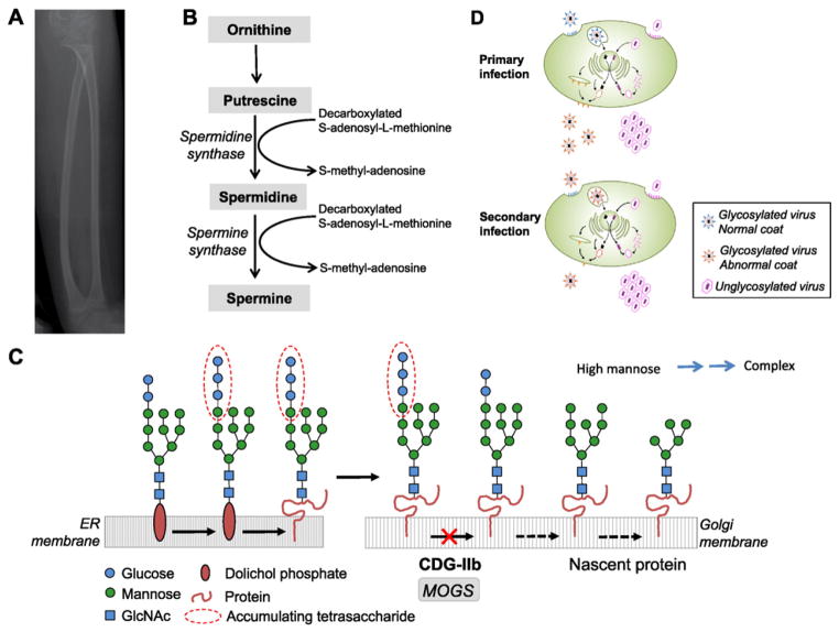
A 15-year-old male had perinatal thrombocytopenia with intraventricular hemorrhage, hypoglycemia, tracheomalacia, aspiration pneumonias, congenital hip dislocations, infantile spasms at 15 months, and severe developmental delay by 2 years [35]. By age 6, he had renal tubular acidosis, nephrocalcinosis, nephrolithiasis, and fractures of the fibula and humerus. At age 15, he was microcephalic and noninteractive, with short stature, facial dysmorphisms, drooling, hearing loss, seizures, flexion contractures, scoliosis, cryptorchidism, retinitis pigmentosa, and cortical blindness. Brain MRI showed decreased white matter volume and delayed myelination in the frontal lobes. Bone age was delayed. Plain radiographs of lower extremities showed osteoporosis (Fig. 1A); DEXA scan Z scores were −2.9 in the spine and −6.5 in the forearm. Bone biopsy showed no trabecular meshwork, low cancellous bone volume, thick cortex, and decreased osteoblast and osteoclast activity. Differentiated bone marrow stem cells exhibited poor calcium phosphate mineralization.
Fig. 1.
Understanding common and complex diseases. (A) Gracile, osteoporotic tibia and fibula. (B) Polyamine synthetic pathway. Addition of a propylamine moiety from decarboxylated S-adenosylmethionine to putrescine produces spermidine. A second propylamine moiety is added to spermidine by spermine synthase, producing spermine. Spermine synthase is deficient in boys with Snyder-Robinson syndrome. Spermidine and spermine are polyamines whose ratio is crucial for cell processes including transcription and translation [39]. (C) Pathway defective in Congenital Disorder of Glycosylation IIb. Glucosidase I, deficient in the disorder, catalyzes the first step in the conversion of high mannose glycoproteins to complex glycoproteins, as part of the endoplasmic reticulum protein quality control process, also known as the unfolded protein response. (D) Viral infections in patient cells. Glycosylated viruses such as HIV can infect these cells, but the cells produce viruses that contain an abnormally glycosylated coat, making them less infective. No differences in terms of infectivity were noted when non-glycosylated viruses were tested. (Courtesy of Dr. Sergio D. Rosenzweig.)
Exome sequencing of the boy and his 9-year-old brother revealed a maternally inherited missense variant in the SMS gene on chromosome X, i.e., c.443A>G (p.Gln148Arg). SMS encodes the enzyme spermine synthase, whose level was decreased 2.6-fold in the proband’s cultured fibroblasts (Fig. 1B). Hemizygous mutations in SMS result in the Snyder-Robinson syndrome, characterized by facial dysmorphisms, neurological deficits, and osteoporosis [39–41].
Osteoporosis is a major public health problem whose mechanism is not completely understood. Snyder-Robinson syndrome can serve as a monogenic disorder in which to study the role of polyamines [42,43] in preventing osteoporosis in humans.
2.1.2. Nephrolithiasis
A 38-year-old man had recurrent episodes of nephrolithiasis [14]; stone analysis revealed calcium phosphate, and the patient had osteopenia and fractures. Serum ionized calcium was 1.32–1.41 mmol/L (normal, 1.12–1.32), the serum parathyroid hormone was suppressed to 3–10 pg/mL (normal, 16–87), urine calcium/creatinine ratio was persistently high (median 0.33; normal, <0.22), and the fractional excretion of phosphate was 34% (normal, <20%). The serum 1α.25 (OH)2D3 was elevated at 83–160 pg/mL (normal, 18–64); specialized studies showed that the ultimate, inactive vitamin D metabolite, 24.25(OH)2D, was 0.33 ng/mL (normal, 1.2–2.6). Ketoconazole therapy normalized the patient’s calcium axis. Fibroblasts showed no metabolites of 1α,25 (OH)2D3 and reduced expression of CYP24A1 protein. Sequencing of the CYP24A1 gene revealed a paternally inherited deletion, p.E143del, and a maternally inherited transition in exon 9, c.1226T>C (p.L409S).
The CYP24A1 gene encodes 1,25(OH)2D-24-hydroxylase, the enzyme responsible for inactivating vitamin D [44]. Biallelic mutations in this gene result in increased active vitamin D levels, hypercalcemia, nephrocalcinosis, and nephrolithiasis. The frequency of deleterious CYP24A1 variants in the general population is estimated at 0.06–0.14. Since 10% of all people have nephrolithiasis, CYP24A1 mutations could account for 4–20% of patients with calcium kidney stones [14]. Individuals at risk for this common disease could be screened for high 1α,25 (OH)2D3 levels for consideration of a low calcium diet, less vitamin D- fortified foods, and minimal sun exposure.
2.1.3. Viral infections
A brother and sister, ages 11 and 6 years, presented with dysmorphic facial features, global developmental delays, hypotonia, optic atrophy, cerebral atrophy, and severe hypogammaglobulinemia with no increase in the number of infections [26]. Plasma levels of IgG were 317 and 142 mg/dL (normal, 574–1474), IgA <7 and 17 mg/dL (normal, 34–305), and IgM 21 and 21 (normal 32–208), respectively. Antibodies to measles, mumps, and varicella were negative or equivocal, despite adequate vaccine administration. Antibodies to tetanus toxoid, diphtheria toxoid, Haemophilus influenza, and Streptococcus pneumonia were adequate after vaccination. Thin layer chromatography of urine revealed a tetrasaccharide, identified on mass spectrometry as glucose3-mannose, making the diagnosis of Congenital Disorder of Glycosylation type IIb, now known as MOGS-CDG; this was confirmed by finding heterozygous mutations in the gene MOGS, encoding glucosidase I. Affected individuals fail to synthesize complex glycans on glycoproteins, creating a devastating, multisystem disease seen in only one prior patient [45] (Fig. 1C).
The children were hypogammaglobulinemic because immune globulins are N-linked glycoproteins stabilized by their glycans; Dr. Sergio Rosenzweig and colleagues demonstrated that the half-life of the patients’ IgG was 6 days compared with 21 days for normal IgG. However, this defect did not significantly increase their susceptibility to infections, because their T cells failed to produce normal amounts of infective envelope-glycosylated virus (Fig. 1D).
MOGS-CDG represents a rare example of a genetic disorder that confers resistance, rather than susceptibility, to an environmental agent. It emphasizes the importance of glycans for immunoglobulin stability as well as infectivity of viruses; interference with viral glycosylation could be a therapeutic strategy for halting viral replication. Moreover, the patient’s cells provide a model system for studying the infectivity of microbes that require glycoprotein synthesis.
2.2. Coincidental rare Mendelian diseases: Dulling Occam’s razor
A law of parsimony in diagnostic medicine, Occam’s razor, is to seek a single explanation for various manifestations of a patient’s condition. However, multiple rare diseases are bound to coexist in some patients with unusual phenotypes, raising the question of how one best defines a disease.
2.2.1. Multiple diagnoses in a consanguineous sibship
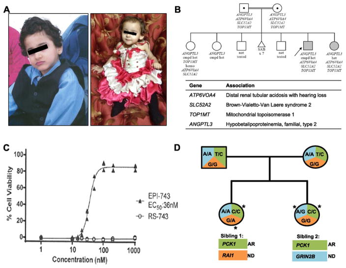
Two sibs were products of a first cousin marriage (Fig. 2A). The older, a 6-year-old boy, was born with microcephaly and by 4 months had failure to thrive, distal renal tubular acidosis, nystagmus, and optic nerve atrophy. He fell frequently following a febrile illness at 2½ years; neurologic evaluation, including a nerve biopsy, revealed a diffuse motor axonal polyneuropathy. A muscle biopsy showed myofiber atrophy. By the time of his UDP evaluation, he had lost all developmental milestones. His 2-year-old sister exhibited a similar course.
Fig. 2.
Coincidental rare Mendelian diseases: Dulling Occam’s razor. (A) Brother (left) and sister (right) with Brown-Vialetto-van Laere syndrome type 2, distal renal tubular acidosis, hearing loss, and mitochondrial topoisomerase 1 deficiency. (B) The family pedigree; double line indicates consanguinity. There were seven previous spontaneous abortions. Below each patient symbol is a list of that individual’s pertinent gene variants. The table lists the genes and the family’s associated diseases, all recessive disorders. Abbreviations: cmpd het = compound heterozygous; homo = homozygous; SAB = spontaneous abortions. (C) Response of the proband’s cultured fibroblasts to EPI-743 compared with inactive compound (RS-743) in the presence of oxidizing agents. The patient’s fibroblasts have increased sensitivity to oxidizing compounds, and EPI-743 rescues cell viability. (Courtesy of Edison Pharmaceuticals, Inc.) (D) Pedigree of a non-consanguineous family with multiple disorders; asterisks denote allele pairs causing disease.
SNP analysis showed 5% homozygosity. Sequencing revealed a homozygous ATP6VOA4 mutation (c.1185delC; p.Y396TfsX12) in both sibs, explaining their renal tubular acidosis and hearing loss [46]. Exome sequencing identified homozygous, predicted deleterious mutations in SLC52A2 (c.1327T>C; p.C443R) in both affected siblings; mutations in this riboflavin transporter are associated with Brown-Vialetto-van Laere syndrome type 2, a progressive neurologic disorder with deafness, bulbar dysfunction, and axial and limb hypotonia [47]; the children were treated with riboflavin. Homozygous, predicted deleterious mutations in TOP1MT (c.1030C>T; p.R344C) were also present in both affected siblings. TOP1MT encodes a mitochondrial DNA topoisomerase not previously associated with human disease. The proband’s cultured fibroblasts responded to the drug EPI-743 (alphatocotrienol quinone; Fig. 2C), rendering the boy eligible for a clinical trial of this drug, which has demonstrated clinical response in Leigh encephalopathy and Leber hereditary optic neuropathy [48,49].
This case illustrates the importance of reconsidering the principle of Occam’s razor, especially in cases of consanguinity.
2.2.2. Multiple diagnoses in a sibship independent of consanguinity
An 11-year-old girl (Fig. 2D, Sibling 1, left) born with microcephaly, had motor delay at 6 months, intellectual disability, easy fatigability, hypotonia, and an unexplained neuropathy [28]. At 20 months, she suffered the first of a dozen episodes of fever, lethargy, fasting hypoglycemia, ketonuria, undetectable insulin and, on one occasion, lactic acidemia. She had coarse facial features, midfacial hypoplasia, hypotelorism, synorphrys, widely spaced teeth, brachydactyly, fifth finger clinodactyly, hypoplastic toenails, aggressive and obstinate behavior, and abdominal striae. BMI, weight, and head circumference were >97th centiles. Her younger sister (Sibling 2) had a similar presentation, but with more severe developmental delay and hypotonia and no dysmorphisms. At age 5 years, milestones ranged from 4 to 15 months. Gait was unsteady, hand movements stereotypic, and pincher grasp absent. There was no consanguinity.
The two sisters displayed a homozygous variant in PCK1 (c.134T>C; p.I45T) on exome sequencing [28]; this variant conferred a short half life on the PCK protein, which is cytosolic phosphoenolpyruvate carboxykinase, a gluconeogenic enzyme that maintains glucose homeostasis. The older sister had a de novo nonsense variant in RAI1 (c.2273G>A; p.W758X), producing reduced mRNA consistent with Smith-Magenis syndrome [50]; this accounted for her dysmorphisms, intellectual disability, and behavior issues. The younger sister had a de novo mutation in GRIN2B (c.1238A>G; p.E413G), the gene for N-methyl-D-aspartic glutamate receptor subunit 2B. This variant accounted for her specific neurological deficits [51].
Even in the absence of consanguinity, multiple rare diseases can occur in the same patient.
2.3. Newly recognized mechanisms of disease
The discovery of novel diseases in undiagnosed patients often reveals a previously unrecognized metabolic or cell biological pathway.
2.3.1. Arterial calcification
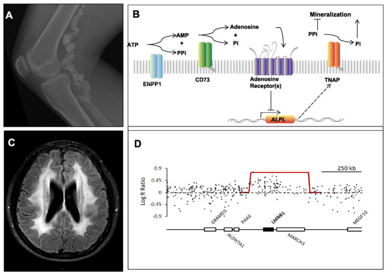
In 2011, the UDP reported nine adults in three families with a rare disease, arterial calcification due to deficiency of CD73 (ACDC) involving joint and arterial calcifications (Fig. 3A) due to biallelic mutations in NT5E [7]. This gene encodes CD73, an ectonucleotidase that converts adenosine monophosphate (AMP) to adenosine and inorganic phosphate within vascular endothelial cells (Fig. 3B).
Fig. 3.
Newly recognized mechanisms of disease. (A) Plain radiograph of a patient showing extensive, irregular calcification and dilatation of the femoral and popliteal arteries [7]. (B) Purinergic pathway at the surface of vascular endothelial cells. Normally, CD73 (encoded by NT5E, defective in ACDC) converts adenosine monophosphate to adenosine and inorganic phosphate (Pi). Adenosine binds to the adenosine receptor, trophically inhibiting alkaline phosphatase (APL) expression. When CD73 is absent, ALP is increased and converts pyrophosphate (PPi, an inhibitor of calcification) to Pi (a stimulator of calcification). In vitro studies showed that NT5E mutant fibroblasts calcified under osteogenic conditions, and the calcification could be reversed by adenosine or an APL inhibitor. (Courtesy of Shira Ziegler, Johns Hopkins University School of Medicine.) (C) Brain MRI showing leukodystrophy in brainstem and middle cerebellar peduncles of a patient with duplication of LMNB1. (D) Log R ratios of fluorescent intensity of SNPs on chromosome 5q. A contiguous group of SNPs has higher intensity, reflecting a duplication, i.e., three total copies rather than two. The gene annotation of the region indicates that LMNB1 is encompassed by the duplication.
The discovery of ACDC showed that adenosine functions in preventing vascular calcification, and suggested that the purinergic pathway is involved in other disorders, such as pseudoxanthoma elasticum [52] and Mönckeberg arteriosclerosis [53]. In addition, cultured fibroblasts provide an in vitro model to investigate therapeutic interventions; based on such studies, a clinical trial of the bisphosphonate etidronate for ACDC is currently being conducted.
2.3.2. Impairment of inner nuclear membrane lamins
A 48-year-old man had nocturnal enuresis and erectile dysfunction at age 40, and back pain, spastic diplegia, leg weakness, poor balance, and dysesthias of his feet at age 43. He progressed to dysautonomia, impaired cognition, drooling, and inappropriate crying. He needed a wheelchair and had spastic tetraparesis, ataxia, and dysarthria. MRI showed leukodystrophy in the brainstem and middle cerebellar peduncles (Fig. 3C).
SNP array analysis revealed a segmental duplication including LMNB1 on chromosome 5q23.3–q31.1 (Fig. 3D). Duplications of LMNB1, encoding lamin B1, cause autosomal dominant leukodystrophy [19,54].
The lamins (A, B, and C) combine to form cells’ inner nuclear membranes. When the ratio of lamins is perturbed, the membrane structure is altered, resulting in seemingly unrelated disorders. LMNA mutations cause progeria, lipodystrophy, Charcot-Marie-Tooth disease, limb-girdle muscular dystrophy, and mandibuloacral dysplasia, among other diseases [55,56]. In contrast, LMNB1 mutations largely affect oligodendrocytes responsible for myelin deposition in the CNS, perturbing nuclear structure and gene expression [57]. Our patient’s induced pluripotent stem cells can be differentiated into oligodendrocytes or neurons to investigate the effects of LMNB1 mutations on transcription, myelin synthesis, and senescence.
2.4. Practice of precision medicine
Nowhere is the opportunity to practice precision medicine greater than in rare and undiagnosed diseases. Determining the pathogenesis of patients’ unique signs and symptoms can point to innovative treatments.
2.4.1. Refractory epilepsy
A 6-year-old boy had refractory seizures starting in infancy, along with axial hypotonia, appendicular hypertonia, hyporeflexia, random multifocal myoclonic movements, and developmental delays [24,27]. Brain MRI showed progressive cerebral atrophy, thin corpus callosum, and hypomyelination of terminal zones and temporal lobes. The electroencephalogram showed a potential right cerebral epileptic focus at 13 months, and exhibited slow disorganized activity with intermittent irregular high amplitude discharges at age 6 years.
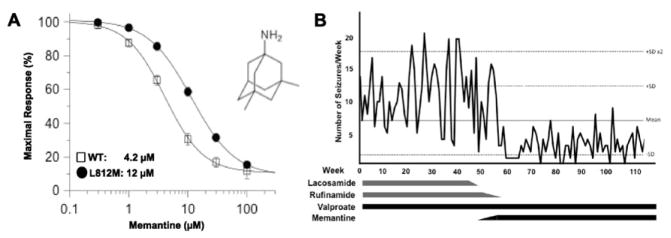
Exome sequencing revealed a de novo missense variant in GRIN2A (c.2434C>A; p.L812M) [27]. After studies in Xenopus oocytes showed that this mutant’s increased responsiveness to glutamate and glycine was attenuated by memantine (Fig. 4A), the boy underwent a trial of this FDA-approved drug that decreased his seizures (Fig. 4B), stopped all myoclonic jerks, and substantially improved his EEG [24].
Fig. 4.
Precision medicine: Treatment based on variant function studies. (A) Patch clamp results showing maximal response to glutamate and glycine in Xenopus oocytes transfected with the normal and mutant GRIN2A. Both the wild type and the mutant receptors were responsive to memantine inhibition. (Courtesy of Drs. Hongjie Yuan and Stephen F. Traynelis.) (B) Seizure frequency of the patient in response to various anti-epileptic medications. Note pronounced decrease in seizures on memantine. (Courtesy of Dr. Tyler Pierson.)
Sophisticated in vitro studies targeting a single nucleotide mutation can change the course of a disease and associate another gene with a beneficial anti-epileptic [58].
2.4.2. Diagnosis-based therapy for a rare pediatric disorder
A 12-year-old girl had painful metatarsal exostoses and a calcified elbow mass; excisional biopsy showed ectopic calcification. At the NIH, calcified exudate extruded from her right first distal metatarsal bone computerized tomography revealed a 9 mm mid-diaphyseal cortical lesion in the left tibia. Serum calcium, parathormone, osteocalcin, and alkaline phosphatase were normal, but serum phosphorus levels were 6.6–8.1 mg/dL (normal, 3.1–5.5), and the tubular reabsorption of phosphate was 94–97% (normal, 85–95%).
Drs. Michael Collins and Rachel Gafni suspected tumoral calcinosis due to deficiency of fibroblast growth factor 23 (FGF23) and mutations in GALNT3, the gene that encodes polypeptide N-acetyl-galactosaminyltransferase 3 (GALNT3), an enzyme that protects FGF23 from degradation [59]. The patient was compound heterozygous for two GALNT3 splice site variants, c.516-2A>T and c.1525+5G>A. The intact FGF23 level was 38 pg/mL (normal, 10–50), but the C-terminal FGF23 level was 892 RU/mL (normal, 20–100). FGF23 acts at the FGFR-1-α Klotho receptor complex to inhibit the action of transporters that reabsorb phosphate; FGF23 also inhibits the formation of 1,25-OH2-vitamin D. Without GALNT3, poorly glycosylated FGF23 is cleaved to inactive fragments, which may compete with intact FGF23, increasing renal phosphate retention and vitamin D-mediated gastrointestinal absorption of calcium and phosphate.
The patient received a low phosphate diet, non-calcium containing phosphate binders, acetazolamide, and probenecid to promote phosphate excretion. The elbow lesion resolved. In this case, targeted sequencing rather than exome sequencing yielded the molecular diagnosis.
3. Conclusions
Nowhere in medicine have technological advances in diagnostics been as revolutionary as in genetics, where the resolution of human genetic information has increased steadily from the level of chromosomes (karyotyping) to the level of molecules (DNA sequencing). Genomic sequencing now provides vast insights into the individual genetic variations that interact with each person’s environment to create health or disease.
Indeed, targeted and agnostic sequencing constitute a huge component of the modern geneticist’s armamentarium [60]. Half of the UDP’s diagnoses were made by exome sequencing combined with SNP analysis. Many diagnoses, however, were made without agnostic sequencing, and all diagnoses required thorough clinical evaluations, specialized medical expertise, and collaborative consultations.
The benefits of discovery seem profound. For 16 of the 20 UDP patients diagnosed and discussed here, the interval from onset of first symptoms or signs to diagnosis ranged from 2 to 54 years (mean and median, 10 years). A molecular genetic diagnosis in the probands permitted diagnoses of at least seven relatives, genetic counseling in the families, and reassurance of low recurrence risk in some couples. The discovery of a role for adenosine in ectopic calcification led to a therapeutic trial of bisphosphonates. One girl’s progressive calcification was reversed by vigorous manipulation of phosphate excretion. Because fibroblasts of the boy with homozygous TOP1MT mutations responded to EPI-743, he became eligible for a clinical trial of that experimental anti-oxidant.
The UDP also provided insights of broad genetic and medical importance. Rare and novel monogenic disorders described here reflect mechanisms and pathways that are likely to be operational in the pathogenesis of common and complex diseases and that could be targets for therapeutic interventions, although none has yet reached fruition. In addition, the UDP cases expanded the clinical spectrum of several syndromes, e.g., Brown-Vialetto-van Laere syndrome 2 and MOGS-CDG. Other cases provided functional studies critical in establishing the relationship of genes and specific variants to disease. Finally, the clinical utility of exhaustive sequence evaluation and whole genome sequencing was highlighted by two disease-causing mutations found in introns. The UDP also provided the hope embodied in the concept of precision medicine to desperate patients with nearly unique disorders and private mutations.
A national network, the UDN, now offers phenotyping, genotyping, environmental exposure analysis, functional studies, model system investigations, and broad data sharing to undiagnosed patients, bringing customized diagnostics closer to home [5]. With at least one other published program in the US [60] and international expansion already underway [61], the UDN paradigm should allow additional patients, some on a diagnostic road longer than that of Odysseus himself, to find answers.
Acknowledgments
We are deeply indebted to all the patients who entrusted their care to the UDP. The authors appreciate the technical assistance and advice of Jessica Albert, Manfred Boehm, Barbara Burton, Hannah Carlson-Donohoe, Michael Collins, Rachel Gafni, Fred Gill, Rena Godfrey, Gretchen Golas, Catherine Groden, Marjan Huizing, Michele Nehrebecky, Galina Nesterova, Tyler Pierson, Sergio Rosenzweig, Dimitre Simeonov, Stephen F. Traynelis, Zaheer Valivullah, Lynne Wolfe, Hongjie Yuan, Shira G. Ziegler, and the entire UDP staff. This work was supported in part by the Intramural Research Program of the National Human Genome Research Institute and the NIH Common Fund, through the Office of Strategic Coordination, Office of the NIH Director. The content is solely the responsibility of the authors and does not necessarily represent the official views of the NIH.
Footnotes
Conflicts of interest statement
The authors declare no conflict of interest.
Web resources
The URLs for information presented here are: Online Mendelian Inheritance in Man, http://www.omim.org/, http://www.ncbi.nlm.nih.gov/omim, Human Phenotype Ontology, http://human-phenotype-ontology.github.io/, and Phenotips, https://phenotips.org/.
References
- 1.Gahl WA, Tifft CJ. The NIH Undiagnosed Diseases Program: lessons learned. JAMA. 2011;305:1904–1905. doi: 10.1001/jama.2011.613. [DOI] [PubMed] [Google Scholar]
- 2.Gahl WA, Markello TC, Toro C, et al. The NIH Undiagnosed Diseases Program: insights into rare diseases. Genet Med. 2012;14:51–59. doi: 10.1038/gim.0b013e318232a005. [DOI] [PMC free article] [PubMed] [Google Scholar]
- 3.Tifft CJ, Adams DR. The National Institutes of Health Undiagnosed Diseases Program. Curr Opin Pediatr. 2014;26:626–633. doi: 10.1097/MOP.0000000000000155. [DOI] [PMC free article] [PubMed] [Google Scholar]
- 4.Collins FS, Varmus H. A new initiative on precision medicine. N Engl J Med. 2015;372:793–795. doi: 10.1056/NEJMp1500523. [DOI] [PMC free article] [PubMed] [Google Scholar]
- 5.Gahl W, Wise A, Ashley EA. The Undiagnosed Diseases Network of the NIH: a national extension. JAMA. 2015;314:1797–1798. doi: 10.1001/jama.2015.12249. [DOI] [PubMed] [Google Scholar]
- 6.Bone WP, Washington BOJ, et al. Computational evaluation of exome sequence data using human and model organism phenotypes improves diagnostic efficiency. Genet Med. 2015 Nov 12; doi: 10.1038/gim.2015.137. http://dx.doi.org/10.1038/gim.2015.137. [DOI] [PMC free article] [PubMed]
- 7.St Hilaire C, Ziegler SG, Markello T, et al. NT5E mutations and arterial calcifications. N Engl J Med. 2011;364:432–442. doi: 10.1056/NEJMoa0912923. [DOI] [PMC free article] [PubMed] [Google Scholar]
- 8.Pierson TM, Tart G, Adams D, et al. Infantile-onset spinal muscular atrophy with respiratory distress-1 diagnosed in a 20-year-old man. Neuromuscul Disord. 2011;21:353–355. doi: 10.1016/j.nmd.2011.02.005. [DOI] [PMC free article] [PubMed] [Google Scholar]
- 9.Pierson TM, Adams D, Bonn F, et al. Whole-exome sequencing identifies homozygous AFG3L2 mutations in a spastic ataxia-neuropathy syndrome linked to mitochondrial m-AAA proteases. PLoS Genet. 2011 Oct;7(10):e1002325. doi: 10.1371/journal.pgen.1002325. [DOI] [PMC free article] [PubMed] [Google Scholar]
- 10.Pierson TM, Simeonov DR, Sincan M, et al. Exome sequencing and SNP analysis detect heterozygosity in fatty acid hydroxylase-associated neurodegeneration. Eur J Hum Genet. 2012;20:476–479. doi: 10.1038/ejhg.2011.222. [DOI] [PMC free article] [PubMed] [Google Scholar]
- 11.Schiffenbauer AI, Wahl C, Pittaluga S, et al. IgG4-related disease presenting as recurrent mastoiditis. Laryngoscope. 2012;122:681–684. doi: 10.1002/lary.22486. [DOI] [PMC free article] [PubMed] [Google Scholar]
- 12.Pierson TM, Adams DA, Markello T, et al. Exome sequencing as a diagnostic tool in a case of undiagnosed juvenile-onset GM1-gangliosidosis. Neurology. 2012;79:123–126. doi: 10.1212/WNL.0b013e31825f047a. [DOI] [PMC free article] [PubMed] [Google Scholar]
- 13.Vanderver A, Tonduti D, Auerbach S, et al. Neurotransmitter abnormalities and response to supplementation in SPG11. Mol Genet Metab. 2012;107:229–233. doi: 10.1016/j.ymgme.2012.05.020. [DOI] [PMC free article] [PubMed] [Google Scholar]
- 14.Nesterova G, Malicdan MC, Yasuda K, et al. 1,25-(OH)2D-24 hydroxylase (CYP24A1) deficiency as a cause of nephrolithiasis. Clin J Am Soc Nephrol. 2013;8:649–657. doi: 10.2215/CJN.05360512. [DOI] [PMC free article] [PubMed] [Google Scholar]
- 15.Holloman CM, Wolfe LA, Gahl WA, Boerkoel CF. Kearns-Sayre syndrome presenting as isolated growth failure. BMJ Case Rep. 2013 doi: 10.1136/bcr-2012-007272. (pii: bcr2012007272) [DOI] [PMC free article] [PubMed] [Google Scholar]
- 16.Dias C, McDonald A, Sincan M, et al. Recurrent subacute post-viral onset of ataxia associated with a PRF1 mutation. Eur J Hum Genet. 2013;21:1232–1239. doi: 10.1038/ejhg.2013.20. [DOI] [PMC free article] [PubMed] [Google Scholar]
- 17.Pierson TM, Markello T, Accardi J, et al. Novel SNP array analysis and exome sequencing detect a homozygous exon 7 deletion of MEGF10 causing early onset myopathy, areflexia, respiratory distress and dysphagia (EMARDD) Neuromuscul Disord. 2013;23:483–488. doi: 10.1016/j.nmd.2013.01.013. [DOI] [PMC free article] [PubMed] [Google Scholar]
- 18.Manoli I, Kwan JY, Wang Q, et al. Chronic myopathy due to immunoglobulin light chain amyloidosis. Mol Genet Metab. 2013;108:249–254. doi: 10.1016/j.ymgme.2013.01.015. [DOI] [PMC free article] [PubMed] [Google Scholar]
- 19.Giorgio E, Rolyan H, Kropp L, et al. Analysis of LMNB1 duplications in autosomal dominant leukodystrophy provides insights into duplication mechanisms and allele-specific expression. Hum Mutat. 2013;34:1160–1171. doi: 10.1002/humu.22348. [DOI] [PMC free article] [PubMed] [Google Scholar]
- 20.Brady J, Trehan A, Landis D, Toro C. Mucopolysaccharidosis type IIIB (MPS IIIB) masquerading as a behavioral disorder. BMJ Case Rep. 2013 May 8; doi: 10.1136/bcr-2013-009592. (pii: bcr2013009592) [DOI] [PMC free article] [PubMed] [Google Scholar]
- 21.Landouré G, Zhu PP, Lourenco CM, et al. Hereditary spastic paraplegia type 43 (SPG43) is caused by mutation in C19orf12. Hum Mutat. 2013;34:1357–1360. doi: 10.1002/humu.22378. [DOI] [PMC free article] [PubMed] [Google Scholar]
- 22.Wu YW, Hess CP, Singhal NS, Groden C, Toro C. Idiopathic basal ganglia calcifications: an atypical presentation of PKAN. Pediatr Neurol. 2013;49:351–354. doi: 10.1016/j.pediatrneurol.2013.06.021. [DOI] [PubMed] [Google Scholar]
- 23.Lim YH, Ovejero D, Sugarman JS, et al. Multilineage somatic activating mutations in HRAS and NRAS cause mosaic cutaneous and skeletal lesions, elevated FGF23, and hypophosphatemia. Hum Mol Genet. 2014;23:397–407. doi: 10.1093/hmg/ddt429. [DOI] [PMC free article] [PubMed] [Google Scholar]
- 24.Yuan H, Hansen KB, Zhang J, et al. Functional analysis of a missense mutation associated with early-onset epileptic encephalopathy. Nat Commun. 2014;5:3251. doi: 10.1038/ncomms4251. [DOI] [PMC free article] [PubMed] [Google Scholar]
- 25.Rice GI, del Toro DY, Jenkinson EM, et al. Gain-of-function mutations in IFIH1 cause a spectrum of human disease phenotypes associated with upregulated type I interferon signaling. Nat Genet. 2014;46:503–509. doi: 10.1038/ng.2933. [DOI] [PMC free article] [PubMed] [Google Scholar]
- 26.Sadat MA, Moir S, Chun TW, et al. Glycosylation hypogammaglobulinemia, and resistance to viral infections. N Engl J Med. 2014;370:1615–1625. doi: 10.1056/NEJMoa1302846. [DOI] [PMC free article] [PubMed] [Google Scholar]
- 27.Pierson TM, Yuan H, Marsh ED, et al. GRIN2A mutation and early-onset epileptic encephalopathy: personalized therapy with memantine. Ann Clin Transl Neurol. 2014;1:190–198. doi: 10.1002/acn3.39. [DOI] [PMC free article] [PubMed] [Google Scholar]
- 28.Adams DR, Yuan H, Holyoak T, et al. Three rare diseases in one sib pair: RAI1, PCK1, GRIN2B mutations associated with Smith-Magenis syndrome, cytosolic PEPCK deficiency and NMDA receptor glutamate insensitivity. Mol Genet Metab. 2014;113:161–170. doi: 10.1016/j.ymgme.2014.04.001. [DOI] [PMC free article] [PubMed] [Google Scholar]
- 29.Shehata L, Simeonov DR, Raams A, et al. ERCC6 dysfunction presenting as progressive neurological decline with brain hypomyelination. Am J Med Genet A. 2014;164:2892–2900. doi: 10.1002/ajmg.a.36709. [DOI] [PMC free article] [PubMed] [Google Scholar]
- 30.Wolf NI, Toro C, Kister I, et al. DARS-associated leukoencephalopathy can mimic a steroid-responsive neuroinflammatory disorder. Neurology. 2015;84:226–230. doi: 10.1212/WNL.0000000000001157. [DOI] [PMC free article] [PubMed] [Google Scholar]
- 31.Markello T, Chen D, Kwan JY, et al. York platelet syndrome is a CRAC channelopathy due to gain-of-function mutations in STIM1. Mol Genet Metab. 2015;114:474–482. doi: 10.1016/j.ymgme.2014.12.307. [DOI] [PMC free article] [PubMed] [Google Scholar]
- 32.Ng BC, Wolfe LA, Ichikawa M, et al. Biallelic mutations in CAD impair de novo pyrimidine biosynthesis and decrease glycosylation precursors. Hum Mol Genet. 2015;24:3050–3057. doi: 10.1093/hmg/ddv057. [DOI] [PMC free article] [PubMed] [Google Scholar]
- 33.Simons C, Griffin LB, Helman G, et al. Loss-of-function alanyl-tRNA synthetase mutations cause an autosomal-recessive early-onset epileptic encephalopathy with persistent myelination defect. Am J Hum Genet. 2015;96:675–681. doi: 10.1016/j.ajhg.2015.02.012. [DOI] [PMC free article] [PubMed] [Google Scholar]
- 34.Trehan A, Brady JM, Golas GA, et al. MED23-associated intellectual disability in a non-consanguineous family. Am J Med Genet A. 2015;167:1374–1380. doi: 10.1002/ajmg.a.37047. [DOI] [PMC free article] [PubMed] [Google Scholar]
- 35.Albert JS, Bhattacharyya N, Wolfe LA, Bone W, et al. Impaired osteoblast and osteoclast function characterize the osteoporosis of Snyder-Robinson syndrome. Orphanet J Rare Dis. 2015 Mar 7;10:27. doi: 10.1186/s13023-015-0235-8. [DOI] [PMC free article] [PubMed] [Google Scholar]
- 36.Lam C, Golas GA, Davids M, et al. Expanding the clinical and molecular characteristics of PIGT-CDG, a disorder of glycosylphosphatidylinositol anchors. Mol Genet Metab. 2015;115:128–140. doi: 10.1016/j.ymgme.2015.04.007. [DOI] [PMC free article] [PubMed] [Google Scholar]
- 37.Marchegiani S, Davis T, Tessadori F, et al. Recurrent mutations in the basic domain of TWIST2 cause ablepharon macrostomia and Barber-Say syndromes. Am J Hum Genet. 2015;97:99–110. doi: 10.1016/j.ajhg.2015.05.017. [DOI] [PMC free article] [PubMed] [Google Scholar]
- 38.Janecke AR, Li B, Boehm M, et al. The phenotype of the musculocontractural type of Ehlers-Danlos syndrome due to CHST14 mutations. Am J Med Genet A. 2015 Sep 16; doi: 10.1002/ajmg.a.37383. http://dx.doi.org/10.1002/ajmg.a.37383. [DOI] [PMC free article] [PubMed]
- 39.Pegg AE, Michael AJ. Spermine synthase. Cell Mol Life Sci. 2010;67:113–121. doi: 10.1007/s00018-009-0165-5. [DOI] [PMC free article] [PubMed] [Google Scholar]
- 40.Snyder RD, Robinson A. Recessive sex-linked mental retardation in the absence of other recognizable abnormalities. Report of a family. Clin Pediatr. 1969;8:669–674. doi: 10.1177/000992286900801114. [DOI] [PubMed] [Google Scholar]
- 41.Albert J, Schwartz CE, Boerkoel CF, Stevenson RE. In: Snyder-Robinson Syndrome. Pagon RA, Adam MP, Ardinger HH, et al., editors. GeneReviews®; Seattle (WA): 1993–2015. (Available from: http://www.ncbi.nlm.nih.gov/books/NBK144284/) [Google Scholar]
- 42.Yeon JT, Ryu BJ, Choi SW, et al. Natural polyamines inhibit the migration of preosteoclasts by attenuating Ca2+-PYK2-Src-NFATc1 signaling pathways. Amino Acids. 2014;46:2605–2614. doi: 10.1007/s00726-014-1797-9. [DOI] [PubMed] [Google Scholar]
- 43.Yamamoto T, Hinoi E, Fujita H, et al. The natural polyamine spermidine and spermine prevent bone loss through preferential disruption of osteoclastic activation in ovariectomized mice. Br J Pharmacol. 2012;166:1084–1096. doi: 10.1111/j.1476-5381.2012.01856.x. [DOI] [PMC free article] [PubMed] [Google Scholar]
- 44.Kusudo T, Sakaki T, Abe D, et al. Metabolism of A-ring diastereomers of 1α,25-dihydroxyvitamin D3 by CYP24A1. Biochem Biophys Res Commun. 2004;321:774–782. doi: 10.1016/j.bbrc.2004.07.040. [DOI] [PubMed] [Google Scholar]
- 45.DePraeter CM, Gerwig GJ, Bause E, et al. A novel disorder caused by defective biosynthesis of N-linked oligosaccharides due to glucosidase I deficiency. Am J Hum Genet. 2000;66:1744–1756. doi: 10.1086/302948. [DOI] [PMC free article] [PubMed] [Google Scholar]
- 46.Yang Y, Zhang J, Chang Q, et al. De novo mutation in ATP6V1B2 impairs lysosome acidification and causes dominant deafness-onychodystrophy syndrome. Cell Res. 2014;24:1370–1373. doi: 10.1038/cr.2014.77. [DOI] [PMC free article] [PubMed] [Google Scholar]
- 47.Green P, Wiseman M, Crow YJ, et al. Brown-Vialetto-Van Laere syndrome, a ponto-bulbar palsy with deafness, is caused by mutations in C20orf54. Am J Hum Genet. 2010;86:485–489. doi: 10.1016/j.ajhg.2010.02.006. [DOI] [PMC free article] [PubMed] [Google Scholar]
- 48.Enns GM, Kinsman SL, Perlman SL, et al. Initial experience in the treatment of inherited mitochondrial disease with EPI-743. Mol Genet Metab. 2012;105:91–102. doi: 10.1016/j.ymgme.2011.10.009. [DOI] [PubMed] [Google Scholar]
- 49.Sadun AA, Chicani F, Ross-Cisneros FN, et al. Effect of EPI-743 on the clinical course of the mitochondrial disease Leber hereditary optic neuropathy. Arch Neurol. 2012;69:331–338. doi: 10.1001/archneurol.2011.2972. [DOI] [PubMed] [Google Scholar]
- 50.Slager RE, Newton TL, Vlangos CN, Finucane B, Elsea SH. Mutations in RAI1 associated with Smith-Magenis syndrome. Nat Genet. 2003;33:466–468. doi: 10.1038/ng1126. [DOI] [PubMed] [Google Scholar]
- 51.Endele S, Rosenberger G, Geider K, et al. Mutations in GRIN2A and GRIN2B encoding regulatory subunits of NMDA receptors cause variable neurodevelopmental phenotypes. Nat Genet. 2010;42:1021–1026. doi: 10.1038/ng.677. [DOI] [PubMed] [Google Scholar]
- 52.Markello TC, Pak LK, St Hilaire C, et al. Vascular pathology of medial arterial calcifications in NT5E deficiency: implications for the role of adenosine in pseudoxanthoma elasticum. Mol Genet Metab. 2011;103:44–50. doi: 10.1016/j.ymgme.2011.01.018. [DOI] [PMC free article] [PubMed] [Google Scholar]
- 53.McCullough PA, Agrawal V, Danielewicz E, Abela GS. Accelerated atherosclerotic calcification and Mönckeberg’s sclerosis: a continuum of advanced vascular pathology in chronic kidney disease. Clin J Am Soc Nephrol. 2008;3:1585–1598. doi: 10.2215/CJN.01930408. [DOI] [PubMed] [Google Scholar]
- 54.Padiath QS, Saigoh K, Schiffmann R, et al. Lamin B1 duplications cause autosomal dominant leukodystrophy. Nat Genet. 2006;38:1114–1123. doi: 10.1038/ng1872. [DOI] [PubMed] [Google Scholar]
- 55.Rankin J, Ellard S. The laminopathies: a clinical review. Clin Genet. 2006;70:261–274. doi: 10.1111/j.1399-0004.2006.00677.x. [DOI] [PubMed] [Google Scholar]
- 56.Capell BC, Collins FS. Human laminopathies: nuclei gone genetically awry. Nat Rev Genet. 2006;7:940–952. doi: 10.1038/nrg1906. [DOI] [PubMed] [Google Scholar]
- 57.Rolyan H, Tyurina YY, Hernandez M. Defects of lipid synthesis are linked to the age-dependent demyelination caused by lamin B1 overexpression. J Neurosci. 2015;35:12002–12017. doi: 10.1523/JNEUROSCI.1668-15.2015. [DOI] [PMC free article] [PubMed] [Google Scholar]
- 58.EpiPM Consortium. A roadmap for precision medicine in the epilepsies. Lancet Neurol. 2015;14:1219–1228. doi: 10.1016/S1474-4422(15)00199-4. [DOI] [PMC free article] [PubMed] [Google Scholar]
- 59.Topaz O, Shurman DL, Bergman R, et al. Mutations in GALNT3, encoding a protein involved in O-linked glycosylation, cause familial tumoral calcinosis. Nat Genet. 2004;36:579–581. doi: 10.1038/ng1358. [DOI] [PubMed] [Google Scholar]
- 60.Bloss CS, Scott-Van Zeeland AA, Topol SE, et al. A genome sequencing program for novel undiagnosed diseases. Genet Med. 2015;17:995–1001. doi: 10.1038/gim.2015.21. [DOI] [PMC free article] [PubMed] [Google Scholar]
- 61.Taruscio D, Groft SC, Cederroth H, et al. Undiagnosed Diseases Network International (UDNI): white paper for global actions to meet patient needs. Mol Genet Metab. 2015;116:223–225. doi: 10.1016/j.ymgme.2015.11.003. [DOI] [PubMed] [Google Scholar]


