Figure 6.
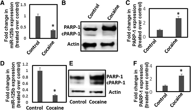
Chronic cocaine exposure reduces miR-125b expression and upregulates PARP-1 expression. A–C, Differentiated SH-SY5Y cells were treated daily with varying concentrations of cocaine for 7 d. Cells were harvested, and miR-125b expression was measured by qPCR (A), whereas PARP-1 expression was detected by Western blotting (B, C). A, miR-125b expression was normalized to 5S rRNA levels and plotted as fold changes in miR-125b expression in cocaine-treated cells relative to untreated control. B, Representative immunoblot of PARP-1 expression in control and cocaine-treated cells. C, Relative PARP-1 expression in cocaine-treated cells and untreated cells as quantified by densitometry from three independent experiments. D-F, Mice were injected (i.p.) with cocaine (20 mg/kg weight of mice) or saline for 7 d. Thereafter, animals were sacrificed and NAc was isolated. RNA and lysates from the NAc tissues were subjected to qPCR and Western blotting, respectively. D, miR-125b expression was normalized to the 5s rRNA levels and relative expression of miR-125b in the NAc of cocaine-treated mice (n = 6) compared to the NAc of saline-treated mice (n = 6) are expressed as fold changes. E, Representative immunoblot showingPARP-1 expression in the NAc of cocaine-treated mice and saline-treated control mice. F, Relative expression of PARP-1 in the NAc of cocaine-treated mice (n = 6) compared to the NAc of saline-treated control mice (n = 6) based on densitometry analysis. *p < 0.05 for the comparison of cocaine-treated samples versus untreated controls.

