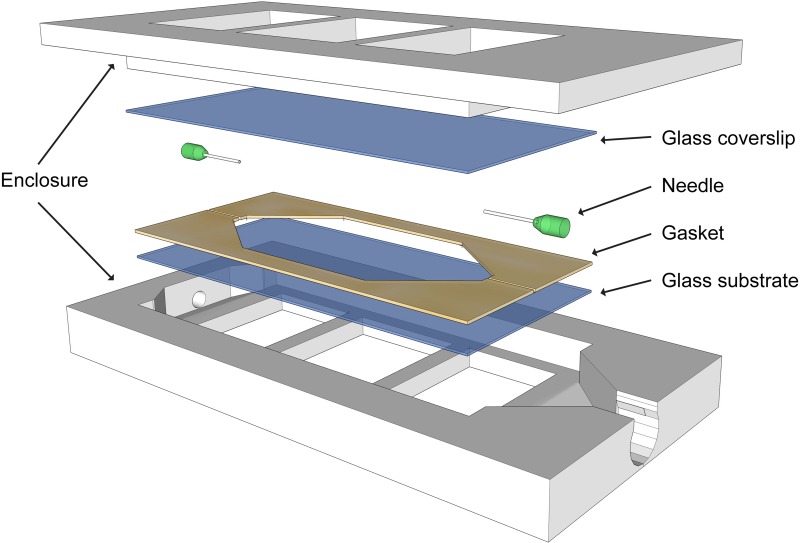
FIG. 2.
3D model of a flow cell shows the sealed enclosure that enables the flow of nutrient media through an imaging chamber. Viewing windows are sandwiched between the two parts of the enclosure. A gasket seals liquid media between the windows. The needle at the inlet supplies liquid media during flow and air bubbles during imaging. The cell supports the growth of the biofilm for extended periods and is tailored for use with white-light interferometry through careful selection of viewing windows with appropriate thickness and group index of refraction.

