Abstract
Gliomas are a malignant tumor group whose patients have survival rates around 12 months. Among the treatments are the alkylating agents as temozolomide (TMZ), although gliomas have shown multiple resistance mechanisms for chemotherapy. Guanosine (GUO) is an endogenous nucleoside involved in extracellular signaling that presents neuroprotective effects and also shows the effect of inducing differentiation in cancer cells. The chemotherapy allied to adjuvant drugs are being suggested as a novel approach in gliomas treatment. In this way, this study evaluated whether GUO presented cytotoxic effects on human glioma cells as well as GUO effects in association with a classical chemotherapeutic compound, TMZ. Classical parameters of tumor aggressiveness, as alterations on cell viability, type of cell death, migration, and parameters of glutamatergic transmission, were evaluated. GUO (500 and 1000 μM) decreases the A172 glioma cell viability after 24, 48, or 72 h of treatment. TMZ alone or GUO plus TMZ also reduced glioma cell viability similarly. GUO combined with TMZ showed a potentiation effect of increasing apoptosis in A172 glioma cells, and a similar pattern was observed in reducing mitochondrial membrane potential. GUO per se did not elevate the acidic vesicular organelles occurrence, but TMZ or GUO plus TMZ increased this autophagy hallmark. GUO did not alter glutamate transport per se, but it prevented TMZ-induced glutamate release. GUO or TMZ did not alter glutamine synthetase activity. Pharmacological blockade of glutamate receptors did not change GUO effect on glioma viability. GUO cytotoxicity was partially prevented by adenosine receptor (A1R and A2AR) ligands. These results point to a cytotoxic effect of GUO on A172 glioma cells and suggest an anticancer effect of GUO as a putative adjuvant treatment, whose mechanism needs to be unraveled.
Keywords: A172 glioma cells, Guanosine, Temozolomide, Cytotoxicity, Glutamate, Adenosine
Introduction
Gliomas are considered the most aggressive type of brain tumors. The origin of this type of cancer can be related to changes in glial cells or their progenitors [1]. The most common therapies involve surgical resection, chemotherapy, and/or radiotherapy; however, the prognosis remains low and the survival rates are around 12 to 15 months. The main approach used as chemotherapy is the administration of alkylating drugs that inhibit DNA replication and evoke the activation of apoptosis cascades [2, 3]. Among the most clinically used chemotherapeutic compounds is the temozolomide (TMZ) that have been showing efficacy in early diagnosed gliomas [4]. TMZ is an alquilant agent that is absorbed and in physiological pH of blood circulation is transformed to its active metabolite, 3-methyl-(trazen-1-yl)imidazole-4-carboxamide (MTIC), which causes DNA damage through DNA methylation and subsequent cell cycle arrestment and cell death [5]. The enzyme N-alkypurine DNA glycosylase (APGN) is able to repair this DNA methylation, thus causing TMZ resistance in glioma cells [6]. This repair mechanism allied to other resistance mechanisms are responsible for the low prognosis observed in gliomas [7] and point to the urgency of developing additional or adjuvant therapies to treat gliomas.
Allied to resistance mechanisms, putative changes in the transmission system evoked by the main excitatory neurotransmitter, the amino acid glutamate, might occur in glioma cells, thus favoring tumor growth. It has been shown that there is increased glutamate release, through cystine-glutamate exchanger (Xc- system) activity and decreased glutamate uptake through reduction of the excitatory amino acid transporter (EAAT) expression in glioma cells [8, 9]. Additionally, it has been shown that glutamate receptor (GluR) antagonists may reduce the proliferation and motility of cancer cells [10]. In this context, the search for molecules that can modulate glutamatergic transmission is considered an interesting treatment strategy.
Guanosine (GUO) is an endogenous guanine-derived nucleoside involved in extracellular signaling that presents the ability to modulate glutamatergic system activity [11, 12]. In this regard, GUO exerts neuroprotective effects against glutamate-induced cell damage or ischemia, through activation of intracellular signaling pathways related to cell survival as the phosphoinositide 3-kinase (PI3K) and mitogen-activated protein kinase/extracellular signal-regulated kinase (MAPK/ERK) [13, 14]. GUO also modulates glutamate transporter activity, by preventing the decrease in glutamate uptake and the increased glutamate release on in vitro models of ischemia [14–17]. A selective receptorial protein to GUO has been suggested, although it has not yet been cloned (for a review see [12]). However, the neuroprotective effect of GUO seems to involve the modulation of adenosine receptors (AdoR) [14] and/or the activation of the large (big) conductance Ca2+-activated K+ channels (BK) [15]. Additionally, the neuroprotective effect of GUO is related to the nuclear factor-kappa B (NF-κB) inhibition and antioxidant and anti-inflammatory effects [17, 18]. There are few studies regarding GUO effects in cancer. A study of an anticancer effect of a purine nucleoside analog, sulfinosine (an oxidized form of 6-thioguanosine), demonstrates an induction of caspase-dependent apoptotic cell death and autophagy in glioma cells. Additionally, sulfinosine increases the reactive oxygen species (ROS) and diminishes the antioxidant peptide glutathione levels [19]. In cancer cells, the induction of differentiation is also related to impairment in malignancy. It has been shown that GUO induces melanoma cell differentiation through protein kinase C (PKC)/ERK pathway, increasing dendritogenesis and melanogenesis and decreasing cell motility [20].
The study of drug combination approach that may enhance the anticancer potential of drugs with different mechanisms of action, improving the survival rates and elucidating better strategies in cancer therapy, is very important. Therefore, this study is testing if GUO shows the potential cytotoxic effect comparing it to TMZ effect, besides analyzing GUO and TMZ combination effects on classical parameters of tumor aggressiveness.
Material and methods
Cell culture
The glioma cell line A172 (the cell line was kind gift from Dr. G. Lenz from Federal University of Rio Grande do Sul, RS, Brazil) was cultured in Dulbecco’s modified Eagle’s medium and nutrient mixture F12 (DMEM-F12, Invitrogen, Carlsbad, CA) supplemented with 10% fetal bovine serum (FBS; Cultilab), in 25-cm2 culture flasks, at 37 °C humidified atmosphere of 5% CO2, as previously reported [21]. For the biochemical analysis, when reached confluence, A172 cells were trypsinized (Trypsin/EDTA, 0.05%; Gibco) and plated in 24- or 96-well plates (3.5 × 10⁵ or 0.5 × 10⁵, respectively).
Cell treatment
A172 cells were plated in 24- or 96-well plates. After confluence, the cells were treated with guanosine (GUO 50–1000 μM in time-course/concentration curves, or 500 μM for other analyses) (Sigma) or TMZ (500 μM) (Tocris) for 48 h. In order to assess GUO mechanism of action, adenosine and glutamate receptor agonists and antagonists, purine nucleosides, and glutamate transporter inhibitors were incubated 30 min before GUO and maintained during 48 h treatment. Drugs used were as follows: the adenosine-metabolizing enzyme, adenosine deaminase (ADA—0.5 U/mL); the equilibrative purine nucleoside transporter inhibitor, dipyridamole (DIPY—10 μM); the broad-spectrum inhibitor of excitatory amino acid transporters (EAATs), dl-threo-β-benzyloxyaspartic acid (DL-TBOA—100 μM); the Xc- system inhibitor, sulfasalazin (SAS—300 μM); the glutamate receptor sub-types α-amino-3-hydroxy-5-methyl-4-isoxazolepropionic acid receptor (AMPAR) and kainate receptor (KAR) antagonist, 6,7-dinitroquinoxaline-2,3-dione (DNQX—1 μM); the glutamate subtype N-methyl-d-aspartate receptor (NMDAR) antagonist, dizocilpine (MK-801—1 μM); KAR antagonist, γ-d-glutamylaminomethylsulphonic acid (GAMS—1 μM); metabotropic glutamate receptor (mGluR) antagonist, (RS)-α-methyl-4-carboxyphenylglycine (MCPG—10 μM); adenosine A1 receptor (A1R) antagonist, dipropylcyclopentylxanthine (DPCPX—100 ηM); adenosine A2A receptor (A2AR) inverse agonist, 4-(2-[7-amino-2-(2-furyl)[1,2,4]triazolo[2,3-a][1,3,5]triazin-5-ylamino]ethyl)phenol (ZM241385—50 ηM); and A2AR agonist, 4-[2-[[6-amino-9-(N-ethyl-β-d-ribofuranuronamidosyl)-9H–purin-2-yl]amino]ethyl]benzenepropanoic acid hydrochloride (CGS 21680—30 ηM). During treatment, cells were incubated in serum-free DMEM-F12, including control groups (CT). Biochemical analyses were carried out after elapsed time of treatment.
Viability analysis
Cell viability was evaluated through cells’ capacity to reduce 3-(4,5-diamethylthiazol-2-yl)-2,5-diphenyltetrazolium bromide (MTT) as previously described [22]. In this assay, viable cells convert water-soluble yellow MTT to water-insoluble blue MTT formazan. Thus, MTT formazan production, identified by optical density, is assumed to be proportional to the number of viable cells. Cells were incubated with MTT (0.2 mg/mL) in PBS for 2 h at 37 °C. The formazan produced was solubilized by replacing the medium with 100 μL of dimethyl sulfoxide (DMSO), resulting in a colored compound from which optical density was measured in an ELISA reader (550 nm).
The Trypan blue exclusion assay was also applied [23]. After time treatment, cells were stained with Trypan blue and counted in a hemocytometer. The percentage of viable cells was calculated as percent of viable cells = (number of viable cells/number of total cells) × 100.
The CellTiter-Glo® Luminescent Cell Viability assay was also used to access the cell viability. ATP quantification was assayed as an indicator of metabolically active cells. According to the manufacturer’s recommendations, the cells were maintained at room temperature for 30 min. Briefly, the CellTiter-Glo® reagents were added and the plate incubated on an orbital shaker for 2 min. The luminescent signal was stabilized for 10 min at room temperature and the total luminescence recorded.
Wound healing assay
In order to analyze changes in cell migration, the wound healing assay was carried out according to previous studies [20, 24]. Briefly, cells were plated in 6-well plates; after 24 h, a Pippete-200 tip was used to scrap the dish surface to generate the “wound” and the medium was replaced by FBS-free medium containing drugs. Phase contrast images were obtained using a ×10 objective lens in an inverted microscope at 0, 24, and 48 h. To each experiment, a scratch was carried out in five replicates and the experiment was repeated three times. The gap width was calculated by 10 measurements from each scratch to obtain the width average. Each measurement was taken from the left edge to the right edge of the scratch, going from the top to the bottom of the image using Image J software. The average of the width was expressed in arbitrary units.
Acridine orange staining
To identify the presence of acidic vesicular organelles (AVOs), cells were plated in 24-well dishes. After 24 h, the cells were treated. Elapsed 48 h of treatment, the cells were washed with PBS and incubated with acridine orange (Sigma) 10 μg/mL for 10 min at room temperature. Then, the solution was replaced by PBS and the cells were analyzed under an inverted fluorescence microscope (Zeiss Axiovert 40 CFL, capture system AxioCamMRc). Depending on their acidity, the autophagic lysosomes appear as orange/red AVOs, while cytoplasm and nuclei are green. The analyses were carried out using a 490-nm band-pass blue excitation and a 515-nm-long pass barrier filters. In order to quantify AVO presence, cells were counted through flow cytometry. After treatment, cells were PBS washed and incubated with acridine orange 1 μg/mL for 15 min. Then, the cells were trypsinized, centrifuged at 220 × g, and resuspended in 500 μL of PBS-EDTA—0.2 μM. The cells were sorted by FACSCanto II flow cytometer and result processed on Flowing Software 2.
Apoptosis and necrosis analysis
To analyze the effect of ATOR and TMZ on apoptosis or necrosis levels in glioma cells, the Annexin V FITC assay kit (Millipore) was used in flow cytometry. The kit detects the phosphatidylserine exposure on the outer layer of the cell membrane. Annexin V FITC conjugated binds to phosphatidylserine exposed in apoptotic cells. Propidium iodide binds to DNA in cells with compromised cell membrane indicating necrotic cells. After treatment, 1 × 10⁵ cells were trypsinized, centrifuged at 1500 rpm, and washed in cold PBS, then resuspended in 200 μL of binding buffer. The cells were stained with FITC-conjugated Annexin V and propidium iodide reagent in concentrations and time recommended by the manufacturer. The cells were sorted by FACSCanto II flow cytometer and result processed on Flowing Software 2.
Measurement of mitochondrial membrane potential
After treatment, the cells were loaded with the mitochondrial selective fluorescent dye, tetramethylrhodamine ethyl ester (TMRE, 100 nM) for 30 min at 37 °C [25], for analyzing the mitochondrial membrane potential after the incubation with GUO and TMZ. Fluorescence was measured using wavelengths of excitation and emission of 550 and 590 nm, respectively. Carbonyl cyanide p-trifluoromethoxy phenylhydrazone (FCCP), a mitochondrial uncoupler, was used as positive control to fluorescence emission.
Glutamine synthetase activity
The assay of the glutamine synthetase enzyme activity has been adapted from Shapiro [26] as Vandresen-Filho et al. [27]. For evaluating drug effects on glutamine synthetase activity in human glioma cell A172 after treatment, the cells were harvested and washed in PBS. Then, the cells were incubated for 15 min in 200 μL of imidazole-HCl buffer (80 mM, pH 7.0) and homogenized. The assay mixture contained 80 mM imidazole-HCl buffer, 30 mM glutamine, 3 mM MnCl2, 30 mM hydroxylamine-HCl, 20 mM sodium arsenate, 0.4 mM ADP, and 50 μL of the cells homogenate. The reaction was stopped after 30 min at 37 °C through the addition of 100 μL of a mixture containing 4/1/0.5/6.5 (v/v/v/v) of 10% (w/v) ferric chloride, 24% (w/v) trichloroacetic acid, 6 M HCl, and water. The reaction product, γ-glutamylhydroxamate, was measured at 540 nm using a microplate reader and converted to the amount of product formed through comparison with a standard curve. The enzyme activity was expressed as a percentage of control.
l-[3H]Glutamate transport
To analyze this parameter, cells were concomitantly plated for transport assays and cell counting, for experiment normalization. After drug incubation, the culture medium was removed and the cells were incubated in Hanks buffer saline solution (HBSS—composition in millimolar: 1.3 CaCl2, 137 NaCl, 5 KCl, 0.65 MgSO4, 0.3 Na2HPO4, 1.1 KH2PO4, 2 glucose, and 5 HEPES) (pH 7.2) for 15 min at 37 °C. Excitatory amino acid release was assessed by adding 0.33 μCi/mL of l-[3 H]glutamate (American Radiolabeled Chemicals—ARC), plus l-glutamate to reach a final concentration of 100 μM and incubated for 7 min. The reaction was stopped with three ice-cold HBSS washes. Cells were incubated in HBSS for 15 min in 300 μL at 37 °C, and the medium was collected to measure l-[3 H]glutamate release. The cells were lysed in the NaOH 0.1 N/SDS 0.1% solution overnight, and the l-[3 H]glutamate content was used to assess the neurotransmitter uptake, determined through scintillation counting [28]. Cells that were concomitantly plated were counted in hemocytometer, and experiment was normalized as femtomole of glutamate per number of viable cells after treatment.
Protein measurement
Protein content was evaluated by using the method of Lowry [29], and bovine serum albumin (Sigma) was used as a standard.
Statistical analysis
Data were analyzed by one-way analysis of variance (ANOVA) when treated groups were compared to the control group only, or two-way ANOVA when the comparisons assessed different times and concentration groups and followed by Student-Newman-Keuls test. The software used were GraphPad Prism 4.0 for one-way analysis or Statistica 7.0 for two-way analysis, considering significance level p < 0.05.
Results
Cellular viability evaluations
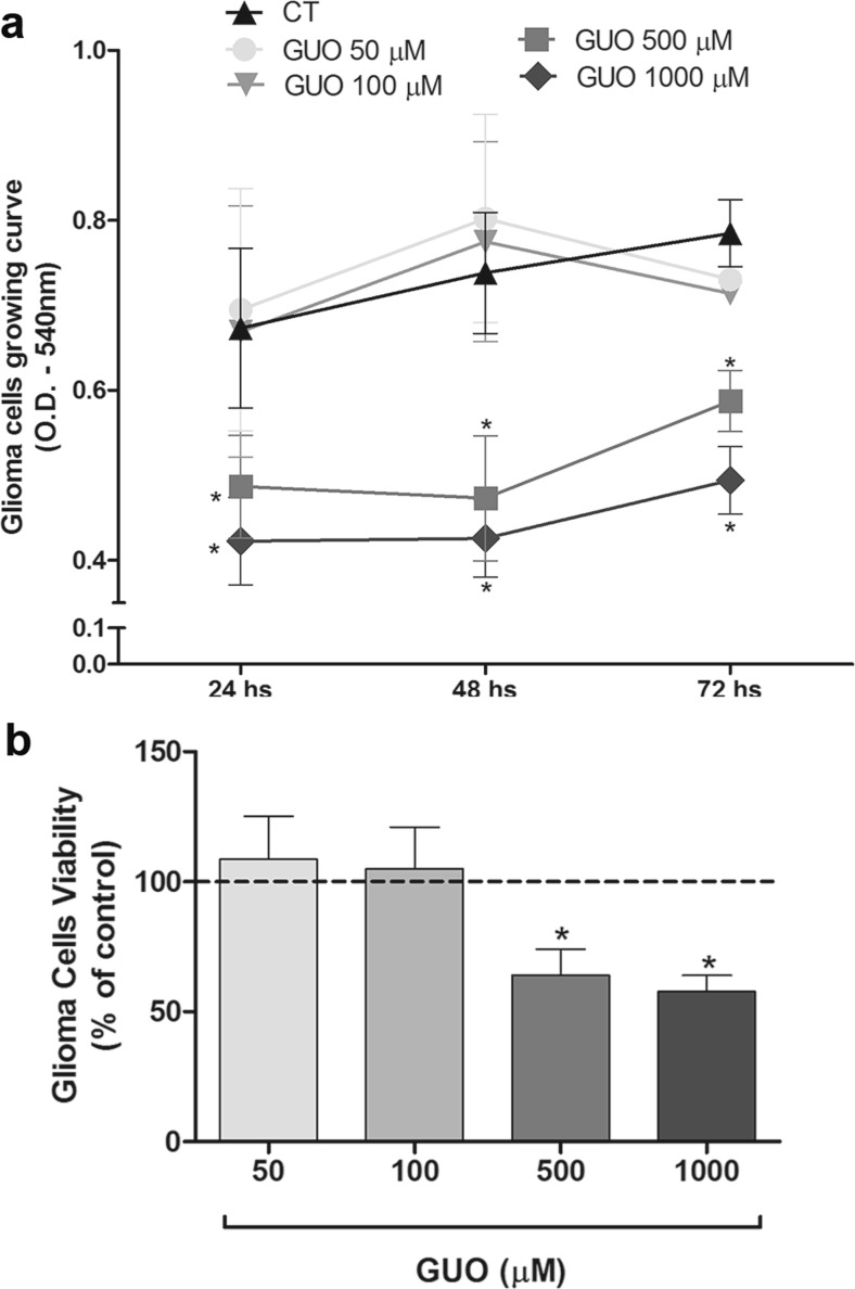
Firstly, GUO time-course and concentration curves were carried out in order to evaluate the nucleoside effect on A172 glioma cell growing curve. The A172 cells were treated with GUO in a concentration range from 50 to 1000 μM for 24, 48, or 72 h, and cell viability was assessed by the MTT assay (Fig. 1a). GUO concentrations of 50 or 100 did not alter glioma cell growing curve. GUO 500 or 1000 decreased A172 cell viability. This pattern of reduction of cell viability was observed also at 48 and 72 h. Therefore, to the subsequent experiments, the concentration of 500 μM GUO at 48 h of treatment was used (Fig. 1b).
Fig. 1.
GUO time-course and concentration-response curves on A172 glioma cells. a GUO time-course (24, 48, and 72 h, hs) and concentration curve (50, 100, 500, and 1000 μM) was performed in A172 glioma cells to assess cellular viability during A172 glioma cell growing curve. b A172 cells were treated with GUO (50 to 1000 μM) for 48 h, and the viability was assessed. Data expressed as optical density (O.D.) at 540 nm in a and as a percentage related to the control group (dotted line) in b. n = 6 of independent experiments for a and b separately. *p < 0.05 compared to the control group
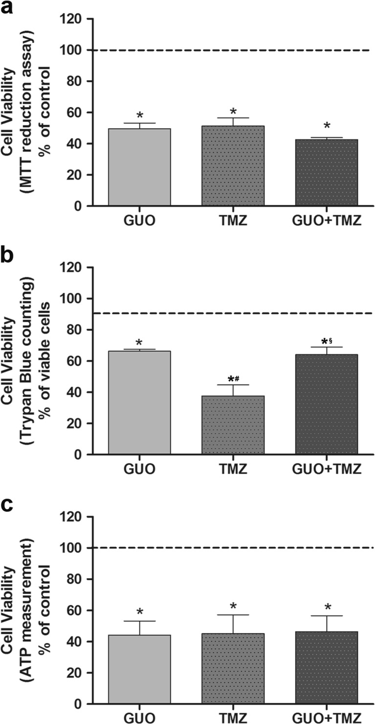
An evaluation of TMZ effect and its association with GUO was carried out in order to compare the effects of these drugs and analyze a possible interaction on cytotoxicity to glioma cells (Fig. 2). Two hundred micromolar TMZ was initially tested to study TMZ effect on A172 cell viability; however, it did not change the glioma cell viability (data not shown). The A172 cells were then treated for 48 h with GUO (500 μM), TMZ (500 μM), or both drugs co-incubated and tested in three different viability methods. In MTT assay, GUO or TMZ treatments showed a similar effect, reducing glioma viability to 49 and 51%, respectively. Similarly, the combined treatment of TMZ and GUO reduced cell viability to 42% and no significant difference was observed among treated groups (Fig. 2a). The Trypan Blue exclusion assay, a cell counting-based method, was also applied. The percentages of viable cells were 66, 38, and 64% to GUO, TMZ, and GUO + TMZ, respectively, being TMZ treatment significantly lower than GUO or GUO + TMZ (Fig. 2b). Cell viability was also assessed through ATP level measurement. GUO, TMZ, or the drug combination showed a decrease in cell viability of 56, 55, and 54% respectively, with no statistical difference among treatments (Fig. 2c). In this way, GUO, TMZ, or GUO + TMZ decreased A172 glioma cell viability similarly, although a higher reduction in cell number was observed in TMZ group than to GUO or GUO + TMZ. No synergistic effect was observed in the combined treatment.
Fig. 2.
GUO and TMZ effects on A172 glioma cell viability. a A172 glioma cells viability assessed by MTT reduction assay after 48 h of GUO (500 μM), TMZ (500 μM), or drug combination treatment. b Cell viability assessed through direct cell counting using Trypan exclusion assay. A172 glioma cells were treated with GUO (500 μM), TMZ (500 μM), or both for 48 h. Values expressed as viable cells, being control group around 90% of viable cells. c A172 cell viability assessed by an ATP-based assay in cells treated with GUO (500 μM), TMZ (500 μM), or both treatments for 48 h. Data expressed as a percentage related to the control group that was considered as 100% (a, b, and c) and expressed as a dotted line. n = 5 of independent experiments for a and n = 3 of independent experiments for b and c.*p < 0.05 compared to the control group, #p < 0.05 compared to the GUO group, and §p < 0.05 compared to the TMZ group in a, b, and c
Cell migration analysis
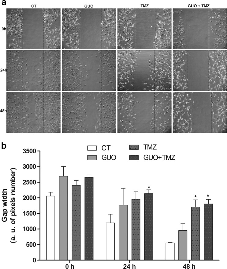
To evaluate the effects of GUO and TMZ on A172 migration capacity, a wound healing assay was carried out, with drug treatment alone or combined (Fig. 3a, b). The distance between the edges of the gap was measured soon after the cells were scratched and 24 and 48 h after scratching. At the 0-h time point, there was no difference among the groups. After 24 h, GUO (500 μM) or TMZ (500 μM) alone did not show any difference in cell migration; however, GUO plus TMZ inhibited the cell migration significantly related to the control group at this same time point. At the last time point, 48 h, GUO did not alter the migration. TMZ reduced migration alone or in combination with GUO in a similar way. Thus, at 24 h evaluation, the combined treatment (GUO + TMZ) reduced cell migration. At 48 h evaluation, TMZ or GUO + TMZ decreased cell migration. GUO treatment alone had no effect on A172 glioma cell migration.
Fig. 3.
GUO and TMZ effects on A172 glioma cell migration. After confluence, cells were treated (GUO—500 μM; TMZ—500 μM; GUO 500 μM + TMZ 500 μM), the scratch assay was performed, and the cells photographed subsequently after 24 and 48 h. a Illustrative images from cells subjected to the scratched protocol and evaluated after 0, 24, and 48 h, scale bar—200 μm. b Gap width was quantified as arbitrary units of pixel number on Image J Software. n = 3 of independent experiments. *p < 0.05 compared to the same time control group
Acidic vesicular organelle assay
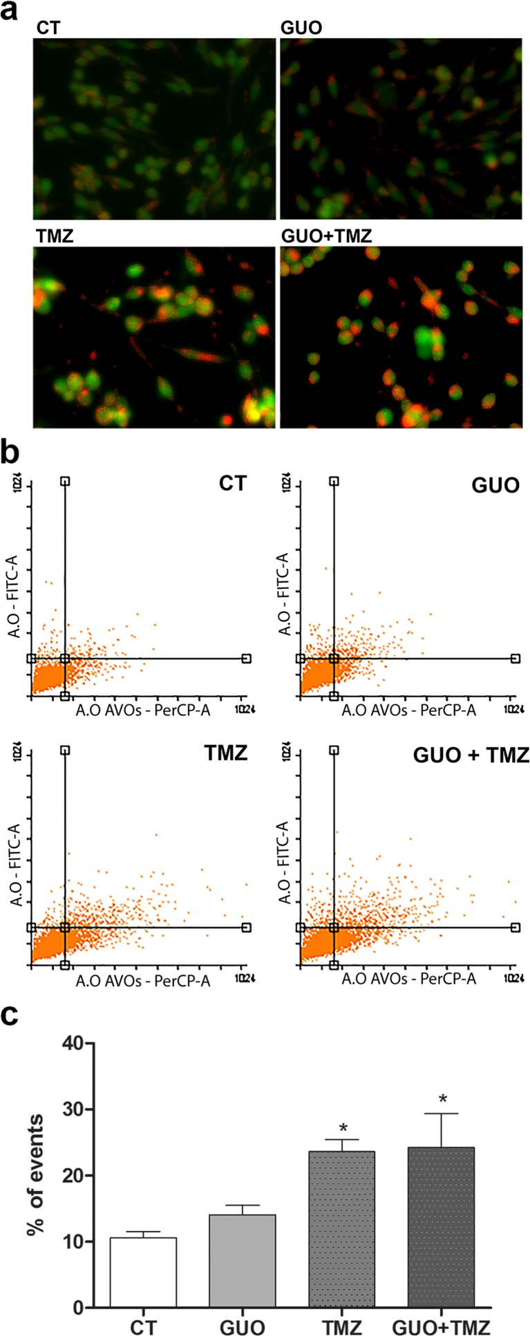
Acridine orange is a cell-permeant cationic fluorescent dye which indicates the acidic vesicular organelle (AVO) presence. AVOs are considered as an indicative of autophagy since the autophagosomes and autophagolysosomes are acidic organelles. In this way, the AVO presence after GUO, TMZ, and GUO + TMZ for 48 h was measured (Fig. 4a–c). Representative photomicrographs show AVO presence and changes in cell number following GUO or TMZ treatment (Fig. 4a). Figure 4b shows representative dot plots obtained through flow cytometry. GUO did not significantly increase the AVO staining in glioma cells. TMZ or TMZ + GUO groups showed an increase, being the percentage of events around 24% for both (Fig. 4c). Therefore, TMZ or GUO + TMZ increased autophagy similarly, with no synergistic effect in combined treatment.
Fig. 4.
GUO and TMZ effects on autophagy in A172 glioma cells. After confluence, A172 cells were treated (GUO—500 μM; TMZ—500 μM; GUO 500 μM + TMZ 500 μM). After 48 h of treatment, the cells were washed and stained with acridine orange and photographed. a Representative images of acridine orange staining in AVOs, scale bar—80 μm. b Dot plot of AVO presence counting in a flow cytometer. c Quantification of AVO presence counting in a flow cytometer. A.O. acridine orange, AVOs acidic vesicular organelles. n = 5 independent experiments. *p < 0.05 compared to the control group
Apoptosis and necrosis analysis
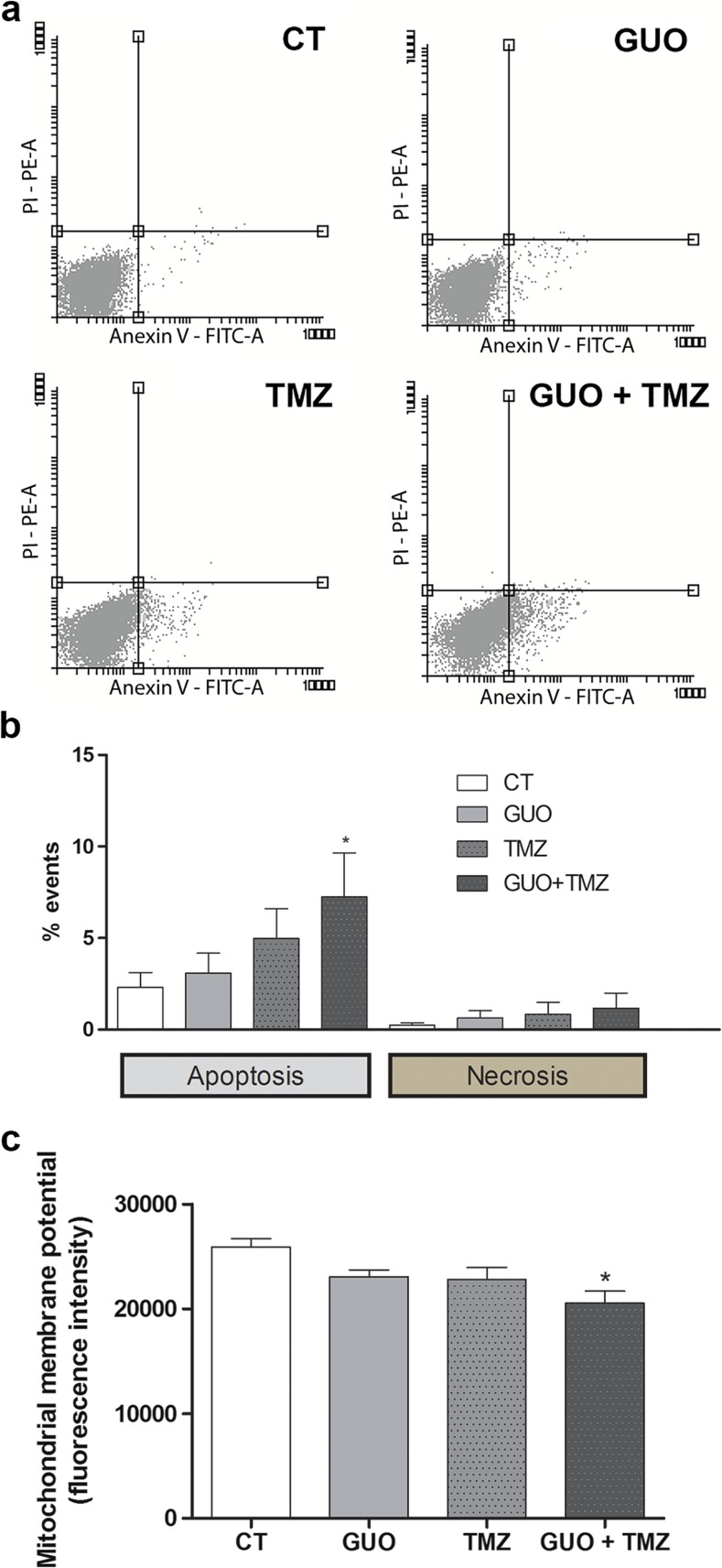
Regarding the type of cell death caused by GUO or TMZ, apoptotic and necrotic cell death in cells treated with GUO, TMZ, or both drugs combined was analyzed, after 48 h of treatment. GUO or TMZ alone did not significantly alter the number of apoptotic cells. GUO + TMZ treatment increased the labeling of apoptotic cells (Figs. 5a, b), suggesting an improvement in antitumor effect in this type of programmed cell death. Regarding the necrotic cell death, there were no significant changes in glioma cells treated with GUO, TMZ, or both compounds (Fig. 5b).
Fig. 5.
GUO and/or TMZ effects on apoptosis levels in A172 glioma cells. After confluence, A172 cells were treated with GUO 500 μM, TMZ 500 μM, or with the drug combination. After 48 h, cells were detached, washed and stained with Annexin V and/or propidium iodide, and counted in a flow cytometer. a Illustrative dot plots of flow cytometry. b Quantification of flow cytometer cells sorted. c Mitochondrial membrane potential (ΔΨm) was assessed and quantified. PI propidium iodide. n = 5 of independent experiments for b and c. *p < 0.05 compared to the control group
Mitochondrial membrane potential evaluation
Mitochondrial dysfunctions have been shown to participate of apoptosis cascade. In this way, we studied GUO, TMZ, or drug combination on mitochondrial membrane potential (ΔΨm) maintenance. GUO or TMZ alone did not show any effect. Nevertheless, GUO + TMZ-combined treatment decreased the ΔΨm in approximately 23% (Fig. 5c). FCCP (10 μM), a mitochondrial uncoupler, decreased 38% the ΔΨm (data not shown). These data corroborate with the increased apoptotic levels observed with GUO + TMZ treatment. In this way, GUO or TMZ was not able to change apoptosis levels and ΔΨm. However, GUO + TMZ-combined treatment increased apoptosis and decreased mitochondrial membrane potential.
Analysis of glutamatergic transmission involvement
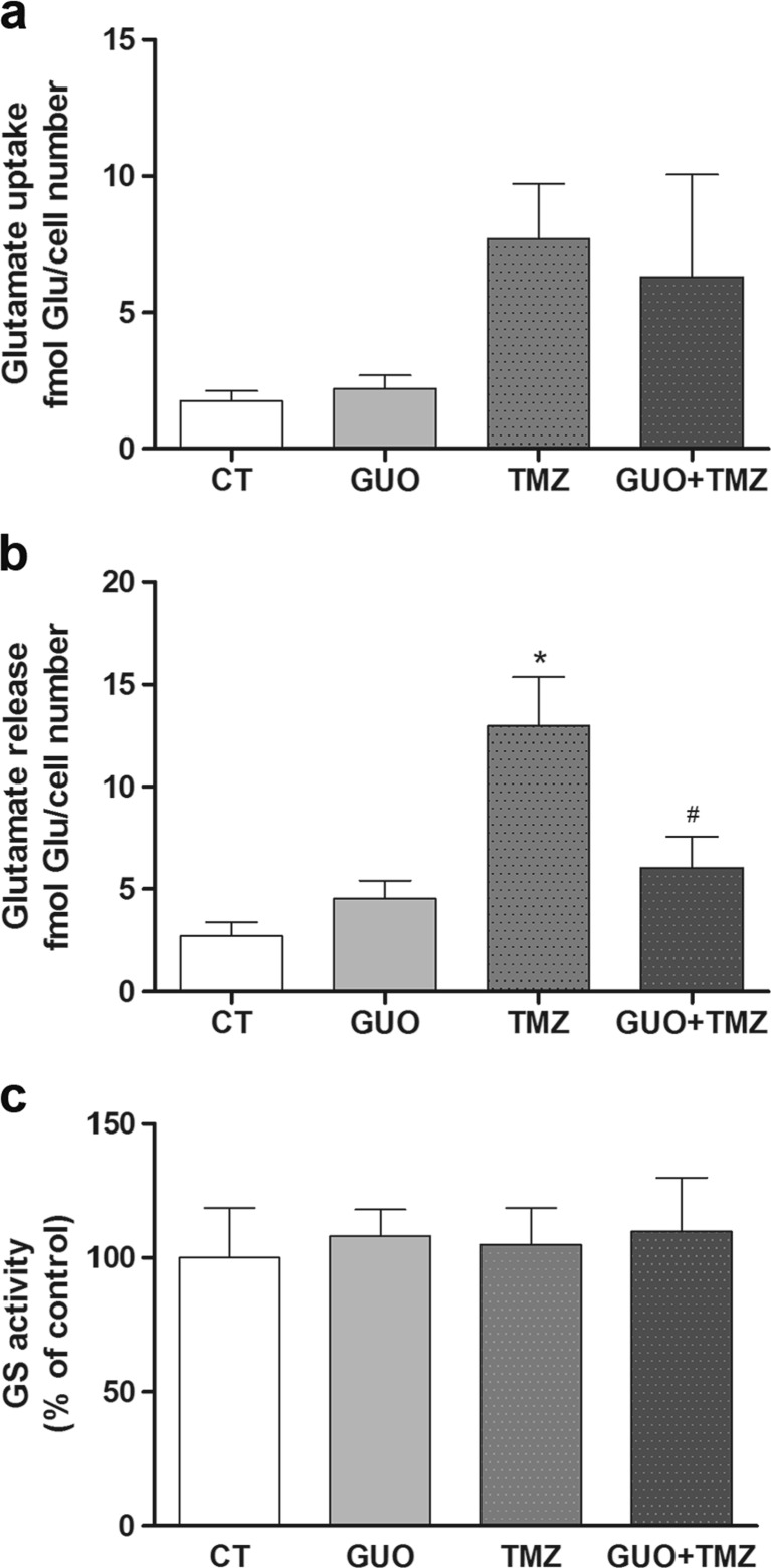
As glioma cells are supposed to release a large amount of glutamate and there is evidence showing GUO is able to modulate glutamate transport, A172 glioma cells treated with GUO and/or TMZ were assayed for glutamate uptake and release evaluation. GUO did not significantly change glutamate uptake (Fig. 6a) or release (Fig. 6b). There was a tendency of increasing glutamate uptake induced by TMZ and GUO + TMZ treatment, although no statistical significance was reached (Fig. 6a). However, TMZ significantly increased glutamate release whereas co-incubation with GUO prevented this effect (Fig. 6b).
Fig. 6.
Effects of treatment with GUO and/or TMZ on glutamate transport and glutamine synthetase (GS) activity in A172 glioma cells. After confluence, A172 glioma cells were treated with GUO 500 μM, TMZ 500 μM, or both during 48 h and then evaluated the levels of a glutamate uptake into A172 cells and b glutamate release from A172 cells. The glutamate content was normalized to viable cell number. c After GUO and/or TMZ treatment (48 h), the A172 cells were detached from the culture bottles and washed and the GS activity was measured. n = 5 of independent experiments for a, b, and c separately. *p < 0.05 compared to the control group and #p < 0.05 compared to TMZ group
The activity of the enzyme responsible for converting glutamate to glutamine, glutamine synthetase (GS), was also assessed in cells treated with GUO, TMZ, or both compounds combined. However, no changes in GS activity were observed in glioma cells treated with GUO, TMZ, or the combination of both drugs (Fig. 6c).
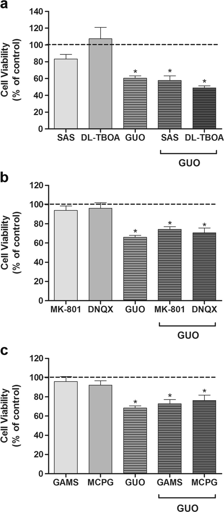
A possible involvement of glutamate (excitatory amino acids) transporters (EAAT) in GUO effects was also investigated through pharmacological intervention and evaluation of glioma cell viability. Taking GUO effect on cell viability into consideration and preventing the increase of glutamate release caused by TMZ, glutamate transporter blockers were incubated for 30 min before GUO. Sulfasalazin (SAS, 300 μM), a Xc- system inhibitor, or DL-TBOA (100 μM), a non-transportable broad-spectrum glutamate transporters inhibitor, was tested. None of the blockers altered GUO effect, suggesting GUO did not decrease cell viability through glutamate transporter modulation (Fig. 7a).
Fig. 7.
Evaluation of glutamate transporters and receptors involvement on GUO induced cytotoxic effect in A172 glioma cells. After confluence, A172 glioma cells were pre-treated with glutamate transporter inhibitors or glutamate receptor antagonists then incubated with GUO for 48 h and cell viability was assayed via MTT reduction method. a SAS (300 μM) or DL-TBOA (100 μM) were used to analyze glutamate transporters or Xc- system involvement on GUO cytotoxic effect. b MK801 (1 μM) or DNQX (1 μM) were used to analyze NMDA or AMPA receptor participation on GUO cytotoxic effect. c GAMS (1 μM) or MCPG (10 μM) were used to analyze kainate or metabotropic glutamate receptor interaction on GUO cytotoxic effect. Data expressed as a percentage related to the control group that was considered as 100% and expressed as a dotted line. n = 6 of independent experiments for a, b, and c separately. *p < 0.05 compared to the control group
The ionotropic glutamate receptors (iGluRs) and the metabotropic glutamate receptors (mGluRs) contribution to GUO cytotoxicity to glioma cells were also evaluated. The NMDAR antagonist MK-801 (1 μM) did not change glioma cell viability. Preincubation of MK-801 did not prevent the reduction in cell viability observed with GUO (Fig. 7b). DNQX (1 μM), a non-selective AMPA and kainate (KA) receptor antagonist also, did not change glioma viability alone, neither co-incubated with GUO (Fig. 7b). A selective KA receptor antagonist, GAMS (1 μM), and a non-selective metabotropic glutamate receptors antagonist, MCPG (10 μM), were also tested (Fig. 7c). None of the antagonists altered GUO effect on decreasing cell viability, discarding the participation of glutamate receptors on GUO effect.
Evaluation of adenosinergic system involvement
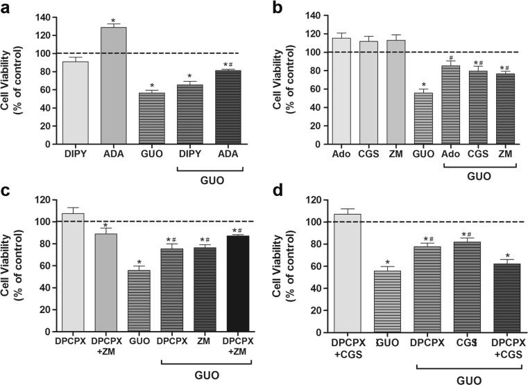
As the nucleoside adenosine regulates glutamatergic transmission and transport, and protective effects of GUO were abolished by modulating adenosine receptors (mainly A1R and A2AR), the involvement of adenosinergic system on GUO effect over A172 cells was also evaluated. The purine nucleoside transport inhibitor, dipyridamole (DIPY, 10 μM), has no effect on cell viability. DIPY did not abolish GUO effect, pointing to an extracellular effect of GUO (Fig. 8a). In order to evaluate a dependence of GUO effect on extracellular endogenous adenosine levels, cell viability was assessed in the presence of the metabolizing adenosine enzyme, adenosine deaminase (ADA, 0.5 U/mL). ADA per se increased A172 cell viability as compared to control cells. The cytotoxic effect of GUO was partially prevented in the presence of ADA (Fig. 8a).
Fig. 8.
GUO exerts cytotoxicity on A172 glioma cells via adenosine receptor interaction. After confluence, A172 glioma cells were pre-treated with transporter inhibitors or receptor ligands and then incubated with GUO for 48 h and cell viability was assayed via MTT reduction method. a DIPY (10 μM) was used to analyze nucleoside transport involvement on GUO effect. ADA (0.5 U/mL) was used to metabolize endogenous adenosine. b Adenosine (500 μM)—AdoR full agonist, CGS 21680 (30 ηM)—A2AR full agonist, and ZM241385 (50 ηM)—A2AR inverse agonist were used to evaluate AdoR and A2AR participation on GUO cytotoxic effect. c DPCPX (100 ηM) was used to evaluate A1R participation on GUO effect. DPCPX (100 ηM) + ZM241385 (50 ηM) were co-incubated to study A1R-A2AR interaction in GUO cytotoxic effect. d DPCPX (100 ηM) + CGS 21680 (30 ηM) were co-incubated to study A1R inactivation and A2AR activation, and receptors interaction in GUO cytotoxic effect. Data expressed as a percentage related to the control group that was considered as 100% and expressed as a dotted line. n = 6 of independent experiments for a, b, c, and d separately. *p < 0.05 compared to the control group and #p < 0.05 compared to GUO group
Concerning to assess a putative effect of GUO on modulating adenosine receptor (AdoR) activity, AdoR ligands were incubated in the presence or absence of GUO and cell viability was evaluated. Adenosine, at the same GUO concentration used (500 μM), did not modify glioma cell viability alone, but it prevented GUO cytotoxicity. The A2AR full agonist (CGS 21680, 30 ηM) or the A2AR inverse agonist also did not change glioma cell viability per se. CGS 21680 (A2AR agonist) or ZM241385 (A2AR inverse agonist) partially prevented GUO effect (Fig. 8b), indicating an A2AR involvement on GUO cytotoxicity to glioma cells.
The involvement of adenosine A1 receptor (A1R) on GUO cytotoxic effect was also evaluated by using an A1R antagonist, DPCPX (100 ηM). DPCPX alone did not change glioma cell viability. However, this A1R antagonist also partially prevented GUO effect on reducing glioma cells viability (Fig. 8c). Considering the partial effect observed with both synthetic AdoR ligands, an association of these compounds on GUO effect was assessed. The incubation of A1R antagonist, DPCPX, plus A2AR inverse agonist, ZM241385, promoted a slight reduction in glioma cell viability (Fig. 8c). In the presence of DPCPX, ZM241385, or DPCPX + ZM241385, GUO still presented a partial cytotoxic effect (Fig. 8c). However, the co-incubation of the A1R antagonist (DPCPX) plus the A2AR full agonist (CGS21680) did not alter glioma cell viability per se, and it did not interfere with GUO cytotoxic effect, pointing to a GUO effect of modulating adenosine A1-A2A receptor interaction (Fig. 9).
Fig. 9.
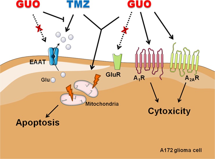
Schematic overview of GUO and GUO plus TMZ association effects on A172 glioma cells. GUO shows cytotoxic effect to glioma cells via adenosine receptor (A1R and A2AR) interaction, but its cytotoxic effect does not depend on glutamate receptors (GluR) or glutamate (excitatory amino acids) transporter (EAAT) interaction. GUO plus TMZ treatment promoted a decreased mitochondrial membrane potential (ΔΨm) and increased apoptosis. TMZ induces an increase in glutamate release, an effect that is prevented by co-treatment with GUO. Additional mechanisms of GUO plus TMZ cytotoxic effects on glioma cells remain to be identified. This figure was produced using Servier Medical Art (http://www.servier.com)
Discussion
Gliomas are a harmful cancer type that exhibit a typical malignant and resistant phenotype, and currently available therapies present several adverse effects and low responsiveness. Therefore, studies concerning adjuvant drugs that may improve the chemotherapy effects over gliomas and decrease the adverse side effects of chemotherapy treatment only are highly desirable [30, 31]. Guanosine is an endogenous non-toxic nucleoside that has been evinced as a neuroprotective agent [11, 12]. In this study, the cytotoxic effect of GUO was compared to the known chemotherapic agent TMZ, as well as their combination, on classical parameters related to glioma malignancy.
The antitumoral effect of GUO was already described to Ehrlich carcinoma, in a study where animals were treated for 10 days with 15 mg/kg/day GUO and it caused a 30% reduction of tumor weight [32]. The association of GUO with acriflavine treatment in vivo demonstrated and enhanced acriflavine antitumoral effect, by decreasing 96% of tumor weight [32]. In the B16F10 melanoma cell line, GUO treatment (500, 1000, or 2000 μM) diminished cell growth after 48 h [20]. And, in leukemia and mastocytoma models, the co-administration of GUO and 5′-deoxy-5-fluorouridine, a chemotherapeutic compound used in solid tumors treatment, showed an improvement of the chemotherapeutic antitumoral effect [33]. Therefore, GUO and GUO plus chemotherapeutic agent treatment have been already evaluated. In this study, we are showing that association of GUO with the alkylating agent TMZ potentiates the apoptotic process in A172 glioma cell line. For the best of our knowledge, it is the first demonstration of a beneficial effect of GUO in a glioma cell line.
In A172 glioma cell line, GUO diminished cell viability in concentrations (500 and 1000 μM) that were previously shown not to be toxic to native brain tissue (0.1–10 mM) [34]. GUO or TMZ alone promoted a decrease in A172 cell viability, and the drug combination does not improve this effect. However, TMZ plus GUO prevented the migration of glioma cells after 24 h of treatment, indicating an improvement in the antitumor effect with this drug association. Tumor cell migration capacity is directly related to the malignancy and the metastasis process. GUO had also been shown to induce melanoma cells to a less malignant feature through inhibition of proliferation and migration [20]. Recently, a purine analog, sulfinosine, has been shown to promote cancer cell death through the induction of apoptosis by activation of caspase-3 [19]. Acute promyelocytic leukemia exposition to GUO causes cell death with classical biochemical changes indicative of apoptosis, such as caspase-3 activation and poly-ADP-ribose polymerase (PARP) cleavage, an enzyme involved in DNA repair [35]. However, GUO treatment in melanoma cells shows that there are no changes in phosphatidylserine exposure related to the control group [20]. Furthermore, in human hepatoma, GUO treatment does not change features related to apoptosis, as soluble Fas ligand (a protein of tumor necrosis factor family) content and caspase-3 activity [36]. Our results also showed no effect on apoptosis activation in glioma cells treated with GUO or TMZ alone. However, the combination of TMZ and GUO increases apoptosis and promotes dissipation of the ΔΨm as observed with the uncoupler agent FCCP. These data suggest that the compound association may alter mitochondrial functionality and facilitate apoptosis, which improves the cytotoxic effect [37].
Autophagy is a normal process related to the self-renewal capacity of cells for the removal of damaged proteins and organelles through the lysosomal action, and apoptosis may be induced by upstream signaling and this way occurs in combination with autophagy [19, 38]. The purine sulfinosine increases autophagy events in U87 human glioma cells [19]. In this context, TMZ is able to induce autophagy in glioma cells and the inhibition of the lysosomes acidification, through bafilomycin treatment, actives apoptosis via caspase-3 activation [39]. In this study, GUO demonstrated no effect on the AVO presence, suggesting its cytotoxic effect may not be related to autophagy. TMZ increased autophagy occurrence significantly as well as the association with GUO.
It has been shown that neuroprotective effects of GUO depend on glutamatergic system modulation, through increasing glutamate uptake or decreasing glutamate release, that may be responsible for increasing glutamine synthetase (GS) activity [13, 14, 16, 18, 40, 41]. Despite all of this evidence, we did not observe GUO effects on glutamate uptake, release, or GS activity, an enzyme that had been considered as a negative growth regulator in glioma cells [42]. Additionally, the pharmacological blockade of glutamate receptors or transporters did not interfere with the cytotoxic effect of GUO. However, TMZ increased glutamate release from glioma cells and the co-incubation with GUO prevented this increase. It is well described that the high extracellular glutamate levels released by glioma cells can lead to excitotoxic neuronal death and improve tumor viability, progression, and aggressiveness [43]. In this sense, the increase in glutamate release could be a possible aggravating factor of TMZ side effects, if it occurs in vivo. In this way, the prevention of glutamate release observed during the combined treatment with GUO may be a desirable effect to efficient glioma treatment.
It is already described that alterations in glutamate transmission may not explain all the complexity of glioma development, thus the purinergic signaling could also interfere with glioma progress [44]. AdoR activation has been shown to promote apoptosis in colorectal cancer cells, carcinoma, and osteosarcoma [45]. Several effects of GUO may be dependent on AdoR interaction, mainly A1R and A2AR (for a review, see [12]), and our group showed that the neuroprotective effect of GUO is abolished by A1R antagonist and A2AR agonist [14, 46], suggesting GUO effects over AdoR may rely on AdoR forming oligomers, a receptorial organization not yet evaluated in glioma cells. Additionally, it was showed that GUO may release adenosine from astrocytes [47]. More recently, it is suggested that GUO increases extracellular adenosine disposition in vascular and endothelial cell cultures [48]. However, this effect was not showed in neural cells and the mechanism is not completely understood, although it seems not to be dependent on nucleoside transporters and adenosine-metabolizing enzyme inhibition [48, 49]. Thus, it is still not clear whether GUO effects depend on increasing adenosine levels or may be due to a direct modulation on AdoR. Through extracellular endogenous adenosine removal with ADA, we showed that GUO cytotoxic effect was partially reduced (and not fully, if one expected GUO effect would rely only on adenosine release). Surprisingly, adenosine alone (in a high concentration, probably activating A2AR) has no cytotoxic effect, as observed to the A2AR full agonist (CGS21680) and also to A2AR inverse agonist (ZM241385), which blocks the constitutive activity of A2AR in the absence of ligands (basal activity). Thus, it seems GUO effect depends on A2AR modulation, although the site of interaction and the exact modulatory effect of GUO at A2AR are not known yet. However, it is still puzzling why adenosine has no cytotoxic effect on A172 glioma cells if GUO cytotoxic effect is partially abolished by AdoR ligands. Regarding ADA effect per se observed in our study, one might suggest that adenosine deamination, and then inosine formation, would be a mechanism for maintenance of glioma viability. Alternatively, these purine nucleosides may compete to each other for AdoR modulation. This is a possibility that needs further investigation.
When evaluating putative GUO interaction with AdoR, here, we showed that the blockade of A1R (with DPCPX), or the reduction of the constitutive activity of A2AR (with ZM241385), and additionally, the activation of A2AR (with CGS21680) interfere with cytotoxic GUO effect. A1R and A2AR blockade per se decreased glioma cell viability, and A1R blockade concomitantly with A2AR activation did not interfere with GUO effect. Taken together, these data suggest that GUO regulates AdoR interaction, probably by modulating an interaction between A1R and A2AR, and the downstream signaling over glioma cell viability, with a mechanism that needs further investigation.
The activation of AdoR has shown dual effects on cell survival and death cascades. A1R activation inhibits proliferation in human metastatic cell line, and A2AR activation promotes human melanoma cell death [50, 51]. AdoR activation also has been shown to sensitize glioma cells to chemotherapic cytotoxic effects [52, 53]. In this way, it has been already described that AdoR activation is capable of modulating apoptotic pathways in colorectal cancer, hepatocellular carcinoma, osteosarcoma, and lung cancer, for example, increasing BAX and BAD levels, promoting alteration in mitochondrial membrane potential and increasing the efflux of cytochrome C and triggering apoptosis [45]. There is also a well-known TMZ effect inducing endoplasmic reticulum stress, increasing caspase-3 activation [54]. Taken together, the AdoR modulation by GUO and TMZ toxicity could lead to an increase in apoptosis levels.
In conclusion, we hereby demonstrated the effects of GUO and TMZ causing cytotoxicity in human A172 glioma cells. The combination of the compounds induces an alteration in mitochondrial membrane potential and triggers an increase in apoptosis. GUO effects may involve the modulation of AdoR interaction, although the exact mechanisms of GUO cytotoxic effect on glioma cells must be better understood, in order to be considered as an adjuvant treatment.
Acknowledgements
This study was supported by grants from the Brazilian funding agencies CNPq (Conselho Nacional de Desenvolvimento Científico e Tecnológico)-(Universal/2012; INCT), FAPESC (Fundação de Amparo à Pesquisa e Inovação do Estado de Santa Catarina)-(NENASC/PRONEX), and CAPES (Coordenação de Aperfeiçoamento de Pessoal de Ensino Superior)-(PVE-052/2012) to C.I.T., and CNPq (Universal/2013) to C.B.N. T.D.-C. is a recipient of a CAPES/PVE post-doctoral fellowship, and C.I.T. is a recipient of CNPq productivity fellowship.
Abbreviations
- A1R
Adenosine A1 receptors
- A2AR
Adenosine A2A receptors
- AdoR
Adenosine receptors
- AMPAR
α-Amino-3-hydroxy-5-methyl-4-isoxazolepropionic acid receptor
- AVOs
Acidic vesicular organelles
- BK
Large (big) conductance Ca2+-activated K+ channels
- CGS 21680
4-[2-[[6-Amino-9-(N-ethyl-β-d-ribofuranuronamidosyl)-9H-purin-2-yl]amino]ethyl]benzenepropanoic acid hydrochloride
- DIPY
Dipyridamole
- DL-TBOA
dl-threo-β-Benzyloxyaspartic acid
- DNQX
6,7-Dinitroquinoxaline-2,3-dione
- DPCPX
Dipropylcyclopentylxanthine
- EAAT
Excitatory aminoacid transporters
- ERK
Extracellular signal-regulated kinase
- FBS
Fetal bovine serum
- FCCP
Carbonyl cyanide p-trifluoromethoxy phenylhydrazone
- GAMS
γ-d-Glutamylaminomethylsulphonic acid
- GluR
Glutamate receptors
- GUO
Guanosine
- KAR
Kainate receptor
- MAPK
Mitogen-activated protein kinase
- MCPG
(RS)-α-Methyl-4-carboxyphenylglycine
- mGluR
Metabotropic glutamate receptor
- MK-801
Dizolcipine
- MTT
3-(4,5-Diamethylthiazol-2-yl)-2,5-diphenyltetrazolium bromide
- NF-κB
nuclear factor-kappa B
- NMDAR
N-methyl-d-aspartate receptor
- PI3K
Phosphoinositide 3-kinase
- PKC
Protein kinase C
- ROS
Reactive oxygen species
- SAS
Sulfasalazin
- TMRE
Tetramethylrhodamine ethyl ester
- TMZ
Temozolomide
- Xc- system
Cystine-glutamate exchanger
- ZM241385
4-(2-[7-Amino-2-(2-furyl)[1,2,4]triazolo[2,3-a][1,3,5]triazin-5-ylamino]ethyl)phenol
Compliance with ethical standards
Conflict of interest
Karen A. Oliveira declares that she has no conflict of interest.
Tharine A. Dal-Cim declares that she has no conflict of interest.
Flávia G. Lopes declares that she has no conflict of interest.
Cláudia B. Nedel declares that she has no conflict of interest.
Carla Inês Tasca declares that she has no conflict of interest.
Ethical approval
This article does not contain any studies with participants or animals performed by any of the authors.
References
- 1.Jiang Y, Uhrbom L. On the origin of glioma. Ups J Med Sci. 2012;117(2):113–121. doi: 10.3109/03009734.2012.658976. [DOI] [PMC free article] [PubMed] [Google Scholar]
- 2.Ohgaki H, Kleihues P. Genetic alterations and signaling pathways in the evolution of gliomas. Cancer Sci. 2009;100(12):2235–2241. doi: 10.1111/j.1349-7006.2009.01308.x. [DOI] [PMC free article] [PubMed] [Google Scholar]
- 3.Liu C, Sage JC, Miller MR, Verhaak RG, Hippenmeyer S, Vogel H, Foreman O, Bronson RT, Nishiyama A, Luo L, Zong H. Mosaic analysis with double markers reveals tumor cell of origin in glioma. Cell. 2011;146(2):209–221. doi: 10.1016/j.cell.2011.06.014. [DOI] [PMC free article] [PubMed] [Google Scholar]
- 4.Desjardins A, Rich JN, Quinn JA, Vredenburgh J, Gururangan S, Sathornsumetee S, Reardon DA, Friedman AH, Bigner DD, Friedman HS. Chemotherapy and novel therapeutic approaches in malignant glioma. Front Biosci. 2005;10:2645–2668. doi: 10.2741/1727. [DOI] [PubMed] [Google Scholar]
- 5.Newlands ES, Stevens MF, Wedge SR, Wheelhouse RT, Brock C. Temozolomide: a review of its discovery, chemical properties, pre-clinical development and clinical trials. Cancer Treat Rev. 1997;23(1):35–61. doi: 10.1016/s0305-7372(97)90019-0. [DOI] [PubMed] [Google Scholar]
- 6.Agnihotri S, Burrell KE, Wolf A, Jalali S, Hawkins C, Rutka JT, Zadeh G. Glioblastoma, a brief review of history, molecular genetics, animal models and novel therapeutic strategies. Arch Immunol Ther Exp. 2013;61(1):25–41. doi: 10.1007/s00005-012-0203-0. [DOI] [PubMed] [Google Scholar]
- 7.Holland EC. Glioblastoma multiforme: the terminator. Proc Natl Acad Sci U S A. 2000;97(12):6242–6244. doi: 10.1073/pnas.97.12.6242. [DOI] [PMC free article] [PubMed] [Google Scholar]
- 8.de Groot J, Sontheimer H. Glutamate and the biology of gliomas. Glia. 2011;59(8):1181–1189. doi: 10.1002/glia.21113. [DOI] [PMC free article] [PubMed] [Google Scholar]
- 9.Ye ZC, Rothstein JD, Sontheimer H. Compromised glutamate transport in human glioma cells: reduction-mislocalization of sodium-dependent glutamate transporters and enhanced activity of cystine-glutamate exchange. J Neurosci. 1999;19(24):10767–10777. doi: 10.1523/JNEUROSCI.19-24-10767.1999. [DOI] [PMC free article] [PubMed] [Google Scholar]
- 10.Ribeiro MP, Custodio JB, Santos AE. Ionotropic glutamate receptor antagonists and cancer therapy: time to think out of the box? Cancer Chemother Pharmacol. 2016 doi: 10.1007/s00280-016-3129-0. [DOI] [PubMed] [Google Scholar]
- 11.Schmidt AP, Lara DR, Souza DO. Proposal of a guanine-based purinergic system in the mammalian central nervous system. Pharmacol Ther. 2007;116(3):401–416. doi: 10.1016/j.pharmthera.2007.07.004. [DOI] [PubMed] [Google Scholar]
- 12.Lanznaster D, Dal-Cim T, Piermartiri TC, Tasca CI. Guanosine: a neuromodulator with therapeutic potential in brain disorders. Aging and disease. 2016;7(5):657–679. doi: 10.14336/AD.2016.0208. [DOI] [PMC free article] [PubMed] [Google Scholar]
- 13.Molz S, Dal-Cim T, Budni J, Martín-de-Saavedra MD, Egea J, Romero A, del Barrio L, Rodrigues AL, López MG, Tasca CI. Neuroprotective effect of guanosine against glutamate-induced cell death in rat hippocampal slices is mediated by the phosphatidylinositol-3 kinase/Akt/ glycogen synthase kinase 3β pathway activation and inducible nitric oxide synthase inhibition. J Neurosci Res. 2011;89(9):1400–1408. doi: 10.1002/jnr.22681. [DOI] [PubMed] [Google Scholar]
- 14.Dal-Cim T, Ludka FK, Martins WC, Reginato C, Parada E, Egea J, Lopez MG, Tasca CI. Guanosine controls inflammatory pathways to afford neuroprotection of hippocampal slices under oxygen and glucose deprivation conditions. J Neurochem. 2013;126(4):437–450. doi: 10.1111/jnc.12324. [DOI] [PubMed] [Google Scholar]
- 15.Dal-Cim T, Martins WC, Santos AR, Tasca CI. Guanosine is neuroprotective against oxygen/glucose deprivation in hippocampal slices via large conductance Ca2+-activated K+ channels, phosphatidilinositol-3 kinase/protein kinase B pathway activation and glutamate uptake. Neuroscience. 2011;183:212–220. doi: 10.1016/j.neuroscience.2011.03.022. [DOI] [PubMed] [Google Scholar]
- 16.Dal-Cim T, Martins WC, Thomaz DT, Coelho V, Poluceno GG, Lanznaster D, Vandresen-Filho S, Tasca CI. Neuroprotection promoted by guanosine depends on glutamine synthetase and glutamate transporters activity in hippocampal slices subjected to oxygen/glucose deprivation. Neurotox Res. 2016;29(4):460–468. doi: 10.1007/s12640-015-9595-z. [DOI] [PubMed] [Google Scholar]
- 17.Thomaz DT, Dal-Cim TA, Martins WC, Cunha MP, Lanznaster D, de Bem AF, Tasca CI. Guanosine prevents nitroxidative stress and recovers mitochondrial membrane potential disruption in hippocampal slices subjected to oxygen/glucose deprivation. Purinergic signalling. 2016;12(4):707–718. doi: 10.1007/s11302-016-9534-3. [DOI] [PMC free article] [PubMed] [Google Scholar]
- 18.Dal-Cim T, Ludka FK, Martins WC, Reginato C, Parada E, Egea J, López MG, Tasca CI. Guanosine controls inflammatory pathways to afford neuroprotection of hippocampal slices under oxygen and glucose deprivation conditions. J Neurochem. 2013;126(4):437–450. doi: 10.1111/jnc.12324. [DOI] [PubMed] [Google Scholar]
- 19.Dačević M, Isaković A, Podolski-Renić A, Isaković AM, Stanković T, Milošević Z, Rakić L, Ruždijić S, Pešić M. Purine nucleoside analog—sulfinosine modulates diverse mechanisms of cancer progression in multi-drug resistant cancer cell lines. PLoS One. 2013;8(1) doi: 10.1371/journal.pone.0054044. [DOI] [PMC free article] [PubMed] [Google Scholar]
- 20.Naliwaiko K, Luvizon AC, Donatti L, Chammas R, Mercadante AF, Zanata SM, Nakao LS. Guanosine promotes B16F10 melanoma cell differentiation through PKC-ERK 1/2 pathway. Chem Biol Interact. 2008;173(2):122–128. doi: 10.1016/j.cbi.2008.03.010. [DOI] [PubMed] [Google Scholar]
- 21.de Saldanha da Gama Fischer J, Costa Carvalho P, da Fonseca CO, Liao L, Degrave WM, da Gloria da Costa Carvalho M, Yates JR, Domont GB. Chemo-resistant protein expression pattern of glioblastoma cells (A172) to perillyl alcohol. J Proteome Res. 2011;10(1):153–160. doi: 10.1021/pr100677g. [DOI] [PMC free article] [PubMed] [Google Scholar]
- 22.Mosmann T. Rapid colorimetric assay for cellular growth and survival: application to proliferation and cytotoxicity assays. J Immunol Methods. 1983;65(1–2):55–63. doi: 10.1016/0022-1759(83)90303-4. [DOI] [PubMed] [Google Scholar]
- 23.Stepanenko AA, Dmitrenko VV. Pitfalls of the MTT assay: direct and off-target effects of inhibitors can result in over/underestimation of cell viability. Gene. 2015;574(2):193–203. doi: 10.1016/j.gene.2015.08.009. [DOI] [PubMed] [Google Scholar]
- 24.Yelskaya Z, Carrillo V, Dubisz E, Gulzar H, Morgan D, Mahajan SS. Synergistic inhibition of survival, proliferation, and migration of U87 cells with a combination of LY341495 and Iressa. PLoS One. 2013;8(5) doi: 10.1371/journal.pone.0064588. [DOI] [PMC free article] [PubMed] [Google Scholar]
- 25.Egea J, Rosa AO, Cuadrado A, García AG, López MG. Nicotinic receptor activation by epibatidine induces heme oxygenase-1 and protects chromaffin cells against oxidative stress. J Neurochem. 2007;102(6):1842–1852. doi: 10.1111/j.1471-4159.2007.04665.x. [DOI] [PubMed] [Google Scholar]
- 26.Shapiro BM. Regulation of glutamine synthetase by enzyme catalyzed structural modification. Angew Chem Int Ed Engl. 1970;9(9):670–678. doi: 10.1002/anie.197006701. [DOI] [PubMed] [Google Scholar]
- 27.Vandresen-Filho S, Martins WC, Bertoldo DB, Mancini G, Herculano BA, de Bem AF, Tasca CI. Atorvastatin prevents cell damage via modulation of oxidative stress, glutamate uptake and glutamine synthetase activity in hippocampal slices subjected to oxygen/glucose deprivation. Neurochem Int. 2013;62(7):948–955. doi: 10.1016/j.neuint.2013.03.002. [DOI] [PubMed] [Google Scholar]
- 28.Molz S, Decker H, Oliveira IJ, Souza DO, Tasca CI. Neurotoxicity induced by glutamate in glucose-deprived rat hippocampal slices is prevented by GMP. Neurochem Res. 2005;30(1):83–89. doi: 10.1007/s11064-004-9689-0. [DOI] [PubMed] [Google Scholar]
- 29.Lowry OH, Rosebrough NJ, Farr AL, Randall RJ. Protein measurement with the Folin phenol reagent. J Biol Chem. 1951;193(1):265–275. [PubMed] [Google Scholar]
- 30.Ohgaki H, Kleihues P. Epidemiology and etiology of gliomas. Acta Neuropathol. 2005;109(1):93–108. doi: 10.1007/s00401-005-0991-y. [DOI] [PubMed] [Google Scholar]
- 31.Westphal M, Lamszus K. The neurobiology of gliomas: from cell biology to the development of therapeutic approaches. Nat Rev Neurosci. 2011;12(9):495–508. doi: 10.1038/nrn3060. [DOI] [PubMed] [Google Scholar]
- 32.Kim SG, Kim CW, Ahn ET, Lee KY, Hong EK, Yoo BI, Han YB. Enhanced anti-tumour effects of acriflavine in combination with guanosine in mice. J Pharm Pharmacol. 1997;49(2):216–222. doi: 10.1111/j.2042-7158.1997.tb06783.x. [DOI] [PubMed] [Google Scholar]
- 33.Iigo M, Miwa M, Ishitsuka H, Nitta K. Potentiation of the chemotherapeutic action of 5′-deoxy-5-fluorouridine in combination with guanosine and related compounds. Cancer Chemother Pharmacol. 1987;19(1):61–64. doi: 10.1007/BF00296258. [DOI] [PubMed] [Google Scholar]
- 34.Molz S, Dal-Cim T, Tasca CI. Guanosine-5′-monophosphate induces cell death in rat hippocampal slices via ionotropic glutamate receptors activation and glutamate uptake inhibition. Neurochem Int. 2009;55(7):703–709. doi: 10.1016/j.neuint.2009.06.015. [DOI] [PubMed] [Google Scholar]
- 35.Flanagan SA, Gandhi V, Meckling KA. Guanosine acts intracellularly to initiate apoptosis in NB4 cells: a role for nucleoside transport. Leuk Lymphoma. 2007;48(9):1816–1827. doi: 10.1080/10428190701528491. [DOI] [PubMed] [Google Scholar]
- 36.Yang SC, Chiu CL, Huang CC, Chen JR. Apoptosis induced by nucleosides in the human hepatoma HepG2. World J Gastroenterol. 2005;11(40):6381–6384. doi: 10.3748/wjg.v11.i40.6381. [DOI] [PMC free article] [PubMed] [Google Scholar]
- 37.Ly JD, Grubb DR, Lawen A. The mitochondrial membrane potential (deltapsi(m)) in apoptosis: an update. Apoptosis. 2003;8(2):115–128. doi: 10.1023/a:1022945107762. [DOI] [PubMed] [Google Scholar]
- 38.Maiuri MC, Zalckvar E, Kimchi A, Kroemer G. Self-eating and self-killing: crosstalk between autophagy and apoptosis. Nat Rev Mol Cell Biol. 2007;8(9):741–752. doi: 10.1038/nrm2239. [DOI] [PubMed] [Google Scholar]
- 39.Kanzawa T, Germano IM, Komata T, Ito H, Kondo Y, Kondo S. Role of autophagy in temozolomide-induced cytotoxicity for malignant glioma cells. Cell Death Differ. 2004;11(4):448–457. doi: 10.1038/sj.cdd.4401359. [DOI] [PubMed] [Google Scholar]
- 40.Moretto MB, Arteni NS, Lavinsky D, Netto CA, Rocha JB, Souza DO, Wofchuk S. Hypoxic-ischemic insult decreases glutamate uptake by hippocampal slices from neonatal rats: prevention by guanosine. Exp Neurol. 2005;195(2):400–406. doi: 10.1016/j.expneurol.2005.06.005. [DOI] [PubMed] [Google Scholar]
- 41.Thomazi AP, Boff B, Pires TD, Godinho G, Battú CE, Gottfried C, Souza DO, Salbego C, Wofchuk ST. Profile of glutamate uptake and cellular viability in hippocampal slices exposed to oxygen and glucose deprivation: developmental aspects and protection by guanosine. Brain Res. 2008;1188:233–240. doi: 10.1016/j.brainres.2007.10.037. [DOI] [PubMed] [Google Scholar]
- 42.Yin Y, Sun W, Xiang J, Deng L, Zhang B, Xie P, Qiao W, Zou J, Liu C. Glutamine synthetase functions as a negative growth regulator in glioma. J Neuro-Oncol. 2013;114(1):59–69. doi: 10.1007/s11060-013-1168-5. [DOI] [PubMed] [Google Scholar]
- 43.Robert SM, Sontheimer H. Glutamate transporters in the biology of malignant gliomas. Cell Mol Life Sci. 2014;71(10):1839–1854. doi: 10.1007/s00018-013-1521-z. [DOI] [PMC free article] [PubMed] [Google Scholar]
- 44.Braganhol E, Wink MR, Lenz G, Battastini AM. Purinergic signaling in glioma progression. Adv Exp Med Biol. 2013;986:81–102. doi: 10.1007/978-94-007-4719-7_5. [DOI] [PubMed] [Google Scholar]
- 45.Antonioli L, Blandizzi C, Pacher P, Haskó G. Immunity, inflammation and cancer: a leading role for adenosine. Nat Rev Cancer. 2013;13(12):842–857. doi: 10.1038/nrc3613. [DOI] [PubMed] [Google Scholar]
- 46.Ciruela F. Guanosine behind the scene. J Neurochem. 2013;126(4):425–427. doi: 10.1111/jnc.12328. [DOI] [PubMed] [Google Scholar]
- 47.Ciccarelli R, Di Iorio P, Giuliani P, D'Alimonte I, Ballerini P, Caciagli F, Rathbone MP. Rat cultured astrocytes release guanine-based purines in basal conditions and after hypoxia/hypoglycemia. Glia. 1999;25(1):93–98. [PubMed] [Google Scholar]
- 48.Jackson EK, Gillespie DG. Regulation of cell proliferation by the guanosine-adenosine mechanism: role of adenosine receptors. Physiol Rep. 2013;1(2) doi: 10.1002/phy2.24. [DOI] [PMC free article] [PubMed] [Google Scholar]
- 49.Jackson EK, Cheng D, Jackson TC, Verrier JD, Gillespie DG. Extracellular guanosine regulates extracellular adenosine levels. Am J Physiol Cell Physiol. 2013;304(5):C406–C421. doi: 10.1152/ajpcell.00212.2012. [DOI] [PMC free article] [PubMed] [Google Scholar]
- 50.Ceruti S, Abbracchio MP. Adenosine signaling in glioma cells. Adv Exp Med Biol. 2013;986:13–30. doi: 10.1007/978-94-007-4719-7_2. [DOI] [PubMed] [Google Scholar]
- 51.Merighi S, Mirandola P, Varani K, Gessi S, Leung E, Baraldi PG, Tabrizi MA, Borea PA. A glance at adenosine receptors: novel target for antitumor therapy. Pharmacol Ther. 2003;100(1):31–48. doi: 10.1016/s0163-7258(03)00084-6. [DOI] [PubMed] [Google Scholar]
- 52.Daniele S, Zappelli E, Natali L, Martini C, Trincavelli ML. Modulation of A1 and A2B adenosine receptor activity: a new strategy to sensitise glioblastoma stem cells to chemotherapy. Cell Death Dis. 2014;5 doi: 10.1038/cddis.2014.487. [DOI] [PMC free article] [PubMed] [Google Scholar]
- 53.D'Alimonte I, Nargi E, Zuccarini M, Lanuti P, Di Iorio P, Giuliani P, Ricci-Vitiani L, Pallini R, Caciagli F, Ciccarelli R. Potentiation of temozolomide antitumor effect by purine receptor ligands able to restrain the in vitro growth of human glioblastoma stem cells. Purinergic Signal. 2015;11(3):331–346. doi: 10.1007/s11302-015-9454-7. [DOI] [PMC free article] [PubMed] [Google Scholar]
- 54.Germano IM, Emdad L, Qadeer ZA, Binello E, Uzzaman M. Embryonic stem cell (ESC)-mediated transgene delivery induces growth suppression, apoptosis and radiosensitization, and overcomes temozolomide resistance in malignant gliomas. Cancer Gene Ther. 2010;17(9):664–674. doi: 10.1038/cgt.2010.31. [DOI] [PMC free article] [PubMed] [Google Scholar]


