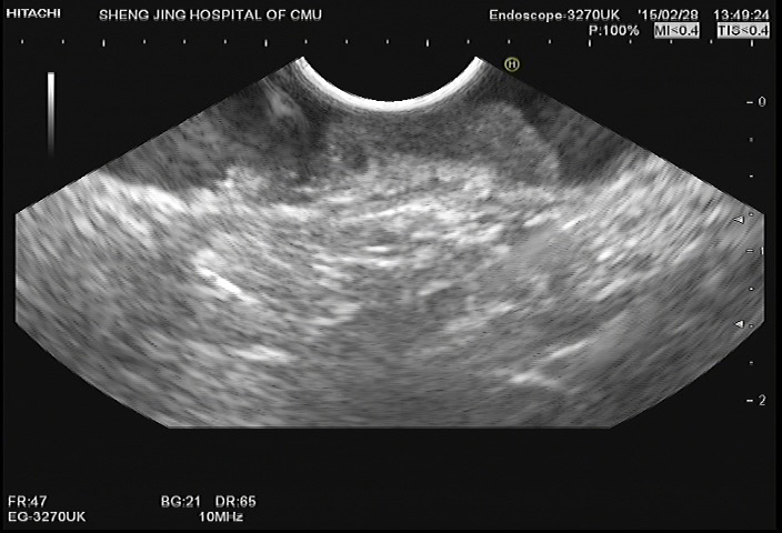
Figure 4. Image from EUS obtained for evaluation of a lesion identified on colonoscopy.

The EUS probe was placed to the lesion. The image demonstrates abnormally decreased echogenicity and thickening of the mucosa and submucosa. The muscularis propria is completely visualized.
