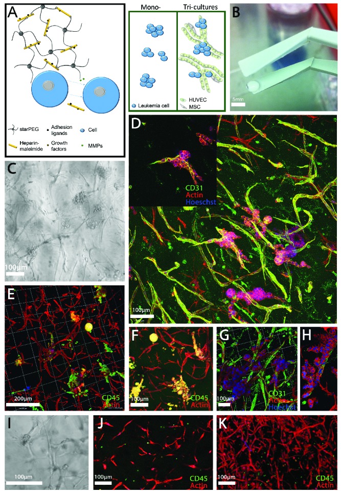
Figure 1.

A three-dimensional culture model of acute myeloid leukemia-vascular interactions. (A) A biohybrid starPEG-heparin hydrogel was utilized which allows for the culture of AML mono-cultures and tri-cultures with HUVEC and bone marrow-derived MSC. B) Macroscopic image of final cast hydrogel before culture. Scale bar = 5 mm. (C) Light microscope and (D,H) confocal images of the OCI-AML3 cell line (as a representative of all cell lines utilized in this study) in tri-culture with HUVEC and MSC after 7 days depicting (D, G, H) CD31 and (E, F) CD45 expression. Images display leukemia cell growth primarily along the vascular endothelial cells. (I) Light microscope and confocal images (J,K) of primary donor cells from a patient with AML in tri-culture with HUVEC and MSC after 7 days. Scale bar = 100 and 200 μm as indicated. Images display the preference of leukemia cells to attach to and grow along vascular structures or within vascular branching.
