Abstract
A sensitive turn-on luminescent sensor for H2O2 based on the silver nanoparticle (AgNP)-mediated quenching of an luminescent Ir(III) complex (Ir-1) has been designed. In the absence of H2O2, the luminescence intensity of Ir-1 can be quenched by AgNPs via non-radiative energy transfer. However, H2O2 can oxidize AgNPs to soluble Ag+ cations, which restores the luminescence of Ir-1. The sensing platform displayed a sensitive response to H2O2 in the range of 0−17 μM, with a detection limit of 0.3 μM. Importantly, the probe was successfully applied to monitor intracellular H2O2 in living cells, and it also showed high selectivity for H2O2 over other interfering substances.
Introduction
H2O2 is widely used in industry and households for rinsing, bleaching and disinfection. For example, in the food industry, H2O2 is used to replace chlorine-containing bleaching and sterilizing agents1. It also plays an important role in many biological processes and enzymatic reactions, particularly those related to intracellular oxidative stress2. In fact, escalated levels of H2O2 can cause irreversible cellular damage through the oxidation of biomolecules, leading to cell death3. Moreover, oxidative damage to cellular proteins, nucleic acids, and lipid molecules are associated with aging and age-related disorder ranging from neurodegeneration to diabetes3, 4. Therefore, a rapid and reliable detection of H2O2 is important in pharmaceutical, clinical, and food industries.
Multiple methods such as spectrophotometry5, 6, chemiluminescence7 and electrocatalysis8 have been developed for the detection of H2O2. Specifically, biosensors have been developed on the basis of electrocatalysis of immobilized enzymes arising from H2O2 reduction9. However, the enzyme-based biosensors are limited by sensitivity to environmental conditions, high cost, short shelf-life and complicated immobilization procedures10–12. Meanwhile, fluorescent strategies have lots of advantages, particularly rapid response, high sensitivity, and simple manipulation13, 14. Various fluorescence probes such as organic molecules15, carbon dots16, 17, metal nanoclusters18, and nanoparticles19–21, have good performance on the determination of H2O2. However, there are still some drawbacks for these reported probes, including poor sensitivity and selectivity, low stability in biological environment, or complicated operation17, 18, 22. Fluorescence turn-on sensors are generally more desirable than fluorescence quenching sensors as the former is less susceptible to false positive signals23, 24.
Luminescent Ir(III) complexes have been employed to detect a variety of analytes25–27. Compared with organic molecules, Ir(III) complexes generally exhibit large Stokes shifts, ease in synthesis and long-lived luminescence which could be distinguished from fluorescence noise in biological matrices26, 27. Meanwhile, silver nanoparticles (AgNPs) form a promising nanomaterial that has been developed in many applications because of their remarkable properties, such as high extinction coefficient and surface plasmon resonance absorption28–30. It has been reported that AgNPs can be oxidized by traces of H2O2, to form Ag+ 31 . In addition, AgNPs can function as excellent quenchers for fluorescent materials, such as organic dyes and quantum dots (QDs)32–35. However, as far as we know, the application of the Ir(III) complexes combined with AgNPs has not yet been reported in the literature for H2O2 sensing. Consequently, taking advantages of the Ir(III) complex (Ir-1, [Ir(tfppy)2(pyphen)]+, where tfppy = 2-[4-(trifluoromethyl)phenyl]pyridine, pyphen = pyrazino[2,3-f][1,10]phenanthroline) and AgNPs, we designed a novel turn-on luminescent probe for rapid and sensitive detection of intracellular H2O2.
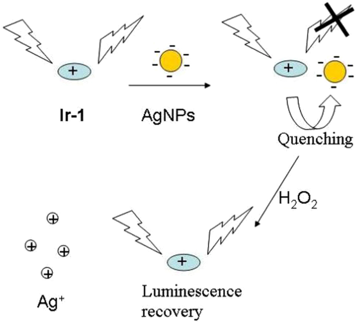
The sensing mechanism of the Ir-1–AgNP probe for H2O2 is illustrated in Fig. 1. In the initial system, the luminescence of Ir-1 was significantly quenched by AgNPs. However, this AgNPs-induced quenching effect can be reversed by H2O2 due to oxidation of AgNPs to Ag+. To our knowledge, the Ir-1–AgNP is the first application of the combination of Ir(III) complexes and AgNPs for H2O2 sensing in both aqueous solutions and living cells.
Figure 1.

Illustration of the design rationale for the detection of H2O2 using a luminescence sensor based on Ir-1–AgNPs system.
Results and Discussion
Sensing Mechanism
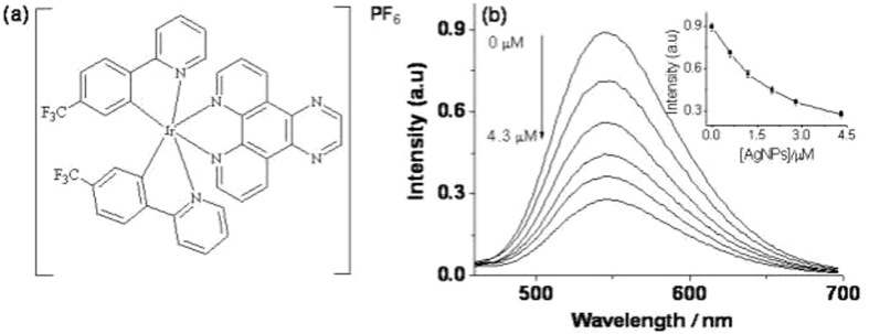
Ir-1, carrying tfppy as its C^N ligand and pyphen as its N^N ligand (Fig. 2a), was characterized by1H-NMR,13C-NMR and HRMS (Figs S1–S3 and Table S1). Ir-1 emits strong luminescence at 545 nm under the excitation of 295 nm in aqueous buffer solution. As expected, the luminescence of Ir-1 decreased gradually with increasing amounts of AgNPs in solution (Fig. 2b). This is because the positively charged Ir-1 could be adsorbed on the surface of the citrate-stabilized AgNPs through electrostatic interactions, which efficiently quenched the luminescence of Ir-1. However, the luminescence could be recovered in the presence of H2O2 attributed to oxidation of AgNPs into soluble Ag+ by H2O2. In order to study the kinetic behavior between the Ir-1–AgNP system and H2O2, the luminescence change was monitored as a function of time. As shown in Fig. S4, the luminescence intensity of the Ir-1–AgNP system increased with time and reached the plateau after 10 min, indicating that the reaction between AgNPs and H2O2 at ambient temperature is rapid. In the absence of AgNPs, H2O2 showed no apparent effect on the luminescence of Ir-1 (Fig. S5). Therefore, the increase in the luminescence of the system should arise primarily from the decomposition of AgNPs by H2O2, which restores the emission of Ir-1.
Figure 2.
(a) Chemical structure of Ir-1. (b) Luminescence emission spectra 0.3 μM Ir-1 in Tris-HNO3 buffer solution (pH 7.0) containing different concentrations of AgNPs. The inset is the luminescence intensity at 545 nm plotted against the AgNPs concentration.
The mechanism involved in the luminescence quenching and recovery process was also demonstrated by transmission electron microscopy (TEM) imaging. In the absence of the Ir-1, the AgNPs were well-dispersed (Fig. 3a). However, after the addition of Ir-1, slight aggregation of AgNPs was observed, suggesting that Ir-1 and AgNPs interacted on the surface of AgNPs (Fig. S6b). The identity of the Ir-1–AgNP complex was further confirmed by energy dispersive X-ray spectroscopy (EDX), which showed strong elemental signals for both Ir and Ag (Fig. S6c). Strikingly, after treatment of AgNPs with H2O2, no AgNPs could be observed in the TEM images (Fig. 3b). This suggests that the AgNPs were decomposed and transformed to Ag+, which is consistent with previously reported36–38. The UV–vis absorbance spectra of AgNPs in the absence and presence of H2O2 are shown in Fig. S7. AgNPs alone showed a strong characteristic surface plasmon resonance peak at around 390 nm39. However, the absorption band of AgNPs gradually decreased upon increasing concentration of H2O2. These phenomena were ascribed to the oxidation of AgNPs to Ag+ by H2O2, leading the decomposition of the AgNPs.
Figure 3.
Transmission electron microscopy images of (a) AgNPs and (b) AgNPs in the presence of H2O2.
Sensitivity
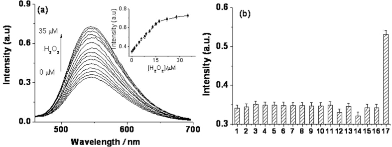
To explore the applicability of the proposed luminescence sensor for H2O2 detection, we studied the luminescence response of the Ir-1–AgNP system toward varying concentrations of H2O2. The luminescence intensity of the system was gradually restored with increasing concentration of H2O2 (Fig. 4a). Meanwhile, a good linear relationship over the range from 0 to 17 μmol L−1 with a correlation coefficient of 0.998 was obtained (Fig. S8). The limit of detection (LOD) was calculated to 0.3 μM according to the signal-to-noise method (S/N = 3). The sensitivity of this method is comparable to other reported methods for H2O2 detection as summarized in Table S2 4, 10–16, 20, 36, 40–44.
Figure 4.
(a) Luminescence emission spectra of 1 μM Ir-1 in Tris-HNO3 buffer solution (pH 7.0) containing 2.8 μM AgNPs and different concentrations of H2O2 (0 μM to 35 μM). The inset is the luminescence intensity plotted against the H2O2 concentration. (b) Luminescence intensity of the Ir-1-AgNP system (0.3 μM Ir-1 and 2.8 μM AgNPs in Tris-HNO3 at pH 7.0) in the presence of interfering species (HSA and BSA 50 μg/L, other interfering species 50 μM) or H2O2 (9 μM) (from 1 to 17: blank, threonine, serine, glycine, ascorbic acid, HSA, BSA, Zn2+, Co2+, Ni2+, Cd2+, Fe3+, Mg2+, Cu2+, K+, Na+, and H2O2).
Selectivity
To assess the selectivity of Ir-1–AgNPs system for H2O2, the influences of metal ions and amino acids were studied. As shown in Fig. 4b, nearly no luminescence changes could be observed with the other substances (Fig. 4b), which demonstrates that the Ir-1–AgNP system is highly selective for H2O2 over other non-target substances.
Cell imaging
Given the promising capability of Ir-1 for sensing H2O2 in aqueous solution, we then investigated the ability of Ir-1 for monitoring H2O2 in living human cells. Ir-1 showed cytotoxicity against HeLa (human cervical cancer) cells with an IC50 value of 5.12 μM (Fig. S9).
In the cell imaging study, the luminescence intensity of HeLa cells was enhanced with increasing concentration of Ir-1 (Fig. 5a), showing that Ir-1 could effectively penetrate into cells. A concentration of 0.3 μM of Ir-1 was chosen for subsequent cell experiments as this concentration was over 10-fold lower than the IC50 value for cytotoxicity, while it still gave a good luminescence signal.
Figure 5.
Confocal luminescence microscopy imaging of HeLa cells. (a) HeLa cells were incubated with the indicated concentration of Ir-1 for 1 h. (b) HeLa cells were pretreated with Ir-1 (0.3 μM) and AgNPs (2.8 μM) for 1 h before incubation with different concentration of H2O2. The upper row is luminescence imaging, and the lower row is bright field imaging. Excitation wavelength = 405 nm.
Next, HeLa cells were pretreated with Ir-1 (0.3 μM) for 1 h before incubation with different concentration of AgNPs. The luminescence intensity of HeLa cells was remarkably reduced with increasing concentration of AgNPs (Fig. S10), which was attributed to AgNPs-mediated quenching of an luminescent Ir-1 as described previously. However, when H2O2 was added into the growth medium for another 1 h, the luminescence of HeLa cells was recovered in a dose-dependent manner (Fig. 5b). Collectively, these results suggest that Ir-1–AgNP can be developed for the monitoring of H2O2 levels in living cells.
Conclusion
Consequently, we have proposed a turn-on luminescence assay for H2O2 detection employing the Ir-1–AgNP system. In this nano-composite system, Ir-1 functioned as a luminescence reporter, while AgNPs were employed both as a luminescence quencher and as a recognition unit for H2O2. Based on the luminescence recovery of the Ir-1–AgNP system triggered by H2O2, this nanoprobe was successfully applied to detect H2O2 at the intracellular level in living cells. In addition, the Ir-1–AgNP probe possesses some superior properties, including label-free, good sensitivity and selectivity, low cost, easy manipulation, low cytotoxicity, and turn-on luminescent response. To our knowledge, the probe is the first combination of Ir(III) and AgNPs applied for the detection of H2O2 in living cells reported in the literature.
Materials and Methods
Chemicals and materials
Iridium chloride hydrate (IrCl3·xH2O) was purchased from Precious Metals Online (Australia). Other reagents were purchased from Sigma Aldrich (St. Louis, MO) and used as received. All of the reagents were of analytical grade and were used as received without further purification. All solutions were prepared in Milli-Q water under ambient conditions. HeLa cell lines were obtained from ATCC (Manassas, VA, USA). Dulbecco’s Modified Eagle’s medium, fetal bovine serum, penicillin and streptomycin were obtained from Sigma-Aldrich Co. LLC (St. Louis, MO, USA).
Synthesis of AgNPs
AgNPs were fabricated according to reported methods with slight modifications28, 45. In a typical procedure, 0.08 mL AgNO3 (0.1 M) and 0.1 mL trisodium citrate (0.1 M) were mixed into 100 mL pure water and stirred under the condition of ice bath. Then, freshly prepared NaBH4 solution was added dropwise into the mixture until it turned yellow. The resulting yellow solution was stirred for another 30 min to form AgNPs quantitatively, which was stored at 4 °C for subsequent use. The diameter of AgNPs prepared was measured to be 8–9 nm by transmission electron microscopy (TEM).
Synthesis of Ir-1
Ir-1 was synthesized based on a reported literature method46–49. [Ir2(tfppy)4Cl2] (0.2 mmol) and pyppy (0.42 mmol) in a mixed solvent of DCM:methanol (1:1.2 (v/v), 36 mL) was refluxed overnight. The reaction mixture was allowed to cool to ambient temperature, and unreacted cyclometallated dimer was removed by filtration. Excess ammonium hexafluorophosphate was then added into the filtrate, and the resulting mixture was stirred for another 30 min. Afterwards, the solution was evaporated under reduced pressure until precipitation was initiated. The precipitate was filtered, and washed by several portions of water and diethyl ether. The crude product was then recrystallized by the acetonitrile/diethyl ether vapor diffusion to obtain the desired compound, which was characterized by1H-NMR,13C-NMR, high resolution mass spectrometry (HRMS) and elemental analysis.
Luminescence response of Ir-1 towards AgNPs
Ir-1 (0.3 μM) was added to varying concentrations of AgNPs in Tris-HNO3 buffer (5 mM Tris-HNO3, pH 7.0), then their emission intensity were measured.
Detection of H2O2
A series of sample solutions of same composition was prepared by mixing Ir-1 (0.3 μM) with AgNPs (2.8 μM) in Tris-HNO3 buffer (5 mM Tris-HNO3, pH 7.0). Upon individual addition of varying concentrations of stock H2O2 solution, the sample solutions were incubated for 10 min at room temperature. Emission spectra were collected in the range of 450–700 nm at the excitation wavelength of 295 nm.
Cell imaging
HeLa cells were pretreated with Ir-1 (0.3 μM) for 1 h at 37 °C, then AgNPs of different concentrations (0 μM, 0.1 μM, 0.3 μM, 1 μM, 3 μM and 5 μM) was added before further incubation for 1 h. After washing with PBS three times, the luminescence intensity of HeLa cells was imaged by a Leica SP8 laser scanning confocal microscope upon excitation at 405 nm.
For H2O2 detection, the experiment was performed as above except that after incubation in the presence of AgNPs (2.8 μM), cells were further treated with H2O2 ranging from 0 to 20 μM for 1 h. After washing with PBS three times, the luminescence intensity of HeLa cells was then imaged as above.
Statistics analysis
One-way analysis of variance (ANOVA) followed by the Dunnett’s method for multiple comparisons by using GraphPad Prism 6.0 was used to analyse the data.
Electronic supplementary material
Acknowledgements
This work is supported by Hong Kong Baptist University (FRG2/15–16/002), the Health and Medical Research Fund (HMRF/14130522, 14150561), the Research Grants Council (HKBU/12301115, HKBU/204612 and HKBU/201913), the National Natural Science Foundation of China (21575121, 21628502), the Guangdong Province Natural Science Foundation (2015A030313816), the Hong Kong Baptist University Century Club Sponsorship Scheme 2016, the Interdisciplinary Research Matching Scheme (RC-IRMS/15–16/03), Innovation and Technology Fund (ITS/260/16FX), Matching Proof of Concept Fund (MPCF-001-2017/18), the Science and Technology Development Fund, Macao SAR (077/2016/A2, 007/2014/AMJ), and the University of Macau (MYRG2015–00137-ICMS-QRCM, MYRG2016–00151-ICMS-QRCM and MRG044/LCH/2015/ICMS).
Author Contributions
Jinshui Liu contributed to the sensing experiments and muniscript draft; Zhen-Zhen Dong prepared the iridium complex, SEM and TEM experiments; Chao Yang and Guodong Li completed the cell imaging and MTT experiments; Chun Wu helped to draw and arrange the Figures 1–5 in the manuscript; Fu-Wa Lee mainly commented and reviesed the manuscript; Chung-Hang Leung and Dik-Lung Ma proposed and instructed the project.
Competing Interests
The authors declare that they have no competing interests.
Footnotes
Jinshui Liu and Zhen-Zhen Dong contributed equally to this work.
Electronic supplementary material
Supplementary information accompanies this paper at doi:10.1038/s41598-017-09478-6
Publisher's note: Springer Nature remains neutral with regard to jurisdictional claims in published maps and institutional affiliations.
References
- 1.Liao Y-X, Li K, Wu M-Y, Wu T, Yu X-Q. A selenium-contained aggregation-induced “turn-on” fluorescent probe for hydrogen peroxide. Organic & biomolecular chemistry. 2014;12:3004–3008. doi: 10.1039/c4ob00206g. [DOI] [PubMed] [Google Scholar]
- 2.Tian J, et al. Preparation of Ag nanoparticle-decorated poly (m-phenylenediamine) microparticles and their application for hydrogen peroxide detection. Analyst. 2011;136:1806–1809. doi: 10.1039/c0an00929f. [DOI] [PubMed] [Google Scholar]
- 3.Wang P, Wang K, Chen D, Mao Y, Gu Y. A novel colorimetric and near-infrared fluorescent probe for hydrogen peroxide imaging in vitro and in vivo. RSC Advances. 2015;5:85957–85963. doi: 10.1039/C5RA16827A. [DOI] [Google Scholar]
- 4.Peng R, et al. A novel SERS nanoprobe for the ratiometric imaging of hydrogen peroxide in living cells. Chemical Communications. 2016;52:8553–8556. doi: 10.1039/C6CC03412H. [DOI] [PubMed] [Google Scholar]
- 5.Wang F, Liu X, Lu C-H, Willner I. Cysteine-mediated aggregation of Au nanoparticles: the development of a H2O2 sensor and oxidase-based biosensors. ACS nano. 2013;7:7278–7286. doi: 10.1021/nn402810x. [DOI] [PubMed] [Google Scholar]
- 6.Dalui A, et al. Insight into the mechanism revealing the peroxidase mimetic catalytic activity of quaternary CuZnFeS nanocrystals: colorimetric biosensing of hydrogen peroxide and glucose. Nanoscale. 2015;7:9062–9074. doi: 10.1039/C5NR01728A. [DOI] [PubMed] [Google Scholar]
- 7.Lee Y-D, et al. Dye/peroxalate aggregated nanoparticles with enhanced and tunable chemiluminescence for biomedical imaging of hydrogen peroxide. ACS nano. 2012;6:6759–6766. doi: 10.1021/nn3014905. [DOI] [PubMed] [Google Scholar]
- 8.Su S, et al. Facile Synthesis of a MoS2–Prussian Blue Nanocube Nanohybrid-Based Electrochemical Sensing Platform for Hydrogen Peroxide and Carcinoembryonic Antigen Detection. ACS Applied Materials & Interfaces. 2017;9:12773–12781. doi: 10.1021/acsami.7b01141. [DOI] [PubMed] [Google Scholar]
- 9.Zhao B, et al. Silver microspheres for application as hydrogen peroxide sensor. Electrochemistry Communications. 2009;11:1707–1710. doi: 10.1016/j.elecom.2009.06.035. [DOI] [Google Scholar]
- 10.Li Z, Zheng X, Zheng J. A non-enzymatic sensor based on Au@Ag nanoparticles with good stability for sensitive detection of H2O2. New Journal of Chemistry. 2016;40:2115–2120. doi: 10.1039/C5NJ02582F. [DOI] [Google Scholar]
- 11.Wen T, Qu F, Li NB, Luo HQ. Polyethyleneimine-capped silver nanoclusters as a fluorescence probe for sensitive detection of hydrogen peroxide and glucose. Analytica chimica acta. 2012;749:56–62. doi: 10.1016/j.aca.2012.08.048. [DOI] [PubMed] [Google Scholar]
- 12.Yan Y, et al. Hydrothermally driven three-dimensional evolution of mesoporous hierarchical europium oxide hydrangea microspheres for non-enzymatic sensors of hydrogen peroxide detection. Environmental Science: Nano. 2016;3:701–706. [Google Scholar]
- 13.Ren M, et al. A Single fluorescent probe for Dual-imaging Viscosity and H2O2 in Mitochondria with Different Fluorescence Signals in Living Cells. Analytical Chemistry. 2016;89:552–555. doi: 10.1021/acs.analchem.6b04385. [DOI] [PubMed] [Google Scholar]
- 14.Wu G, Zeng F, Yu C, Wu S, Li W. A ratiometric fluorescent nanoprobe for H2O2 sensing and in vivo detection of drug-induced oxidative damage to the digestive system. Journal of Materials Chemistry B. 2014;2:8528–8537. doi: 10.1039/C4TB01432D. [DOI] [PubMed] [Google Scholar]
- 15.Weinstain R, Savariar EN, Felsen CN, Tsien RY. In vivo targeting of hydrogen peroxide by activatable cell-penetrating peptides. Journal of the American Chemical Society. 2014;136:874–877. doi: 10.1021/ja411547j. [DOI] [PMC free article] [PubMed] [Google Scholar]
- 16.Zhou J, et al. Co3O4-cored carbon dots for chemiluminescence analysis of intracellular hydrogen peroxide. RSC Advances. 2016;6:39480–39483. doi: 10.1039/C6RA04029B. [DOI] [Google Scholar]
- 17.Wei J, et al. Fluorescence turn-off detection of hydrogen peroxide and glucose directly using carbon nanodots as probes. Analytical Methods. 2014;6:1922–1927. doi: 10.1039/c3ay41837e. [DOI] [Google Scholar]
- 18.Zhao Q, et al. Colorimetric and ultra-sensitive fluorescence resonance energy transfer determination of H2O2 and glucose by multi-functional Au nanoclusters. Analyst. 2014;139:1498–1503. doi: 10.1039/c3an01906c. [DOI] [PubMed] [Google Scholar]
- 19.Yang L, et al. Fluorescent Nanocomposite for Visualizing Cross-Talk between MicroRNA-21 and Hydrogen Peroxide in Ischemia-Reperfusion Injury in Live Cells and In Vivo. Analytical Chemistry. 2016;88:11886–11891. doi: 10.1021/acs.analchem.6b03701. [DOI] [PubMed] [Google Scholar]
- 20.Qiao J, Liu Z, Tian Y, Wu M, Niu Z. Multifunctional self-assembled polymeric nanoprobes for FRET-based ratiometric detection of mitochondrial H2O2 in living cells. Chemical Communications. 2015;51:3641–3644. doi: 10.1039/C4CC09120E. [DOI] [PubMed] [Google Scholar]
- 21.Tao Y, Ju E, Ren J, Qu X. Polypyrrole nanoparticles as promising enzyme mimics for sensitive hydrogen peroxide detection. Chemical Communications. 2014;50:3030–3032. doi: 10.1039/C4CC00328D. [DOI] [PubMed] [Google Scholar]
- 22.Huang X, Lan T, Zhang B, Ren J. Gold nanoparticle–enzyme conjugates based FRET for highly sensitive determination of hydrogen peroxide, glucose and uric acid using tyramide reaction. Analyst. 2012;137:3659–3666. doi: 10.1039/c2an35503e. [DOI] [PubMed] [Google Scholar]
- 23.Zhang Q, Kong D-M. A general fluorescent sensor design strategy for “turn-on” activity detection of exonucleases and restriction endonucleases based on graphene oxide. Analyst. 2013;138:6437–6444. doi: 10.1039/c3an01447a. [DOI] [PubMed] [Google Scholar]
- 24.Liu J, Liu G, Liu W, Wang Y. Turn-on fluorescence sensor for the detection of heparin based on rhodamine B-modified polyethyleneimine–graphene oxide complex. Biosensors and Bioelectronics. 2015;64:300–305. doi: 10.1016/j.bios.2014.09.023. [DOI] [PubMed] [Google Scholar]
- 25.Lu L, et al. Detection of nicking endonuclease activity using a G-quadruplex-selective luminescent switch-on probe. Chemical Science. 2014;5:4561–4568. doi: 10.1039/C4SC02032D. [DOI] [Google Scholar]
- 26.Wang M, Wang W, Kang T-S, Leung C-H, Ma D-L. Development of an Iridium (III) complex as a G-quadruplex probe and its application for the G-quadruplex-based luminescent detection of picomolar insulin. Analytical chemistry. 2015;88:981–987. doi: 10.1021/acs.analchem.5b04064. [DOI] [PubMed] [Google Scholar]
- 27.Leung K-H, et al. Label-free luminescence switch-on detection of hepatitis C virus NS3 helicase activity using a G-quadruplex-selective probe. Chemical Science. 2015;6:2166–2171. doi: 10.1039/C4SC03319A. [DOI] [PMC free article] [PubMed] [Google Scholar]
- 28.Bhattacharjee Y, Chakraborty A. Label-free cysteamine-capped silver nanoparticle-based colorimetric assay for Hg (II) detection in water with subnanomolar exactitude. ACS Sustainable Chemistry & Engineering. 2014;2:2149–2154. doi: 10.1021/sc500339n. [DOI] [Google Scholar]
- 29.Liu F, Bing T, Shangguan D, Zhao M, Shao N. Ratiometric Fluorescent Biosensing of Hydrogen Peroxide and Hydroxyl Radical in Living Cells with Lysozyme–Silver Nanoclusters: Lysozyme as Stabilizing Ligand and Fluorescence Signal Unit. Analytical Chemistry. 2016;88:10631–10638. doi: 10.1021/acs.analchem.6b02995. [DOI] [PubMed] [Google Scholar]
- 30.Zhu D, et al. Coordination-Mediated Programmable Assembly of Unmodified Oligonucleotides on Plasmonic Silver Nanoparticles. ACS applied materials & interfaces. 2015;7:11047–11052. doi: 10.1021/acsami.5b03066. [DOI] [PubMed] [Google Scholar]
- 31.Chen S, Hai X, Chen X-W, Wang J-H. In situ growth of silver nanoparticles on graphene quantum dots for ultrasensitive colorimetric detection of H2O2 and glucose. Analytical chemistry. 2014;86:6689–6694. doi: 10.1021/ac501497d. [DOI] [PubMed] [Google Scholar]
- 32.Cao X, Shen F, Zhang M, Sun C. Rapid and highly-sensitive melamine sensing based on the efficient inner filter effect of Ag nanoparticles on the fluorescence of eco-friendly ZnSe quantum dots. Sensors and Actuators B: Chemical. 2014;202:1175–1182. doi: 10.1016/j.snb.2014.06.056. [DOI] [Google Scholar]
- 33.Ran X, Sun H, Pu F, Ren J, Qu X. Ag nanoparticle-decorated graphene quantum dots for label-free, rapid and sensitive detection of Ag+ and biothiols. Chemical Communications. 2013;49:1079–1081. doi: 10.1039/c2cc38403e. [DOI] [PubMed] [Google Scholar]
- 34.Li Y, et al. A homogeneous assay for highly sensitive detection of CaMV35S promoter in transgenic soybean by förster resonance energy transfer between nitrogen-doped graphene quantum dots and Ag nanoparticles. Analytica Chimica Acta. 2016;948:90–97. doi: 10.1016/j.aca.2016.10.031. [DOI] [PubMed] [Google Scholar]
- 35.Li J, et al. Fluorescence turn-on detection of glucose via the Ag nanoparticle mediated release of a perylene probe. Chemical Communications. 2015;51:6354–6356. doi: 10.1039/C4CC10381E. [DOI] [PubMed] [Google Scholar]
- 36.Zhang Y, et al. One-pot green synthesis of Ag nanoparticles-graphene nanocomposites and their applications in SERS, H2O2, and glucose sensing. Rsc Advances. 2012;2:538–545. doi: 10.1039/C1RA00641J. [DOI] [Google Scholar]
- 37.Han D-M, Zhang QM, Serpe MJ. Poly (N-isopropylacrylamide)-co-(acrylic acid) microgel/Ag nanoparticle hybrids for the colorimetric sensing of H2O2. Nanoscale. 2015;7:2784–2789. doi: 10.1039/C4NR06093H. [DOI] [PubMed] [Google Scholar]
- 38.Zhang Q, et al. Dissolving Ag from Au− Ag Alloy Nanoboxes with H2O2: A Method for Both Tailoring the Optical Properties and Measuring the H2O2 Concentration. The Journal of Physical Chemistry C. 2010;114:6396–6400. doi: 10.1021/jp100354z. [DOI] [PMC free article] [PubMed] [Google Scholar]
- 39.Kumar A, Vyas G, Bhatt M, Bhatt S, Paul P. Silver nanoparticle based highly selective and sensitive solvatochromatic sensor for colorimetric detection of 1, 4-dioxane in aqueous media. Chemical Communications. 2015;51:15936–15939. doi: 10.1039/C5CC06744H. [DOI] [PubMed] [Google Scholar]
- 40.Bai J, Jiang X. A facile one-pot synthesis of copper sulfide-decorated reduced graphene oxide composites for enhanced detecting of H2O2 in biological environments. Analytical chemistry. 2013;85:8095–8101. doi: 10.1021/ac400659u. [DOI] [PubMed] [Google Scholar]
- 41.Wu S, Tan SY, Ang CY, Luo Z, Zhao Y. Oxidation-triggered aggregation of gold nanoparticles for naked-eye detection of hydrogen peroxide. Chemical Communications. 2016;52:3508–3511. doi: 10.1039/C5CC09447J. [DOI] [PubMed] [Google Scholar]
- 42.Zhang Y, et al. Highly sensitive graphene–Pt nanocomposites amperometric biosensor and its application in living cell H2O2 detection. Analytical chemistry. 2014;86:9459–9465. doi: 10.1021/ac5009699. [DOI] [PubMed] [Google Scholar]
- 43.Narayanaswamy N, et al. Stimuli-responsive colorimetric and NIR fluorescence combination probe for selective reporting of cellular hydrogen peroxide. Chemical Science. 2016;7:2832–2841. doi: 10.1039/C5SC03488D. [DOI] [PMC free article] [PubMed] [Google Scholar]
- 44.Lu W, Luo Y, Chang G, Sun X. Synthesis of functional SiO2-coated graphene oxide nanosheets decorated with Ag nanoparticles for H2O2 and glucose detection. Biosensors and Bioelectronics. 2011;26:4791–4797. doi: 10.1016/j.bios.2011.06.008. [DOI] [PubMed] [Google Scholar]
- 45.Wang G-L, Zhu X-Y, Jiao H-J, Dong Y-M, Li Z-J. Ultrasensitive and dual functional colorimetric sensors for mercury(II) ions and hydrogen peroxide based on catalytic reduction property of silver nanoparticles. Biosensors and Bioelectronics. 2012;31:337–342. doi: 10.1016/j.bios.2011.10.041. [DOI] [PubMed] [Google Scholar]
- 46.Zhao Q, et al. A Highly Selective and Multisignaling Optical− Electrochemical Sensor for Hg2+ Based on a Phosphorescent Iridium(III) Complex. Organometallics. 2007;26:2077–2081. doi: 10.1021/om061031r. [DOI] [Google Scholar]
- 47.Liu, J.-B. et al. A long lifetime iridium(III) complex as a sensitive luminescent probe for bisulfite detection in living zebrafish. Sensors and Actuators B: Chemical243, 971–976 (2017).
- 48.Ko, C.-N. et al. A long-lived phosphorescence iridium(III) complex as a switch on-off-on probe for live zebrafish monitoring of endogenous sulfide generation. Biosensors and Bioelectronics94, 575–583 (2017). [DOI] [PubMed]
- 49.Liu, J.-B. et al. An Aldol Reaction-Based Iridium(III) Chemosensor for the Visualization of Proline in Living Cells. Scientific Reports6(1) (2016). [DOI] [PMC free article] [PubMed]
Associated Data
This section collects any data citations, data availability statements, or supplementary materials included in this article.


