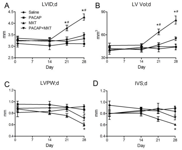
Figure 3.
Serial echocardiographic measures of left ventricular structure. (A–D) Treatment with MXT significantly increased LVID;d and LV Vol;d while significantly decreasing LVPW;d and IVS;d. PACPA reversed the increases in VLID;d and LV Vol;d and attenuated the decreases in IVS;d (A, B, D), but not the decrease in LVPW;d (C). LV; left ventricle, LVID;d, LV diameter during diastole, LV Vol;d, LV volume during diastole, LVPW;d, LV posterior wall thickness during diastole, IVS;d, intraventricular septal thickness during diastole. n = 7 mice per group, except for the MXT group (n = 5–7). * p < 0.05 versus the saline group. # p < 0.05 MXT group versus the PACAP+MXT group.

