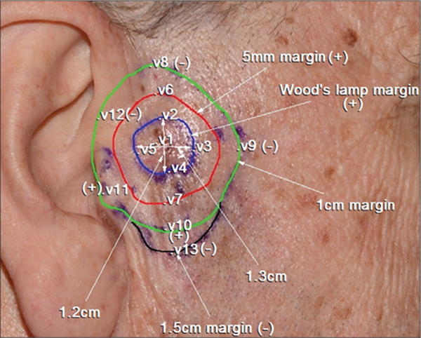
Figure 2.

Confocal mapping of lentigo maligna melanoma on the right preauricular area. The inner blue line demarcates Wood lamp margins. The red line shows the 5-mm surgical margin, which was positive throughout. The green line shows the 10-mm surgical margin, which showed positive reflectance confocal microscopy findings (dendritic atypical cells invading hair follicles, junctional thickening, and nonedged papillae) suggestive of subclinical lentigo maligna at the area close to the tragus (v11) and at the 6-o’clock position (v10). The black line indicates the 15-mm margin where disease was not detected (v13). The lesion was removed guided by this confocal mapping with clear margins. V indicates sites where stacks of images were taken in the vertical direction.
