Abstract
Background
The aim of this study was to evaluate the chronic systemic and local toxicity of a copper intrauterine device in a rat model.
Material/Methods
Cu-IUD fragments were inserted into the uterine horns of female Wistar rats for 26 weeks (equivalent 15.42–17.33 years in humans). We tested copper at 20, 40, and 60 times the clinical dosages to study copper toxicity under severe conditions. Classic chronic systemic toxicity evaluations, including body weight, hematology, organ coefficient, and tissue reactions, were performed.
Results
The body weight of the rats increased during the 26 weeks, which showed that the Cu-IUD implant had no effect on animal growth. Histological examinations and organ coefficients of the uterus and other major organs indicated no obvious systemic or local impairment. Blood tests confirmed that the hepatic and renal functions remained normal.
Conclusions
These results obtained at different dosages and long-term implantation provide solid data confirming the safety of long-term use of Cu-IUDs. However, the elevated leucocyte levels found in this study warrant further investigation.
MeSH Keywords: Intrauterine Devices, Copper; Rats, Wistar; Toxicity Tests, Acute
Background
The copper intrauterine device (Cu-IUD) was first marketed in the early 1970s. It is widely used due to its great efficacy, low cost, and long-lasting action [1]. Women living in China constitute about two-thirds of global users. Cu-IUDs do not cause hormone-related adverse effects such as weight gain, altered libido, or mood changes [2]. Generally, Cu-IUDs are made of Cu wires/tubes around T- or U-shaped flexible polymer material. Up to 60% of users choose, for various reasons, to have their IUDs removed within 5 years after insertion [3].
Sustained release of the corrosion products of Cu ions from a Cu-IUD is quite efficient for contraception through release of uterine inflammatory products – leukocytes and prostaglandins – by the endometrium in response to the inserted Cu-IUD. They also lead to changes in the composition of cells and uterine fluid in tissue, which reduces not only the viability of sperms, but also the receptivity of endometrium to implantation of embryos [4,5]. Copper is an essential trace element for living organisms; however, the tissue surrounding the Cu-IUD is exposed to toxic Cu ion levels. Cu-IUDs also induce adverse effects such as pelvic inflammatory disease, bleeding, and pain [6]. Cu ion toxicity has drawn much attention recently, especially regarding the chronic systemic exposure of the rest of the body [7,8]. Once the Cu ions uptake across a threshold limit, the cells become apoptotic and die [9,10]. Previous studies have focused on copper concentration in serum or in uterine cavity irrigation fluid. A study at the National University of La Platain, Argentina [8] demonstrated that Cu ions released from an inserted Cu-IUD have cytotoxic and genotoxic effects on mammalian cells in vitro. Reduction of mitochondrial activity was observed when the concentration Cu ions was more than 7.42 mg/L, and a significant decrease in cell viability was observed at 10.85 mg/L. The genital tract was exposed to 25–80 μg/day of Cu ions [5], resulting in uterine tissue damage. Furthermore, the sustained high concentration of Cu ions released can lead to accumulation of Cu, and also gives rise to chronic systemic exposure [11]. Most importantly, Cu ions released from Cu-IUDs produce cytotoxicity to the cells, not only in the implant site, but also in remote tissues such as liver, kidney, spleen, and lungs. There are surprisingly little data on the potential toxicity of Cu-IUDs because of the difficulty collecting specimens from humans, and these reports do not reveal the effect of copper at the organ level. In general, data on the potential systemic toxicity of Cu-IUDs are sparse, and more information is needed. The safety evaluation of Cu-IUDs is also expected to help its acceptance worldwide. Thus, the goal of our study was to evaluate the chronic systemic and local toxicity of the copper intrauterine device in different concentrations of Cu ions released, as well as to demonstrate the effect of copper at different organ levels.
In this study, an in situ rat model, which we previously described [12], was used to investigate the chronic systemic toxicity of the Cu-IUD. Uterine implantation was performed rather than ectopic implantation or liquid extraction so as to mimic the clinical application. According to ISO 10993.11, 3 dosages were used [13]. The implantation lasted for 26 weeks, corresponding to 15.42–17.33 years in humans [14], which is longer than the normal service life of the device. Implantation in situ at sufficiently high dosages and a sufficiently long duration thus ensured accurate evaluation of the toxicity of the Cu-IUD.
Material and Methods
Animals
A total of 120 healthy female Wistar rats aged 8 weeks and weighing 200–250 g were used in this study. The animals were supplied by the Laboratory for Animal Breeding and Supply of the Chinese National Institutes for Food and Drug Control (CNIFDC). The experimental protocols were reviewed and approved by the Experimental Animal Management Committee of CNIFDC in compliance with Chinese government regulations. Prior to use, the animals were conditioned for at least 7 days at the CNIFDC animal breeding facility in temperature- and humidity-controlled animal quarters with a 12-h light/dark cycle. Standard diet and water were provided ad libitum. The body weight of each rat was recorded weekly during the initial 4 weeks and then every 4 weeks up to 26 weeks after the procedure. At the end of the pre-scheduled 26 weeks, the rats were sacrificed. The weights of the brain, heart, liver, spleen, lungs, kidneys, and adrenal glands of each group rats were measured to calculate the organ coefficients.
Cu-IUD samples
Copper sleeves obtained from TCu220Cs (Tianjing Medical Equipment, Tianjing, China) were used in this study. The surface area of each copper implant was 37.71 mm2. A titanium alloy rod that was physically similar to the Cu-IUD specimen (CNIFDC Standard reference, length: 5.0 mm, diameter: 2.0 mm) was used as the control.
Operations
The animals were randomly divided into 6 groups with 20 animals in each group: group 1: Blank, no operation; group 2: Sham operation, no implant; group 3: Implantation of titanium alloy; group 4: Low dosage, implantation of one TCu220C segment; group 5: Medium dosage, implantation of 2 TCu220C segments; group 6: High dosage, implantation of 3 TCu220C segments.
Groups 1, 2, and 3 served as the controls, while groups 4, 5, and 6 were designated as the experimental groups. The copper dosage in one TCu220C segment implanted in a 250-g rat was estimated as being 20 times that of the TCu380A used in a 50-kg woman; thus, the amount of copper in the low-, medium-, and high-dosage groups was 20, 40, and 60 times greater, respectively, than that used for contraception in women.
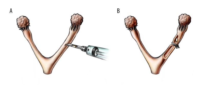
All implants were autoclaved before being placed. After shaving and disinfection, a small incision was made in the middle of the abdomen of the rat under pentobarbital anaesthesia and aseptic conditions. The implant was introduced into 1 uterine horn by means of an injector. To ensure that the uterine horn was patent and to maintain the implant in the appropriate position, a knot with a 4/0 silk suture was loosely made distal to the implant (Figure 1) prior to closing the incision and returning the rats to their housing.
Figure 1.
Insertion of the TCu220C copper sleeves. (A) After an incision was made, the IUD fragment was introduced into 1 uterine horn by means of an injector. (B) Then, a thread was loosely tied over the uterine horn, distally to the IUD, to prevent it from moving while keeping the horn open.
Hematology tests
At the end of the pre-scheduled 26 weeks and prior to sacrifice, blood samples were collected from each group for serum and blood biochemistry analyses, including routine blood tests, blood coagulation time, and blood biochemistry.
Histo-pathology
The rats were then sacrificed and dissected. The brain, heart, liver, spleen, lungs, kidneys, and adrenal glands were weighed prior to macro and histological examinations. The organs were then fixed in buffered formalin, embedded in paraffin wax, sectioned transversely in 2-mm steps at 4 μm, and finally stained with hematoxylin and eosin (HE). The specimens were observed under an optical microscope (Leica Microsystems, GMS GmbH, DM6000 B, Germany).
Statistical analysis
Significance testing was carried out using SPSS. The difference in animal body weight and organ coefficients among the groups was compared using the t test and analysis of variance. Significance was assumed at p<0.05.
Results
Changes in rat body weight
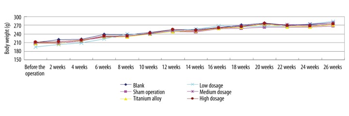
Figures 2 and 3 showed the change in body weight observed at the aforementioned intervals, from immediately before the operation until 26 weeks thereafter. Figure 2 indicates that the body weight of the rats in each group increased normally, with no significant differences among groups from 4 weeks to the end of the experiment. However, in groups 2–6, body weight dropped in the first week, and then slowly recovered in the first month (Figure 3). The body weight of rats in groups 2–6 was significantly different from weights in group 1 during that period. This phenomenon was interpreted as a normal reaction to surgery because group 2 showed the same tendency.
Figure 2.
Change in body weight from baseline to 26 weeks post-intervention. The body weight of the rats in each group increased normally.
Figure 3.
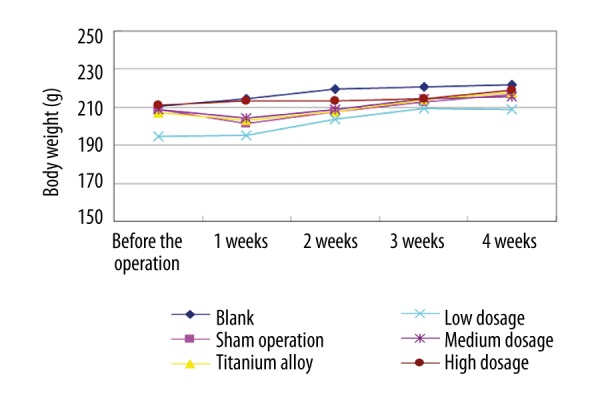
Change in body weight during the first 4 weeks post-intervention. Weight loss was observed shortly after the procedure in the surgical groups, but it gradually increased thereafter. After 2 to 3 weeks, the rats had regained their weight completely.
Organ coefficient
Table 1 presents the organ coefficients of all groups at 26 weeks post-operation. It indicated that none of the data pertaining to the experimental groups significantly differed from that of group 1, meaning that they were within normal range. No malformations or changes in morphology or color were observed in any organ.
Table 1.
Organ coefficient in the 26th week post-operation.
| Group 1 | Group 2 | Group 3 | Group 4 | Group 5 | Group 6 | |
|---|---|---|---|---|---|---|
| Brain | 0.584±0.073 | 0.620±0.048 | 0.585±0.071 | 0.599±0.054 | 0.604±0.029 | 0.611±0.043 |
| Heart | 0.360±0.030 | 0.360±0.033 | 0.316±0.024 | 0.330±0.024 | 0.345±0.030 | 0.341±0.035 |
| Liver | 2.681±0.172 | 2.762±0.183 | 2.612±0.263 | 2.865±0.181 | 2.674±0.175 | 2.689±0.229 |
| Spleen | 0.177±0.017 | 0.184±0.015 | 0.171±0.016 | 0.269±0.433 | 0.184±0.028 | 0.183±0.018 |
| Lung | 0.397±0.038 | 0.412±0.059 | 0.383±0.051 | 0.372±0.049 | 0.392±0.043 | 0.412±0.047 |
| Kidney | 0.649±0.026 | 0.673±0.045 | 0.650±0.049 | 0.651±0.047 | 0.642±0.023 | 0.654±0.033 |
| Adrenal | 0.016±0.004 | 0.016±0.002 | 0.018±0.003 | 0.018±0.003 | 0.018±0.004 | 0.018±0.004 |
Group 1 – Blank; Group 2 – Sham operation; Group 3 – Titanium alloy; Group 4 – Low dosage; Group 5 – Middle dosage; Group 6 – High dosage. Measurements are given as mean ±SD. In the 26th week after operation, the organ coefficients of all the groups were within the normal range.
Hematology tests
Tables 2–4 summarize the results of blood analyses, including blood coagulation time, routine blood tests, and blood biochemistry. The data from each group were within normal range, except for white blood cell (WBC) counts, which were higher in groups 4, 5, and 6 compared to the controls.
Table 2.
Blood coagulation time in the 26th week post-operation.
| Group 1 | Group 2 | Group 3 | Group 4 | Group 5 | Group 6 | |
|---|---|---|---|---|---|---|
| Blood coagulation time (s) | 1.27±0.45 | 1.35±0.52 | 1.36±0.51 | 1.29±0.52 | 1.27±0.42 | 1.19±0.28 |
Group 1 – Blank; Group 2 – Sham operation; Group 3 – Titanium alloy; Group 4 – Low dosage; Group 5 – Middle dosage; Group 6 – High dosage. Measurements are given as mean ±SD. In the 26th week after operation, the blood coagulation times of all the groups were within the normal range.
Table 3.
Blood routine examination in the 26th week post-operation.
| Group 1 | Group 2 | Group 3 | Group 4 | Group 5 | Group 6 | |
|---|---|---|---|---|---|---|
| WBC (109/L) | 10.13±1.35 | 7.51±2.02 | 10.67 ±3.90 | 13.50±3.37* | 13.56±3.21* | 13.20±3.34* |
| RBC (1012/L) | 7.63±0.76 | 7.76 ±0.54 | 7.84±0.67 | 7.21±0.61 | 7.97±0.75 | 7.61±+0.47 |
| HGB (g/L) | 142.80±11.86 | 150.70±7.64 | 149.20±11.45 | 145.85±8.28 | 149.75±14.00 | 143.05±8.73 |
| HCT (%) | 40.58±2.81 | 42.17±2.54 | 41.49±3.18 | 41.16±3.26 | 42.25±3.80 | 40.30±2.59 |
| MCV (fL) | 53.34±2.34 | 54.40±2.07 | 53.01±1.80 | 57.16±2.52 | 53.08±1.43 | 52.97±1.94 |
| MCH (pg) | 18.76±0.63 | 19.45±0.77 | 19.06±0.75 | 20.30±0.92 | 18.82±0.58 | 18.81±0.52 |
| MCHC (g/L) | 351.60±6.29 | 357.75±8.81 | 359.65±6.82 | 355.15±15.39 | 354.40±4.62 | 355.20±9.37 |
| RDW (%) | 11.68±0.20 | 11.07±0.49 | 11.30±0.50 | 12.15±0.83 | 11.38±0.39 | 11.42±0.46 |
| PLT (109/L) | 510.70±57.27 | 510.35±88.28 | 517.35±57.38 | 467.10±67.01 | 499.20±76.85 | 458.55±67.27 |
| PCT (%) | 0.50±0.06 | 0.51± 0.09 | 0.51±0.06 | 0.44±0.07 | 0.49±0.09 | 0.45 ±0.08 |
| MPV (fL) | 9.88±0.34 | 10.14±0.38 | 9.88±0.29 | 9.51±0.53 | 9.96±0.42 | 9.94±0.60 |
| PDW (%) | 13.02±0.56 | 12.95±0.45 | 12.83±0.28 | 12.92±0.61 | 12.92±0.38 | 12.85±0.66 |
| LYM (109/L) | 7.01±1.10 | 5.95±1.89 | 8.07±3.13 | 9.74±2.83 | 9.78±2.36 | 9.12±2.92 |
| MID (109/L) | 0.82±0.24 | 0.37±0.12 | 0.71±0.37 | 1.57±0.61 | 0.97±0.39 | 1.02±0.32 |
| GRN (109/L) | 2.30±0.44 | 1.20±0.56 | 1.90±0.88 | 1.93±1.18 | 2.81±1.45 | 2.85±1.23 |
| LYM (%) | 69.19±4.50 | 78.63±6.21 | 75.28±7.77 | 73.70±5.51 | 72.66±9.28 | 69.75±8.32 |
| MID (%) | 8.01±1.97 | 4.56±0.76 | 6.25±1.79 | 11.79±3.92 | 7.02±1.75 | 7.89±1.95 |
| GRN (%) | 22.80±3.26 | 16.82±5.91 | 18.47±7.17 | 14.52±5.78 | 20.33±8.89 | 22.36±7.85 |
Group 1 – Blank; Group 2 – Sham operation; Group 3 – Titanium alloy; Group 4 – Low dosage; Group 5 – Middle dosage; Group 6 – High dosage. Measurements are given as mean ±SD.
P<0.05.
In the 26th week after operation, the blood routines of all the groups were in the normal range, except the WBC in group 4, 5, 6. WBC – white blood cell; RBC – red blood cell; HGB – hemoglobin; HCT – haematocrit; MCV – mean corpuscular volume; MCH – mean corpuscular hemoglobin; MCHC – mean corpuscular hemoglobin concentration, RDW – red cell distribution width; PLT – platelet; PCT – plateletocrit; MPV – mean platelet volume; PDW – platelet distribution width; LYM – lymphocyte; GRN – gigantocellular reticular nucleus.
Table 4.
Blood biochemistry in the 26th week post-operation.
| Group 1 | Group 2 | Group 3 | Group 4 | Group 5 | Group 6 | |
|---|---|---|---|---|---|---|
| ALT (U/L) | 42.90±7.46 | 43.25±7.97 | 38.35±7.09 | 53.40±15.54 | 41.70±9.23 | 35.00±7.77 |
| AST (U/L) | 175.10±14.67 | 164.55±36.32 | 161.00±32.39 | 108.00±36.71 | 157.05±16.63 | 162.05±22.75 |
| TP (g/L) | 78.00±4.22 | 74.82±6.08 | 75.26±3.79 | 64.07±9.26 | 74.65±5.80 | 75.30±4.21 |
| ALB (g/L) | 34.42±1.30 | 34.19±1.99 | 33.81±1.13 | 30.78±2.93 | 33.78±1.71 | 34.54±1.82 |
| BIL (μmol/L) | 0.49±0.34 | 0.60±0.40 | 0.61±0.33 | 0.78±0.36 | 0.69±0.37 | 0.79±0.42 |
| ALP (U/L) | 74.40±6.06 | 86.85±12.34 | 83.70±10.85 | 85.25±12.20 | 88.50±14.45 | 91.75±14.20 |
| GLU (mmol/L) | 4.08±0.58 | 4.27±0.62 | 4.32±0.48 | 6.22±1.06 | 4.65±0.59 | 4.57±0.88 |
| BUN (mmol/L) | 8.25±1.45 | 6.75±0.83 | 7.51±1.20 | 6.50±1.05 | 7.85±1.20 | 7.99±1.12 |
| CREz (μmol/L) | 56.03±4.02 | 55.64±3.40 | 55.80±5.22 | 59.89±8.13 | 56.93±3.77 | 58.15±6.36 |
| CHOz (mmol/L) | 1.85±0.29 | 1.71±0.30 | 1.83±0.22 | 1.12±0.12 | 1.84±0.28 | 1.65±0.25 |
| A/G | 0.81±0.03 | 0.85±0.07 | 0.83±0.05 | 0.96±0.13 | 0.83±0.07 | 0.85±0.05 |
Group 1 – Blank; Group 2 – Sham operation; Group 3 – Titanium alloy; Group 4 – Low dosage; Group 5 – Middle dosage; Group 6 – High dosage. Measurements are given as mean ±SD. In the 26th week after operation, the blood biochemistries of all the groups were in the normal range. ALT – alanine transarninase; AST – aspartate aminotransferase; TP – total protein; ALB– albumin; BIL – bilirubin; ALP – alkaline phosphatase; GLU – glutamic acid; BUN – blood urea nitrogen; CREz – creatinine; CHOz – cholesterol; A/G – albumin/globin.
Histological and pathological examinations
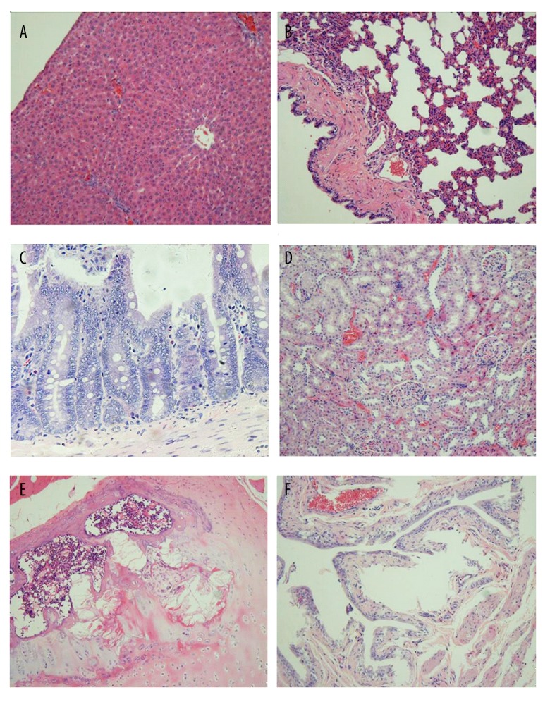
Figure 4 shows the representative light microscopic photographs of the histological slides of the liver, lungs, intestines, kidneys, and bone, as well as that of the oviduct, in group 6 (high dosage). Following intensive comparisons of the experimental groups with group 1, no structural impairment was observed in these organs.
Figure 4.
Histopathology of some major organs in group 6 (high-dosage group) 26 weeks after insertion of the TCu220C copper sleeves. (A) liver; (B) lung; (C) intestine; (D) kidney; (E) bone; (F) oviduct. No structural anomalies were observed (×100, hematoxylin-eosin stain).
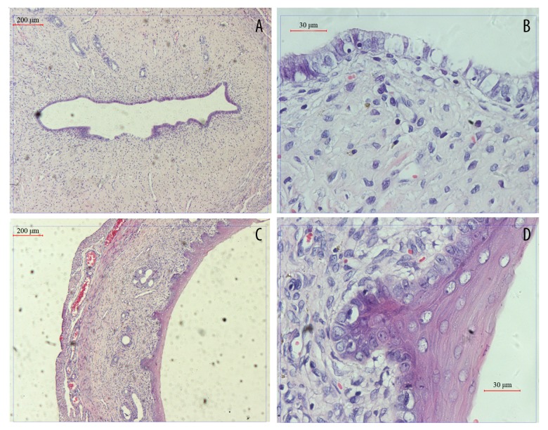
Figure 5 shows the pathological reactions of the uterine tissue after 3 segments of TCu220C (high dosage) had been present in the uterine horn for 26 weeks. The main pathological reaction of the uterus was the chronic inflammatory response. Macrophage- and lymphocyte-based chronic inflammation reaction was observed, as well as the squamous metaplasia phenomenon of the uterine endothelial cells, which more closely resembles the phenomena observed in a clinical setting. No necrosis or atypia of the endometrial cells was seen. There was no malignant or precancerous lesion in any of the endometrium cells.
Figure 5.
Histopathology of the endometrium in groups 1 (blank group) and 6 (high-dosage group) at 26 weeks. (A, B): Blank (A: ×50; B: ×400); (C, D): 3 TCu220C copper sleeves in situ for 26 weeks (C: ×50; D: ×400). The presence of these 3 fragments of TCu220C for 26 weeks elicited a macrophage- and lymphocyte-based chronic inflammatory reaction and squamous metaplasia of the endometrial cells. Neither necrosis nor atypia was observed in the endometrium.
Discussion
Findings and interpretation
Despite its extensive use worldwide, little is known about the toxicity, if any, of Cu-IUDs. The cytotoxicity of copper can be significant [3]. It stimulates a pronounced inflammatory reaction or foreign-body response in the uterus, which destroys sperm, impedes ovum development, and prevents fertilization and implantation.
While the copper ion plays an essential role in a variety of biological processes, it can become toxic at high concentration [15,16]. Exposure of cells and tissues to excessive amounts of copper can result in acute damage to the cell membrane and a leakage of internal enzymes, such as lysosomal enzymes, which lead to a loss of cell integrity and ultimately to cell death [17]. Furthermore, copper is capable of displacing other essential metal ions such as calcium and magnesium [18]. Moreover, in theory as well as in the laboratory, as an oxidizable active metal, its ions and complexes may generate free hydroxyl radicals and reactive oxygen species which in turn may damage biomolecules, including unsaturated lipids and DNA, resulting in DNA strand breaks and DNA base modifications [19]. The question raised is whether this is likely to occur in vivo, and if so, under which conditions and whether it can be tolerated by the host, particularly long-term.
Logically, after entering the circulation, copper arrives at other organs, including the liver, kidneys, brain, and bones. Copper accumulation is progressive, thus large amounts of copper could be detected in these tissues; copper concentration in blood, on the other hand, is relatively less important, which may explain why no difference was reported in the urinary copper excretions [20,21]. Copper toxicity primarily affects the liver, which is the first deposition site once copper has entered the blood. Toxicity is typically manifested by the development of liver cirrhosis, with episodes of hemolysis. The tissue damage may eventually lead to hepatic necrosis, vascular collapse, coma, and even death. Again, the question raised is just how toxic the Cu-IUD is and whether the released copper can damage target organs.
To answer these 2 important questions, i.e., chronic toxicity at both cellular and organ levels, we sought to investigate the chronic systemic toxicity, if any, of the Cu-IUD by using rats as the animal model. As regards the comparative lifespan of humans and rats, 26 weeks for rats corresponds to 15.42–17.33 years for humans, the latter length of time being longer than the duration of approved use of most Cu-IUDs [22]. Furthermore, the lowest copper dosage used in our study was much higher than that resorted to in utero for contraceptive purposes in women. Therefore, considering the long period of time during which the fragments of TCu220C remained in the uterine horns of the rats and the large amount of copper on the aforementioned foreign bodies, the current experimental conditions, which to the best of our knowledge have not been reported before, are much more severe than those characterizing clinical use, making the observed absence of toxic effects more reliable.
Body weight, organ coefficient, and blood biochemistry data are commonly used to reflect the physiologic and pathologic changes in chronic toxicity studies because of their sensitivity, effectiveness, and economy. Body weight indicates the effects, if any, on growth and development. Organ coefficient is used to signal impairment in an organ; it rises or falls depending on the level of toxicity. An increase may result from hyperemia, edema, or hypertrophy, while a decrease may signify organ atrophy or degeneration. Finally, organ function and immune reactions can be ascertained from blood tests.
In our study, following placement of 1, 2 or 3 TCu220C copper sleeves in the uterine horn for 26 weeks, the body weight of the rats increased normally in each group, suggesting that neither the intervention to insert the IUD fragments nor the presence of copper in the uterus had any toxic effect on the animals’ growth and development, even though the quantity of copper was far greater than that used for contraceptive purposes in women. During the first month following implantation, the body weight of the rats that underwent either the sham operation or the placement of low-dosage titanium alloy increased slower than that of group 1 (animals not operated on). However, no significant difference in body weight was found between the medium dosage, high dosage, and blank groups. This indicates that neither the presence of Cu-IUD fragments nor the surgical procedure to insert these had an impact of any magnitude on animal growth. Moreover, neither an abdominal nor a uterine incision is required to fit an IUD in the clinical setting, meaning that the difference in body weight observed in this study is irrelevant in the context of contraception in humans.
Certain organ coefficients significantly differed between the groups. The heart coefficients of the medium- and high-dosage groups differed from that of the titanium alloy group, but were the same as those of the blank and sham operation groups, suggesting that even the highest dosage of copper had no toxic effect on the heart. The different heart coefficient in the titanium alloy group may be related to the material itself. The liver coefficients of the low-, medium-, and high-dosage groups, though not identical, did not differ significantly from those of the controls, including the blank, sham operation, and titanium alloy groups. Based on the organ coefficients, this study confirms that the Cu-IUD lacks toxicity for any of the vital organs tested in this experiment.
Furthermore, the local reaction of the endometrium to the Cu-IUD in the late stage is a chronic inflammatory reaction which closely resembles clinical observations. The absence of necrosis or mutation of endometrial cells in the high-dosage conditions demonstrates that the toxicity of the Cu-IUD is well tolerated by the uterus.
Strengths and weaknesses of the study
We performed the experiments in rats instead of humans because of the difficulties in human studies. Although animal experimentation has its limits due to biological differences, the results of this study still suggest that the long-term use of Cu-IUDs is safe.
Differences in results and conclusions
Our work demonstrates for the first time that the cytotoxicity of long-term high-dosage Cu-IUD fragments in the uterine horn is well tolerated by female Wistar rats. Neither necrosis nor atypia was observed in the endometrium. Growth of major organ functions was unaffected. Thus, Cu-IUD displays neither obvious systemic toxicity nor local toxicity. Considering the severe conditions in this experiment, the clinical use of Cu-IUD is deemed safe. However, the increased leucocytosis warrants further investigation to ascertain the long-term safety of the Cu-IUD in clinical use [23,24].
Data in the literature are contradictory. Some studies showed that the copper concentration in the blood increased in Cu-IUD users, while others revealed no statistically significant difference in the serum levels of copper before and after insertion of the device. Furthermore, no difference was found between the 24-h urinary copper excretion [25,26] of a control group and that of a group of Cu-IUD users.
Relevance of the findings: implications for clinicians and policymakers
Several decades of widespread use of Cu-IUD by women may be assumed to have shown that these most efficacious contraceptives lack any significant toxicity. Our animal experiments confirmed this. However, because we found increased numbers of leucocytes in our study, the long-term use of Cu-IUD should be monitored for inflammation.
Unanswered questions and future research
Blood biochemistry tests and histological examinations of the major organs and uterus provided further proof of the safety of the Cu-IUD. The observed increase in leucocytes in the Cu-IUD groups was indeed due to the inflammatory reaction of the body to the foreign material and copper. While an aseptic inflammatory process is an essential feature of the contraceptive activity of the Cu-IUD, we believe that this observation must be confirmed in human subjects, and further investigation is needed to assess the clinical significance of these leucocyte counts in terms of long-term health of Cu-IUD users [27].
Conclusions
Our work demonstrates for the first time that the cytotoxicity of long-term high-dosage Cu-IUD fragments in the uterine horn is well tolerated by female Wistar rats. Neither necrosis nor atypia was observed in the endometrium. Most importantly, anatomical examinations of vital organs were conducted, and also found that major organ functions were unaffected. Thus, the Cu-IUD displays neither obvious systemic toxicity nor local toxicity. Considering the severe conditions in this experiment, the clinical use of Cu-IUD is deemed safe. However, the elevation of the leucocytosis warrants further investigation to ascertain the long-term safety of the Cu-IUD in clinical use.
Footnotes
Source of support: Departmental sources
References
- 1.Arrowsmith ME, Aicken CR, Saxena S, Majeed A. Strategies for improving the acceptability and acceptance of the copper intrauterine device. Cochrane Database Syst Rev. 2012;(3):CD008896. doi: 10.1002/14651858.CD008896.pub2. [DOI] [PubMed] [Google Scholar]
- 2.Andersson K, Odlind V, Rybo G. Levonorgestrel-releasing and copper-releasing (Nova T) IUDs during five years of use: A randomized comparative trial. Contraception. 1994;49(1):56–72. doi: 10.1016/0010-7824(94)90109-0. [DOI] [PubMed] [Google Scholar]
- 3.Xu XX, Ding MH, Zhang JX, et al. A novel copper/polydimethiylsiloxane nanocomposite for copper-containing intrauterine contraceptive devices. J Biomed Mater Res B Appl Biomater. 2013;101(8):1428–36. doi: 10.1002/jbm.b.32962. [DOI] [PubMed] [Google Scholar]
- 4.Ortiz ME, Croxatto HB. Copper-T intrauterine device and levonorgestrel intrauterine system: biological bases of their mechanism of action. Contraception. 2007;75(6 Suppl):29. doi: 10.1016/j.contraception.2007.01.020. [DOI] [PubMed] [Google Scholar]
- 5.Arancibia V, Peña C, Allen HE, Lagos G, et al. Characterization of copper in uterine fluids of patients who use the copper T-380A intrauterine device. Clin Chim Acta. 2003;332(1–2):69–78. doi: 10.1016/s0009-8981(03)00124-4. [DOI] [PubMed] [Google Scholar]
- 6.Hubacher D, Chen PL, Park S. Side effects from the copper IUD: Do they decrease over time? Contraception. 2009;79(5):356–62. doi: 10.1016/j.contraception.2008.11.012. [DOI] [PMC free article] [PubMed] [Google Scholar]
- 7.Cao B, Xi T, Zheng Y. Release behavior of cupric ions for TCu380A and TCu220C IUDs. Biomed Mater. 2008;3(4):044114. doi: 10.1088/1748-6041/3/4/044114. [DOI] [PubMed] [Google Scholar]
- 8.Grillo CA, Reigosa MA, de Mele MA. Does over-exposure to copper ions released from metallic copper induce cytotoxic and genotoxic effects on mammalian cells? Contraception. 2010;81(4):343–49. doi: 10.1016/j.contraception.2009.12.003. [DOI] [PubMed] [Google Scholar]
- 9.Grillo CA, Reigosa MA, Lorenzo de Mele MF. Effects of copper ions released from metallic copper on CHO-K1 cells. Mutat Res. 2009;672(1):45–50. doi: 10.1016/j.mrgentox.2008.09.012. [DOI] [PubMed] [Google Scholar]
- 10.Hayashi M, Fuse S, Endoh D, et al. Accumulation of copper induces DNA strand breaks in brain cells of Long-Evans Cinnamon (LEC) rats, an animal model for human Wilson disease. Exp Anim. 2006;55(5):419–26. doi: 10.1538/expanim.55.419. [DOI] [PubMed] [Google Scholar]
- 11.De la Cruz D, Cruz A, Arteaga M, et al. Blood copper levels in Mexican users of the T380A IUD. Contraception. 2005;72(2):122–25. doi: 10.1016/j.contraception.2005.02.009. [DOI] [PubMed] [Google Scholar]
- 12.Hua Z, Zhaoxu W. [Local reaction of endometrium after the implantation of Cu-intrauterine device into the urterine horn: Early-and late-stage evaluation]. Chinese Journal of Tissue Engineering Research. 2012;16(8):1407–12. [in Chinese] [Google Scholar]
- 13.International Organization for Standardization, 2006 Biological evaluation of medical devices, in Part 11: Tests for systemic toxicity2006: Geneva
- 14.Andreollo NA, Santos EF, Araújo MR, Lopes LR, et al. Rat’s age versus human’s age: What is the relationship? Arq Bras Cir Dig. 2012;25(1):49–51. doi: 10.1590/s0102-67202012000100011. [DOI] [PubMed] [Google Scholar]
- 15.Wildemeersch D, Goldstuck ND, Hasskamp T. Intrauterine systems: A frameless future? Expert Opin Drug Deliv. 2016;13(6):911–18. doi: 10.1517/17425247.2016.1162153. [DOI] [PubMed] [Google Scholar]
- 16.Whitaker AK, Johnson LM, Harwood B, et al. Adolescent and young adult women’s knowledge of and attitudes toward the intrauterine device. Contraception. 2008;78(3):211–17. doi: 10.1016/j.contraception.2008.04.119. [DOI] [PubMed] [Google Scholar]
- 17.Godfrey EM, Folger SG, Jeng G, et al. Treatment of bleeding irregularities in women with copper-containing IUDs: A systematic review. Contraception. 2013;87(5):549–66. doi: 10.1016/j.contraception.2012.09.006. [DOI] [PubMed] [Google Scholar]
- 18.Morris AW. Nephrotic syndrome: PCSK9: A target for hypercholesterolaemia in nephrotic syndrome. Nat Rev Nephrol. 2016;12(9):18. doi: 10.1038/nrneph.2016.111. [DOI] [PubMed] [Google Scholar]
- 19.Galanis A, Karapetsas A, Sandaltzopoulos R. Metal-induced carcinogenesis, oxidative stress and hypoxia signalling. Mutat Res. 2009;674(1–2):31–35. doi: 10.1016/j.mrgentox.2008.10.008. [DOI] [PubMed] [Google Scholar]
- 20.Haliloglu B, Celik A, Ilter E, et al. Comparison of uterine artery blood flow with levonorgestrel intrauterine system and copper intrauterine device. Contraception. 2011;83(6):578–81. doi: 10.1016/j.contraception.2010.09.001. [DOI] [PubMed] [Google Scholar]
- 21.Lowe RF, Prata N. Hemoglobin and serum ferritin levels in women using copper-releasing or levonorgestrel-releasing intrauterine devices: A systematic review. Contraception. 2013;87(4):486–96. doi: 10.1016/j.contraception.2012.09.025. [DOI] [PubMed] [Google Scholar]
- 22.Grimes DA, Lopez LM, Manion C, Schulz KF. Cochrane systematic reviews of IUD trials: Lessons learned. Contraception. 2007;75(6 Suppl):S55–59. doi: 10.1016/j.contraception.2006.12.004. [DOI] [PubMed] [Google Scholar]
- 23.Sonalkar S, Kapp N. Intrauterine device insertion in the postpartum period: A systematic review. Eur J Contracept Reprod Health Care. 2015;20(1):4–18. doi: 10.3109/13625187.2014.971454. [DOI] [PubMed] [Google Scholar]
- 24.Imani S, Moghaddam-Banaem L, Roudbar-Mohammadi S, Asghari-Jafarabadi M. Changes in copper and zinc serum levels in women wearing a copper TCu-380A intrauterine device. Eur J Contracept Reprod Health Care. 2014;19(1):45–50. doi: 10.3109/13625187.2013.856404. [DOI] [PubMed] [Google Scholar]
- 25.Cao B, Xi T, Zheng Y. Release behavior of cupric ions for TCu380A and TCu220C IUDs. Biomed Mater. 2008;3(4):044114. doi: 10.1088/1748-6041/3/4/044114. [DOI] [PubMed] [Google Scholar]
- 26.Shin DG, Kim TN, Lee W. Intrauterine device embedded into the bladder wall with stone formation: Laparoscopic removal is a minimally invasive alternative to open surgery. Int Urogynecol J. 2012;23(8):1129–31. doi: 10.1007/s00192-011-1632-8. [DOI] [PMC free article] [PubMed] [Google Scholar]
- 27.O’Brien PA, Kulier R, Helmerhorst FM, et al. Copper-containing, framed intrauterine devices for contraception: A systematic review of randomized controlled trials. Contraception. 2008;77(5):318–27. doi: 10.1016/j.contraception.2007.12.011. [DOI] [PubMed] [Google Scholar]


