G&H What is the difference between fine-needle aspiration and fine-needle biopsy?
RM The primary goal of fine-needle aspiration is to acquire individual cells as opposed to tissue, which is the aim of fine-needle biopsy. Cells may not necessarily provide the stroma, or the associated architecture, of the surrounding tissue. Thus, if the surrounding architecture is needed to make a diagnosis, a fine-needle biopsy, which typically uses a core biopsy needle, should provide that.
G&H What is the history of the development of fine-needle biopsy?
RM The original fine-needle biopsy needle (Quick-Core Biopsy Needle, Cook Medical) was an attempt at designing a Tru-Cut needle (Medline Industries) that could be utilized with echoendoscopes, and was introduced in the early 2000s. However, technical issues included challenges in deploying the spring-loaded tray or, when it did deploy, not always having the specimen be retained when the needle was pulled back. Additionally, a certain track length within the pancreas was needed in order to safely deploy the needle and avoid traversing the pancreatic duct, which can increase the risk of developing pancreatitis. Therefore, adoption was limited given the challenges in performing the procedure as well as the frequency at which adequate tissue could be obtained. In 2012, a nonspring-loaded core biopsy needle (ProCore, Cook Medical) was developed and was followed shortly by other needles specifically designed to acquire histology. These needles are now widely available in a variety of sizes, including 19-, 20-, 22-, and 25-gauge.
G&H What are the indications for endoscopic ultrasound–guided fine-needle aspiration or biopsy?
RM Endoscopic ultrasound is increasingly being used to identify and stage a wide variety of malignancies, such as endobronchial cancer, luminal gastrointestinal cancers (eg, esophagus, stomach, rectum), and cancer of the pancreas. It is also indicated for evaluating and diagnosing liver lesions and fluid in the abdomen, among other conditions. Fine-needle aspiration and fine-needle biopsy appear relatively equivalent for diagnosing pancreatic tumors, whereas adenopathy and subepithelial lesions in the gastrointestinal tract appear to achieve an increased diagnostic yield from undergoing fine-needle biopsy. In addition, fine-needle biopsy allows the endoscopist to receive information regarding tissue architecture, perform immunohistochemistry of lesions such as gastrointestinal stromal tumors, and detect autoimmune and chronic pancreatitis.
G&H How are these procedures performed?
RM Both endoscopic ultrasound–guided fine-needle aspiration and fine-needle biopsy are performed during an endoscopic ultrasound procedure using an echoendoscope; this device is slightly larger than a standard-sized upper endoscope. Echoendoscopes are equipped with ultrasound processors on the tip that allow for ultrasonic evaluation not just within the gastrointestinal tract but also across the luminal wall into adjacent structures. Subsequently, needles (either a fine-needle aspiration needle, in 19-, 22-, or 25-gauge sizes, or a special core biopsy needle, in 19-, 20-, 22-, or 25-gauge sizes) are passed under ultrasound guidance into the target lesion. Several specimens are then acquired and sent for analysis, which can be performed in the room or at the pathology laboratory.
G&H What are the benefits and limitations associated with these procedures?
RM The main benefit of both techniques is that they allow an endoscopist to acquire tissue via the patient’s natural orifices as opposed to a percutaneous or surgical biopsy. Patients are undergoing these procedures as part of an endoscopy that was often already needed, so additional radiologic or surgical procedures may be able to be avoided.
G&H Are there any patients in whom these procedures are contraindicated?
RM Patients who cannot safely undergo sedation should in general avoid these techniques. In addition, patients with significant disorders of blood coagulation or who are on medications that inhibit blood-clotting and that cannot be temporarily discontinued should not receive fine-needle aspiration or biopsy, as both procedures carry a small risk of bleeding. There have been reports of hemorrhage after endoscopic ultrasound–guided tissue acquisition in patients taking certain anticoagulation medications, so core needles used for biopsy in particular should be used carefully around major vessels to avoid possible injury.
G&H How does the diagnostic accuracy for malignancy compare between the 2 methods?
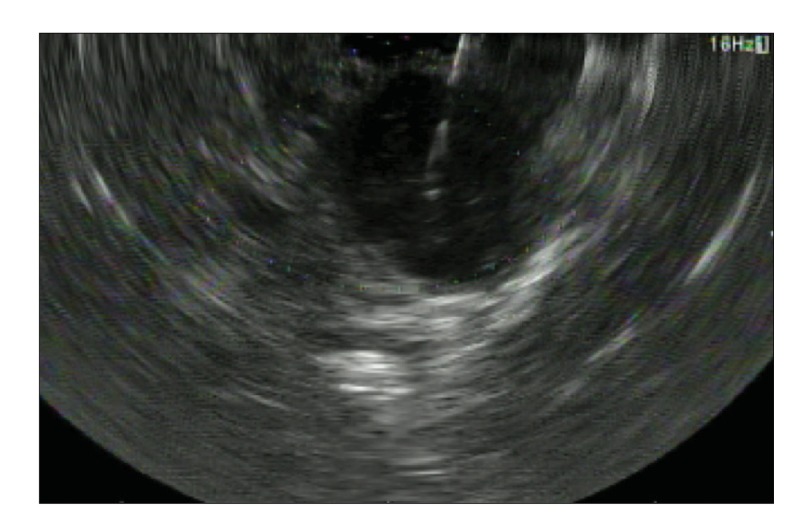
RM The diagnostic accuracy for malignancy of pancreatic lesions with fine-needle aspiration is excellent, in the order of approximately 90% or higher. A randomized, crossover trial comparing fine-needle aspiration vs fine-needle biopsy found that fine-needle biopsy was superior, but only because it achieved improved results in a subset of patients with nonpancreatic indications for tissue acquisition. Fine-needle biopsy had a better diagnostic accuracy when sampling lymph nodes (Figure), subepithelial lesions of the gastrointestinal tract, and other non-pancreatic lesions (eg, liver). Overall, the diagnostic accuracy for fine-needle aspiration and fine-needle biopsy is fairly comparable within the pancreas.
G&H Does the size or the type of the needle affect the diagnostic yield?
RM For fine-needle aspiration, it appears that smaller needles are better for pancreatic lesions. A meta-analysis showed that 25-gauge needles offer some advantage over 22- and 19-gauge needles in such settings. Additionally, 19-gauge needles are typically larger and stiffer, which make navigating these needles through angulated portions of the gastrointestinal tract more difficult. Because core needles are relatively new, there are not a lot of solid data as to which size needle is better for different indications; however, it appears that the 22-gauge is becoming the standard for nonhepatic indications.
G&H What is the number of passes required for diagnosis for each method?
RM Initial studies published in the early to mid-2000s suggested that 7 fine-needle aspiration passes would be necessary for solid pancreatic lesions, which remain the most common indication for endoscopic ultrasound–guided tissue acquisition. However, these studies found that the yield of acquiring adequate material for diagnosis on the first pass was as low as 14%. Due to a variety of advances, current data suggest that the first-pass diagnostic yield is approximately 60% to 80%, and the currently accepted number of passes needed to diagnose most solid pancreatic lesions is 4 to 5. It is believed these numbers are similar for fine-needle biopsy, but specific per pass data for fine-needle biopsy are lacking. Typically, 5 or fewer passes are needed for adequate tissue acquisition from within lymph nodes.
G&H What is the role of rapid onsite evaluation for diagnosis?
RM The answer to this question has evolved. The first fine-needle aspiration was performed in 1994, and in the 10 to 15 years that followed, rapid evaluation from an onsite cytopathologist provided valuable real-time feedback for detecting specimen adequacy and aided in fine-needle aspiration targeting within a lesion. Within the last few years, studies have suggested that, due to advancement in the 4 factors discussed above, the role of the cytopathologist seems to have diminished. A randomized, controlled trial showed that for pancreatic lesions, onsite cytopathology did not improve the diagnostic yield vs simply obtaining 7 passes (as this study was conducted before the most recent data recommending 4-5 passes). Furthermore, it appears that onsite evaluation does not provide a speed advantage, as the endosonographer often waits for the result to be provided from each pass before continuing on to the next pass, rather than just obtaining specimens sequentially without delay. Preliminary studies suggest that onsite evaluation is not beneficial in the setting of fine-needle biopsy as well; in one study, cytotechnologists evaluating biopsy specimens on slides underestimated the frequency of which adequate material was obtained. However, data for onsite evaluation of fine-needle biopsy–acquired specimens are limited.
G&H Have any studies compared the cost-effectiveness of the 2 methods?
RM Research on this topic is ongoing. There is some increase in cost for the core biopsy needles compared with the standard fine-needle aspiration needles, but a clear answer is not available, and detailed analyses on this issue are needed. Certainly, if the diagnostic yield for fine-needle biopsy is higher, the slight cost increment would be justified by avoiding the need for further tissue sampling procedures in nondiagnostic cases.
G&H What training is necessary to perform these procedures? How significant is the learning curve?
RM The training for endoscopic ultrasound is primarily visual, as it deals with lesion identification and characterization. Essential components and techniques in tissue acquisition include lesion identification and assessment, determining the optimal area to target, and understanding which type of needle and suction technique should be used. Tissue acquisition has a learning curve, not just for the endosonographers who acquire the tissue, but also for the technicians and nurses who prepare the specimen and for the cytopathologists who assess and interpret the acquired material.
G&H What are the priorities of research in this field?
RM One of the top priorities is the expansion of the field of endoscopic ultrasound–guided liver biopsy. Patients who require endoscopic procedures (eg, ERCP, upper endoscopy, endoscopic ultrasound) for the management of their biliary diseases or in the posttransplant setting can undergo a biopsy to assess liver tissue at the same time as the endoscopic procedure.
Of note, successful endoscopic ultrasound–guided tissue acquisition is a 4-part process involving (1) the appropriate choice of needle with regards to gauge and type (fine-needle aspiration vs fine-needle biopsy), (2) optimal endoscopic ultrasound tissue acquisition technique used by the endosonographer, (3) proper specimen preparation, and (4) appropriate training and experience of the cytopathologist responsible for interpreting the endoscopic ultrasound–acquired specimen.
Another priority is precision/personalized medicine. Increasingly, clinicians are interested in knowing whether they can use the information obtained from acquired tissue to optimize the chemotherapeutic agents used to treat an individual patient’s cancer. Research is ongoing in this area to determine whether fine-needle biopsy is needed or if the amount of cells acquired through endoscopic ultrasound–guided fine-needle aspiration may be enough. In addition, given advances in amplification techniques, it is uncertain whether tissue from the primary site is even needed or if tumor cells can be acquired from blood or saliva.
Figure.
A large, 3-cm peripancreatic lymph node was identified in a patient with esophageal adenocarcinoma and is shown undergoing endoscopic ultrasound–guided fine-needle biopsy with a 22-gauge needle. Pathology confirmed the presence of adenocarcinoma consistent with the patient’s primary esophageal lesion.
Biography

Suggested Reading
- Aadam AA, Wani S, Amick A, et al. A randomized controlled cross-over trial and cost analysis comparing endoscopic ultrasound fine needle aspiration and fine needle biopsy. Endosc Int Open. 2016;4(5):E497–E505. doi: 10.1055/s-0042-106958. [DOI] [PMC free article] [PubMed] [Google Scholar]
- Bang JY, Hawes R, Varadarajulu S. A meta-analysis comparing ProCore and standard fine-needle aspiration needles for endoscopic ultrasound-guided tissue acquisition. Endoscopy. 2016;48(4):339–349. doi: 10.1055/s-0034-1393354. [DOI] [PubMed] [Google Scholar]
- DiMaio CJ, Kolb JM, Benias PC, et al. Initial experience with a novel EUS-guided core biopsy needle (SharkCore): results of a large North American multicenter study. Endosc Int Open. 2016;4(9):E974–E979. doi: 10.1055/s-0042-112581. [DOI] [PMC free article] [PubMed] [Google Scholar]
- Khan MA, Grimm IS, Ali B, et al. A meta-analysis of endoscopic ultrasound-fine-needle aspiration compared to endoscopic ultrasound-fine-needle biopsy: diagnostic yield and the value of onsite cytopathological assessment. Endosc Int Open. 2017;5(5):E363–E375. doi: 10.1055/s-0043-101693. [DOI] [PMC free article] [PubMed] [Google Scholar]
- Lee YN, Moon JH, Kim HK, et al. Core biopsy needle versus standard aspiration needle for endoscopic ultrasound-guided sampling of solid pancreatic masses: a randomized parallel-group study. Endoscopy. 2014;46(12):1056–1062. doi: 10.1055/s-0034-1377558. [DOI] [PubMed] [Google Scholar]
- Mohamadnejad M, Mullady D, Early DS, et al. Increasing number of passes beyond 4 does not increase sensitivity of detection of pancreatic malignancy by endoscopic ultrasound-guided fine-needle aspiration. Clin Gastroenterol Hepatol. 2017;15(7):1071–1078.e2. doi: 10.1016/j.cgh.2016.12.018. [DOI] [PubMed] [Google Scholar]
- Wang J, Wu X, Yin P, et al. Comparing endoscopic ultrasound (EUS)-guided fine needle aspiration (FNA) versus fine needle biopsy (FNB) in the diagnosis of solid lesions: study protocol for a randomized controlled trial. Trials. 2016;17:198. doi: 10.1186/s13063-016-1316-2. [DOI] [PMC free article] [PubMed] [Google Scholar]
- Wani S, Muthusamy VR, Komanduri S. EUS-guided tissue acquisition: an evidence-based approach (with videos) Gastrointest Endosc. 2014;80(6):939–959.e7. doi: 10.1016/j.gie.2014.07.066. [DOI] [PubMed] [Google Scholar]


