

Abstract
The International Caries Detection and Assessment System (ICDAS) is a clinical scoring system which allows detection and assessment of caries activity. ICDAS was developed for use in clinical research, clinical practice and for epidemiological purposes. A recent review of caries detection criteria systems found that there were inconsistencies among the research criteria for caries measuring systems. There is a need to an uniform system which allows comparison of data collected in different researches. ICDAS allows detection of caries process at every stage and characterization of the caries activity status of lesion. Later, the criteria were modified and ICDAS II created. The aim of this review is to inform about the ICDAS II and make a comparison between ICDAS II criteria and other caries detection systems.
Keywords: Dental caries, detection, assessment, ICDAS II
Introduction
Description of ICDAS
ICDAS is a clinical scoring system that is used to detect and assess dental caries. It is generated to be used in dental education, clinical applications, researches and epidemiological studies (1, 2, 3, 4, 5). This scoring system can be used on curonal surfaces and root surfaces and can be applied for enamel caries, dentine caries, non-cavitated lesions (contrary to many systems) and cavitated lesions to detect and assess these lesions (2, 4, 6). The aim of planning ICDAS is to gain better quality information to make decisions about appropriate diagnosis, prognosis and clinical management of dental caries at both the individual and public health levels. Thus, ICDAS can provide a worksheet that can allow management of caries which is needed to achieve longterm healtful results (5, 7).
Development of ICDAS
A systematic review of all literatures on clinical caries detection found that there were inconsistencies in how the caries process was measured and among the research criteria for measuring dental caries. The future of research, practice, and education in cariology requires to eliminate these inconsistencies. Therefore, it was decided that there is a need to develop an uniform system for measuring the caries process. An integrated and uniform system that can be developed by combining the evidences recieved from all studies can provide comparability of the caries data obtained from different studies. So, the current situation of the disease can be assessed effectively and the most appropriate diagnosis, prognosis and treatment can be decided (8, 9, 10, 11). Moreover, important treatment options can be developed as a result of these accurate assessments (9, 11). As mentioned above, a systematic review of literatures found that there were inconsistencies among the research criteria. In addition, sytematic review of literatures showed that present systems can be applied only for cavitated lesions (12, 13). However, recording only cavitated lesions as an outcome measure is becoming outmoded (12, 14). When these lesions can be detected at an early stage, they can be controlled by preventive treatments. Thus, cost of treatment will decrease (13, 14, 15, 16, 17). New and integrated system should lead to broader information. So, caries can be detected at each stage, activity of them can be monitored and the effect of an material on the lesion can be determined clearly (11). As a result of all these necessities, there is a need to address the answers to the following questions. “What stage of the caries process should be measured?”, “what are the definitons for each selected stage?”, “what is the best clinical approach to detect each stage on different tooth surfaces?” and “what protocols of examiners’ training can provide the highest degree of examiner reliability?” These were the initial questions that initiated the discussion of the ICDAS (18, 19). Initial step of this process was the first convention that was done in Scotland in 2002. In this convention, problems of the current systems and properties that the new system should have were identified. A second session was done in Michigan to develop criteria of the integrated system that was identified in the first session. In the end of the session, ICDAS criteria (ICDAS I) was formed. These criteria are divided into two groups: caries detection criteria and caries activity criteria. The committee reported that while ICDAS caries detection criteria are ready to wider use, caries activity criteria are still part of an expanding research agenta. A third meeting was held in Indiana in 2003 to evaluate mentioned criteria and closely a forth convention was done in Denmark. Thus, the ICDAS II workshop was held in 2005 to share the progress in the ICDAS criteria and seek the input of a wider international expertise. The invitations were mailed to over 60 researchers and those who accepted the invitation convened to revise and agree on the ICDAS II version of the criteria. While caries detection criteria of ICDAS I was modified and accepted entirely, activity criteria was developed more. It’s reported that ICDAS activity criteria should be still part of an expanding research agenda (4, 19).
ICDAS Criteria
Since dental caries is a dynamic process, categorization of them is hard. The process is continuous and could be measured as stages representing minute loss of tooth structure that is currently not detectable using currrent technology available for in vivo use. Therefore; we rely on visual signs that reflect caries process relatively (18, 19). ICDAS measures the surface changes and potential histological depth of carious lesions by relying on surface characteristics (18, 19, 20). ICDAS criteria are divided into two categories: coronal primary caries and root caries. Caries detection coding and caries activity coding should be done separately. ICDAS II system have two digit coding for detection criteria of primary coronal caries. The first one is related to the restoration of teeth and has a coding that ranges from 0 to 9. This coding is presented in Table 1 (18, 19).
Table 1.
| 0 | Surface not restored or sealed | 5 | Stainless steel crown |
| 1 | Sealant, partial | 6 | Porcelain or gold or PFM crown or veneer |
| 2 | Sealant, full | 7 | Lost or broken restoration |
| 3 | Tooth colored restoration | 8 | Temporary restoration |
| 4 | Amalgam restoration | 9 | Used for the following conditions: |
| 96:Tooth surface cannot be examined | |||
| 97: Tooth missing because of caries | |||
| 98: Tooth missing for reasons other than caries | |||
| 99: Unerupted |
The second digit ranges from 0 to 6 that is used for coding the caries. There are minor variations between the visual signs associated with each code depending on a number of factors. Therefore; a detailed description of each of the codes is given under the following headings: Pits and fissures, smooth surface (mesial or distal), free smooth surfaces and caries associated with restorations and sealants (CARS). However, the basis of the codes is essentially the same that is presented in Table 2 (21). Smooth surfaces describe mesial and distal surfaces that have contact with adjacent teeth and requires visual inspection from the occlusal, buccal and lingual directions. Free smooth surfaces describe buccal and lingual surfaces and mesial and distal surfaces that have no adjacent teeth and requires direct examination of buccal, lingual, mesial and distal surfaces (18, 19). CARS (Caries Associated with Restorations and Sealants) describe caries adjacent to the restorations or sealants. The description of the new caries that occurs in restored teeth is so different in the literature (secondary caries, residual caries, recurrent caries). It’s recommended that the term of CARS should be used to prevent complication. Altough CARS is analogous with primary caries histologically, some characteristics of them can lead to diagnostic problems. Main problem is the difficulties in differentiation among restoration margin discrepancies, secondary caries and residual caries. If there is only margin discrepancies, it is also important to record them since these discrepancies show increased caries risk. However, it is important to have a treshold at which the deficiency is recorded as presen tor absent. Two categories could be recorded according to whether or not a ball-ended probe can be admitted into gap between tooth and restoration. If marginal discrepancies and caries are seen together, caries should be coded firstly (19, 22). Detailed coding of all mentioned headings is presented in Table 3. Figure 1 shows one example of each ICDAS II coding.
Table 2.
| 0 | Sound |
| 1 | First visual change in enamel |
| 2 | Distinct visual change in enamel |
| 3 | Localized enamel breakdown (without clinical visual signs of dentinal involvement) |
| 4 | Underlying dark shadow from dentin |
| 5 | Distinct cavity with visible dentin |
| 6 | Extensive distinct cavity with visible dentin |
Table 3.
| Pits and fissures | Sound tooth surface-0: | |
|---|---|---|
| There should be no change in enamel translucency after 5 seconds air drying. | ||
| First visual change in enamel-1: | ||
| When seen wet there is no evidence of any change in color but after 5 seconds air drying a carious opacity or discoloration is visible that is not consistent with the clinical appearance of sound enamel and is limited to the confines of the pit and fissure area. | ||
| Distinct visual change in enamel-2: | ||
| When wet there is a carious opacity and/or brown carious discoloration which is wider than fissure (the lesion is still visible when dry) | ||
| Localized enamel breakdown due to caries with no visible dentin or underlying shadow-3: | ||
| When wet there is a carious opacity and/or brown carious discoloration which is wider than fissure. | ||
| Once dried for approximately 5 seconds there is carious loss of tooth structure at the entrance to, or within, the pit or fissure/fossa but dentin is not visible in the walls or base of the discontinuity. | ||
| Underlying dark shadow from dentin with or without enamel breakdown-4: | ||
| This lesion appears as a shadow of discolored dentin visible through an apparently intact enamel surface which may or may not show signs of localized breakdown. | ||
| The darkened area may appear as grey, blue or brown in color and is seen more easily when the tooth is wet | ||
| Distinct cavity with visible dentin-5: | ||
| Cavitation in opaque or discolored enamel exposing the dentin beneath. | ||
| Extensive distinct cavity with visible dentin-6: | ||
| The cavity is deep and wide and dentin is clearly visible. | ||
| Smooth surface | Sound tooth surface-0: | |
| There should be no change in enamel translucency after 5 seconds air drying. | ||
| First visual change in enamel-1: | ||
| When seen wet there is no evidence of any change in color but after air drying a carious opacity is visible that is not consistent with the clinical appearance of sound enamel and is seen from the buccal or lingual surface. | ||
| Distinct visual change in enamel-2: | ||
| When wet there is a carious opacity and/or brown carious discoloration and the lesion is still visible when dry. Lesion may be seen when viewed from the buccal or lingual direction. | ||
| When viewed from the occlusal direction, this opacity may be seen as a shadow confined to enamel, seen through the marginal ridge. | ||
| Initial enamel breakdown due to caries with no visible dentin-3 | ||
| Once dried for approximately 5 seconds there is distinct loss of enamel integrity viewed from the buccal or lingual direction. | ||
| Underlying dark shadow from dentin with or without enamel breakdown-4 | ||
| This lesion appears as a shadow of discolored dentin visible through an apparently intact marginal ridge, buccal or lingual walls of enamel. | ||
| This shadow may appear as grey, blue or brown in color and is often seen more easily when tooth is wet. | ||
| Distinct cavity with visible dentin-5: | ||
| Cavitation in opaque or discolored enamel with exposed dentin. | ||
| Extensive distinct cavity with visible dentin-6: | ||
| Obvious loss of tooth structure, extensive cavity may be deep or wide and dentin is clearly visible on both walls and at the base. The marginal ridge may or may not be present. | ||
| Free smooth surfaces | Sound tooth surface-0: | |
| There should be no change in enamel translucency after 5 seconds air drying. | ||
| First visual change in enamel-1: | ||
| When seen wet there is no evidence of any change in color but after air drying a carious opacity is visible that is not consistent with the clinical appearance of sound enamel . | ||
| Distinct visual change in enamel when viewed wet-2: | ||
| When wet there is a carious opacity and/or brown carious discoloration and the lesion is still visible when dry. | ||
| The lesion is located in close proximity of the gingival margin. | ||
| Localized enamel breakdown due to caries with no visible dentin -3: | ||
| Once dried for 5 seconds there is carious loss of surface integrity without visible dentin. | ||
| Underlying dark shadow from dentin with or without enamel breakdown-4 | ||
| This lesion appears as a shadow of discolored dentin which may or may not show signs of localized breakdown. | ||
| This shadow may appear as grey, blue or brown in color and is often seen more easily when tooth is wet. | ||
| Distinct cavity with visible dentin-5: | ||
| Cavitation in opaque or discolored enamel with exposed dentin | ||
| Extensive distinct cavity with visible dentin-6: | ||
| Obvious loss of tooth structure, extensive cavity may be deep or wide and dentin is clearly visible on both walls and at the base. | ||
| An extensive cavity involves at least half of a tooth surface or possibly reaching the pulp. | ||
| CARS | Sound tooth surface with restoration or sealant-0: | |
| A sound tooth surface adjacent to a restoration/sealant margin. There should be no evidence of caries | ||
| First visual change in enamel-1: | ||
| When seen wet there is no evidence of any change in color but after air drying a carious opacity or discoloration is visible that is not consistent with the clinical of sound enamel appearance | ||
| Distinct visual change in enamel/dentin adjacent to a restoration/sealant margin-2: | ||
| If the restoration margin is placed on enamel tooth must be viewed wet. When wet there is an opacity consistent with demineralisation thatis not consistent with the clinical appearance of sound enamel . The lesion is still visible when dry. | ||
| If the restoration margin is placed on dentin, discoloration can be seen that is not consistent with the clinical appearance of sound dentin. | ||
| Caries defect of <0.5mm (with the signs of code 2) -3 : | ||
| Cavitation at the margin of the restoration/sealant less than 0.5mm, in addition to either an opacity or discoloration consistent with demineralisation. | ||
| Marginal caries in enamel/dentin/cementum adjacent to restoration/sealant with underlying dark shadow from dentin-4: | ||
| Tooth has a shadow of discolored dentin which is visible through an apparently intact enamel surface or with localized breakdown in enamel but no visible dentin. | ||
| This shadow may appear as grey, blue, orange or brown in color and is often seen more easily when tooth is wet. | ||
| Distinct cavity adjacent to restoration/sealant-5: | ||
| Visible dentin in the interfacial space with signs of caries as described in code 4 in addition to a gap >0.5 mm in width. | ||
| Distinct cavit adjacent to restoration/sealant-6: | ||
| With visible dentin in interfacial space with signs of caries as described in code 4, in addition to a gap >0.5mm. |
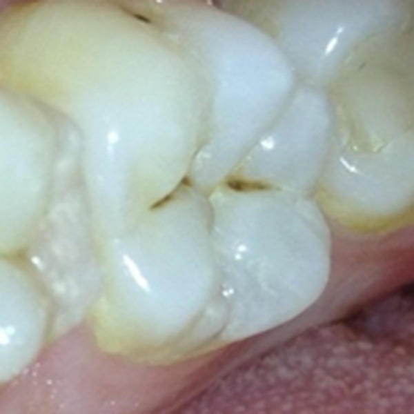
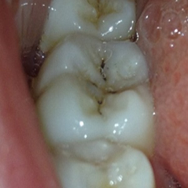
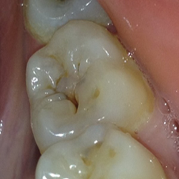
Figure 1.
Clinical examples of each ICDAS II coding.
a.
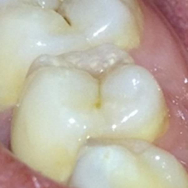
Code 0
b.
Code 1
c.
Code 2
d.
Code 3
e.
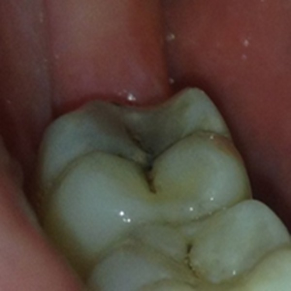
Code 4
f.
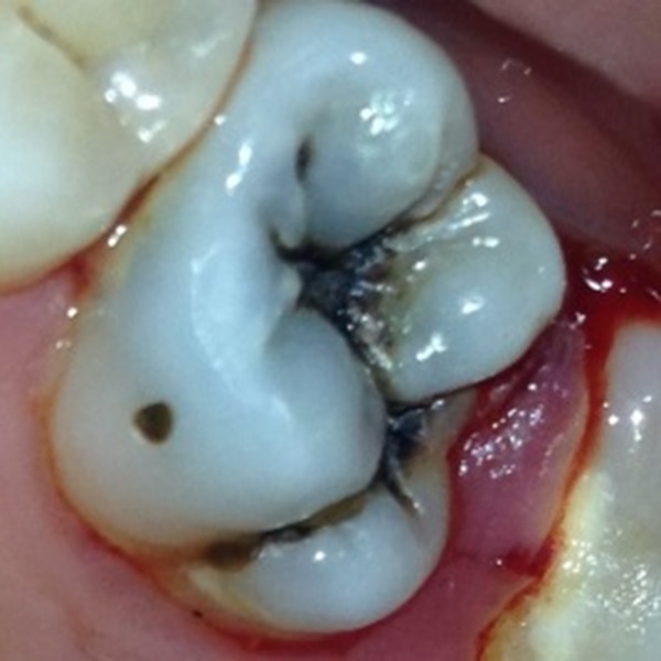
Code 5
g.
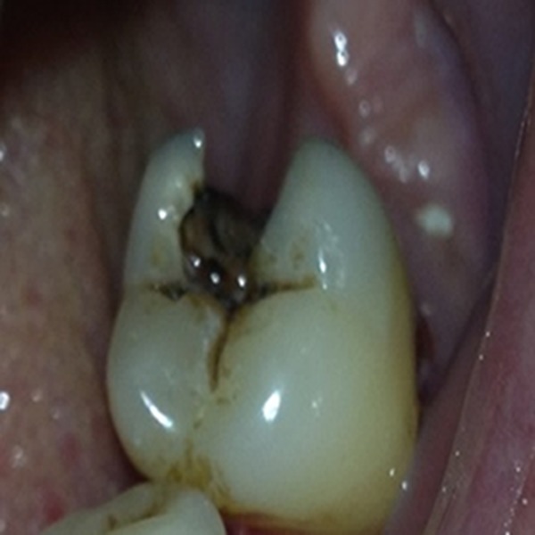
Code 5
Detecting caries lesion is important. However; it only represents part of diagnostic process necessary to properly assess the caries disease status (24). ICDAS criteria can predict progress rate of caries. This situation can provide high quality information that can be used to determine prognosis and treatment (25). Assessment of caries activity can prove to be best way to determine caries risk status and identify patients who require intensive preventive intervention (26). Identifying teeth with passive caries can change the decision of treatment. Moreover, identifying subjects that have teeth with active disease for studies designed to test treatments intended to arrest or reverse caries (27). Nyvad criteria which is designated in 1999 combine severity scoring with lesion activity assessment. ICDAS caries activity assessment are largely based on the Nyvad criteria. However; ICDAS differs in a number of ways from the Nyvad criteria. The comparison of Nyvad and ICDAS criteria is presented in Table 4 (28). Activity criteria of coronal primary caries are presented in Table 5. Actually, differentiating between active and inactive caries lesions is difficult. Since a certain measure of uncertainty must be expected and the clinician should have an ability to detect changes in enamel that can be seen hardly. There is also the possibility of lesions being in a transitional stage, either going from active to inactive or inactive to active. The future holds promise for the development of clinically useful tools to assist dentists and researchers in making decisions about activity status of caries lesions (18, 19).
Table 4.
| Nyvad criteria | ICDAS criteria |
|---|---|
| Lesion severity and activity are determined as one score. | Severity score and activity assessment are provided as two separate scores. |
| These criteria are applied to plaque-covered teeth. | These criteria are initiated on cleaned teeth. |
| A sharp probe is used. | The use of a ball-ended probe is recommended. |
Table 5.
Activity Criteria of coronal primary caries according to ICDAS II (18).
| ICDAS code | Characteristics of Lesion | |
|---|---|---|
| 1, 2 or 3 | Active Lesion | Inactive Lesion |
| Surface of enamel is whitish/yellowish opaque with loss of luster; feels rough when the tip of the probe is moved | Surface of enamel is whitish, brownish or black. | |
| Lesion is in a plaque stagnation area, i.e.:pits and fissures, near the gingival and approximal surface below the contact point. | Enamel may be shiny and feels hard and smooth when the tip of the probe is moved. | |
| For smooth surfaces, caries lesion is typically located at some distance from the gingival margin. | ||
| 4 | Probably active | |
| 5 or 6 | Cavity feels soft or leathery on gently probing the dentin | Cavity may be shiny and feels hard on gently probing the dentin. |
The ICDAS I and II criteria incorporate concepts from the research conducted by Ekstrand et al. (1995). Ekstrand et al (1995) has correlated between the severity of carious lesions and their histological depth . White spot lesions that are seen after airdrying are limited to the outer ½ of enamel. The depth of a white or brown lesion that is seen without air-drying is located some place between the inner ½ of the enamel and the outer ⅓ of dentin. Localized enamel breakdown with no visible dentin indicates that the lesion extends to the middle ⅓ of dentin and a greyish, brownish or bluish shadow of dentin also indicates that the lesion extends to the middle ⅓ of dentin, Cavities with visible dentin extend to the inner ⅓ of dentin (19, 29). Researchers identified ICDAS I criteria in a meeting that was held in 2012. In that meeting, occlusal surfaces of fiftyseven extracted teeth were examined. Teeth were classified according to the ICDAS I and histological scoring of them were identified too. Thus, histological depth of caries of teeth that are classified clinically with code 3 is more than teeth that are classified clinically with code 4. These data support the decision of the ICDAS II workshop to switch the original codes 3 and 4 (ICDAS I) to portray a sequential progression of dental caries (18, 19). Bakhshandeh et al. (2011) reported that there is a strong relationship between ICDAS scores and the real depth of the lesion that is identified histologically (30). Another study done in 2011 also found that there is a strong relationship between ICDAS criteria and histologic depth (31). Because of the strong relationship between ICDAS scores and histologic depth that was supported by previous studies, ICDAS II criteria was used as gold standard in a study in which laser fluorescence was evaluated in occlusal caries detection (32). Root caries detection criteria are divided into two groups according to the ICDAS II: root surfaces with restoration and root surfaces without restoration. However, the basis of the codes is essentially the same for both of groups and presented in Table 6 (18, 19).
Table 6.
| Code E | If the root surface cannot be visualized directly, then it is excluded. |
|---|---|
| Code 0 | The root surface does not exhibit any unusual discoloration that distinguishes it from the surrounding root areas nor does it exhibit a surface defect at the cementoenamel junction or root surface. The root surface has a natural anatomical contour. |
| Code 1 | There is a demarcated area on the root surface or at the cementoenamel junction that is discoloured but there is no cavitation (loss of anatomical contour less than 0.5mm) present. |
| Code 2 | There is a demarcated area on the root surface or at the cementoenamel junction discoloured and there is cavitation (loss of anatomical contour more than 0.5mm) present. |
The characteristics of the discolored area on the root surface can be used to determine the activity status of the lesion. These characteristics are color, texture, appearance, perception on probing, presence of cavitation and location. These criteria that evaluate the activity of root caries are presented in Table 7 (18, 19).
Table 7.
| Color | Active lesions are yellowish or light brown in color whereas arrested lesions appear darkly stained. |
|---|---|
| Perception on probing | Active lesions have been described as soft or leathery compared to arrested lesions that have a hard texture. |
| Appearance | While matte lesions are thought to be active, shiny lesions are thought to be passive. |
| Texture | Rough surfaces are thought to be active whereas smooth surfaces are thought to be arrested. |
| Cavitation | Noncavitated lesions have been described as active whereas cavitated lesions can be either active or arrested. |
| Location | Root caries that occur closely adjacent to the crest of the gingival are considered to be active whereas lesions that occur on the root surface more distant from the gingival crest are more likely to be arrested. |
Honkala et al. (2011) reported that ICDAS criteria can give proper information for the presence of caries lesion (33). In an another study that is done in 2010, three researchers examined 50 permenant teeth according to the ICDAS II criteria. Same teeth are examined 1 day, 1 week and 4 week after first examination by the same researchers again. Thus, the time span didn’t have any effect on the intra- and interexaminer reproducibilty (34).
The Comparison of ICDAS with Other Systems
There are many studies which found that ICDAS and WHO criteria give similar results for detection of occlusal caries. Main advantage of ICDAS is the ability to evaluate noncavitated lesions. However, it also have a disadvantage of long application time when it is compared with WHO criteria (2, 23, 35). Nyvad and ICDAS II were compared for in vivo detection and assessment of occlusal caries of primary teeth in a study. Thus, both of the systems presented good reproducibilty and validity to detect caries lesions and estimate their severities. There were strong relationship between two systems for detection and assessment of caries. However; ICDAS seems to overestimate the caries activity assessment of cavitated lesions compared to the Nyvad (36). In an another study done in 2009, occlusal caries of primary teeth were examined using Nyvad and ICDAS II criteria. The reliability and accuracy of two methods for detection of caries were good. The relationship between two systems were strong. However; there were some differences between two systems when lesion activity was assessed. It is reported that as a reference method is not available, it is not possible to determine which assessed lesion activity more accurately (37). Diniz et al. (2009) examined occlusal surfaces of one hundred sixty-three teeth according to the ICDAS and three weeks later same teeth were assessed by the same researchers again according to the ICDAS criteria (38). Reproducibility was assessed by kappa and the results were compared with the results of the the similar systems that were used previous studies. The systems that were compared with ICDAS were eight point scoring system that were used by Ekstrand et al. in 1995 (29), five point scoring system that were used by Ekstrand et al. in 1997 (39) and four point scoring system that were used by Souza-Zaroni et al. in 2006 (40). Thus, ICDAS II has similar reproducibility for occlusal caries detection compared with other visual scoring systems that were used previous studies (38). In an another study, ICDAS were compared with different diagnostic tools in detecting occlusal caries. Two examiners evaluated 119 occlusal surfaces using LF (Laser fluorescence, DIAGNOdent 2095, KaVo, Germany), LFpen (Laser fluoresans, DIAGNOdent 2190, Kavo, Germany), FC (Fluorescence camera, VistaProof, Durr Dental, Germany), ICDAS II and BW (Bitewing radyografi, HDX, Dental EZ, USA). Then, sensitivity, specificity and accuracy of the methods were compared. Each method has different characteristics and specific modes of functioning and thus varies in sensitivity and specificity. LFpen, FC and ICDAS have better sensitivity whereas LF and BW have better specificity. ICDAS II combined with BW showed the best performance for detecting occlusal caries (41). Diniz et al. (2011) reported that ICDAS showed better performance than bitewing radiography in detecting occlusal caries (42). When the studies that evaluated ICDAS II criteria for detecting approximal caries are examined, it is found that ICDAS II criteria are more successful in detecting approximal caries in vitro (43, 44). However; when these criteria are evaluated in vivo for detecting approximal caries, they are more unsuccessful (45, 46). The reason of this situation is the limitation of in vitro studies that is the difficulty in simulating contact areas of teeth. Can the good results of the in vitro studies be depended on the limitation of the in vitro studies? Or do ICDAS II criteria have really good performance for detection of approximal caries in vitro? Further studies are needed to make right desicion. In an study, approximal surfaces of 20 teeth were examined by two researcher to compare ICDAS II criteria with radiography in detecting caries. While sensitivity of ICDAS II is better, specificity of it is worse than radiography. Thus, they reported that ICDAS II should be preferred as caries diagnostic method in populations at high risk of caries whereas radigraphy should be preferred in population at low risk of caries (47). Ekstrand et al. (2011) revealed that ICDAS can detect and assess approximal caries that can be seen with intraoral examination more successful than radiography (48). The performance of different methods were compared in detecting approximal caries of primary teeth and influence of the discomfort reported by children on the performance of these methods was evaluated in a study done in 2010. Approximal surfaces of 592 primary molars were evaluated by two examiners using LFpen, ICDAS and radiography and patients were asked to assess discomfort. The results of visual inspection is better than other methods in detecting noncavitated lesions but worse than others in detecting cavitated lesions. Discomfort of patients during visual inspection and LFpen application affected the performance of these methods adversely (49). Gomez et al. (2013) compared different methods for the diagnosis of occlusal caries in vitro. ICDAS, digital photographs scored with ICDAS, fibre-optic transillumination (FOTI), optical coherence tomography (OCT), SoproLife(®) camera and quantitative light-induced fluorescence (QLF) system gave similar results for diagnosis of occlusal caries. However, examiners reported that visual examination can be more useful for detection and assessment of the depth of lesion in clinical applications (50). Mitropoulos et al. (2012) evaluated the impact of magnification on occlusal caries diagnosis with implementation of the ICDAS II criteria. They reported that ICDAS criteria has good reproducibility for detection of noncavitated lesions in clinical applications. However, they notified that magnification does not improve the detective performance of visual examination (14).
Conclusion
ICDAS criteria is a coding system that loom large in detecting and assessing dental caries recently. However; these criteria have some limitations too. It is believed that ICDAS criteria will complete their development with the studies that will be done in time. Thus, it is thought that these criteria will be an uniform system that can detect and assess caries without any doubt.
Footnotes
Source of funding: None declared.
Conflict of interest: None declared.
References
- 1.Banerjee A, Watson TF. Pickard’s Manual of Operative Dentistry. 9th ed. Oxford University Press; 2011. p. 19. [Google Scholar]
- 2.Braga MM, Oliveira LB, Bonini GA, Bonecker M, Mendes FM. Feasibility of the international caries detection and assessment system (icdas-ii) in epidemiological surveys and comparability with standard world health organization criteria. Caries Res. 2009;43(4):245–249. doi: 10.1159/000217855. [DOI] [PubMed] [Google Scholar]
- 3.Chu CH, Chau AM, Lo EC. Current and future research in diagnostic criteria and evaluation of caries detection methods. Oral Health Prev Dent. 2013;11(2):181–189. doi: 10.3290/j.ohpd.a29365. [DOI] [PubMed] [Google Scholar]
- 4.Ismail AI, Sohn W, Tellez M, Amaya A, Sen A, Hasson H, Pitts NB. The international caries detection and assessment system (icdas): An integrated system for measuring dental caries. Community Dent Oral Epidemiol. 2007;35(3):170–178. doi: 10.1111/j.1600-0528.2007.00347.x. [DOI] [PubMed] [Google Scholar]
- 5.Pitts N. Icdas--an international system for caries detection and assessment being developed to facilitate caries epidemiology, research and appropriate clinical management. Community Dent Health. 2004;21(3):193–198. [PubMed] [Google Scholar]
- 6.Topping GV, Pitts NB, International Caries D, Assessment System C. Clinical visual caries detection. Monogr Oral Sci. 2009;21:15–41. doi: 10.1159/000224210. [DOI] [PubMed] [Google Scholar]
- 7.Pitts NB. How the detection, assessment, diagnosis and monitoring of caries integrate with personalized caries management. Monogr Oral Sci. 2009 Jun 3;21:1–14. doi: 10.1159/000224208. [DOI] [PubMed] [Google Scholar]
- 8.Altarakemah Y, Al-Sane M, Lim S, Kingman A, Ismail AI. A new approach to reliability assessment of dental caries examinations. Community Dent Oral Epidemiol. 2013;41(4):309–316. doi: 10.1111/cdoe.12020. [DOI] [PubMed] [Google Scholar]
- 9.Ismail AI. Visual and visuo-tactile detection of dental caries. J Dent Res. 2004;83 Spec No C doi: 10.1177/154405910408301s12. [DOI] [PubMed] [Google Scholar]
- 10.Pitts NB, Richards D, International Caries D, Assessment System C. Personalized treatment planning. Monogr Oral Sci. 2009;21:128–143. doi: 10.1159/000224217. [DOI] [PubMed] [Google Scholar]
- 11.Pitts NB, Stamm JW. International consensus workshop on caries clinical trials (icw-cct)--final consensus statements: Agreeing where the evidence leads. J Dent Res. 2004;83 Spec No C doi: 10.1177/154405910408301s27. [DOI] [PubMed] [Google Scholar]
- 12.Agustsdottir H, Gudmundsdottir H, Eggertsson H, Jonsson SH, Gudlaugsson JO, Saemundsson SR, Eliasson ST, Arnadottir IB, Holbrook WP. Caries prevalence of permanent teeth: A national survey of children in iceland using icdas. Community Dent Oral Epidemiol. 2010;38(4):299–309. doi: 10.1111/j.1600-0528.2010.00538.x. [DOI] [PubMed] [Google Scholar]
- 13.Pitts NB, Fyffe HE. The effect of varying diagnostic thresholds upon clinical caries data for a low prevalence group. J Dent Res. 1988;67(3):592–596. doi: 10.1177/00220345880670031401. [DOI] [PubMed] [Google Scholar]
- 14.Mitropoulos P, Rahiotis C, Kakaboura A, Vougiouklakis G. The impact of magnification on occlusal caries diagnosis with implementation of the icdas ii criteria. Caries Res. 2012;46(1):82–86. doi: 10.1159/000335988. [DOI] [PubMed] [Google Scholar]
- 15.Assaf AV, de Castro Meneghim M, Zanin L, Tengan C, Pereira AC. Effect of different diagnostic thresholds on dental caries calibration - a 12 month evaluation. Community Dent Oral Epidemiol. 2006;34(3):213–219. doi: 10.1111/j.1600-0528.2006.00278.x. [DOI] [PubMed] [Google Scholar]
- 16.Fyffe HE, Deery C, Nugent ZJ, Nuttall NM, Pitts NB. Effect of diagnostic threshold on the validity and reliability of epidemiological caries diagnosis using the dundee selectable threshold method for caries diagnosis (dstm) Community Dent Oral Epidemiol. 2000;28(1):42–51. doi: 10.1034/j.1600-0528.2000.280106.x. [DOI] [PubMed] [Google Scholar]
- 17.Ismail AI, Brodeur JM, Gagnon P, Payette M, Picard D, Hamalian T, Olivier M, Eastwood BJ. Prevalence of non-cavitated and cavitated carious lesions in a random sample of 7-9-year-old schoolchildren in montreal, quebec. Community Dent Oral Epidemiol. 1992;20(5):250–255. doi: 10.1111/j.1600-0528.1992.tb01693.x. [DOI] [PubMed] [Google Scholar]
- 18.International Caries Detection and Assessment System (ICDAS) Coordinating Committee. Rationale and Evidence for the International Caries Detection and Assessment System (ICDAS II) [Internet] Scotland: Dental Health Services Research Unit; 2005. Available from: http://www.icdas.org. . [Google Scholar]
- 19.International Caries Detection and Assessment System (ICDAS) Coordinating Committee. Criteria Manual– International Caries Detection and Assessment System (ICDAS II).[Internet] Scotland: Dental Health Services Research Unit; 2005. Available from: http://www.icdas.org. . [Google Scholar]
- 20.Ricketts D, Bartlett D. Advanced Operative Dentistry, A Practical Approach. Elsevier: 2011. p. 3. [Google Scholar]
- 21.Jablonski-Momeni A, Stachniss V, Ricketts DN, Heinzel-Gutenbrunner M, Pieper K. Reproducibility and accuracy of the icdas-ii for detection of occlusal caries in vitro. Caries Res. 2008;42(2):79–87. doi: 10.1159/000113160. [DOI] [PubMed] [Google Scholar]
- 22.Mjor IA, Toffenetti F. Secondary caries: A literature review with case reports. Quintessence Int. 2000;31(3):165–179. [PubMed] [Google Scholar]
- 23.Kuhnisch J, Berger S, Goddon I, Senkel H, Pitts N, Heinrich-Weltzien R. Occlusal caries detection in permanent molars according to who basic methods, icdas ii and laser fluorescence measurements. Community Dent Oral Epidemiol. 2008;36(6):475–484. doi: 10.1111/j.1600-0528.2008.00436.x. [DOI] [PubMed] [Google Scholar]
- 24.Ekstrand KR, Ricketts DN, Kidd EA. Occlusal caries: Pathology, diagnosis and logical management. Dent Update. 2001;28(8):380–387. doi: 10.12968/denu.2001.28.8.380. [DOI] [PubMed] [Google Scholar]
- 25.Ferreira Zandona A, Santiago E, Eckert GJ, Katz BP, Pereira de Oliveira S, Capin OR, Mau M, Zero DT. The natural history of dental caries lesions: A 4-year observational study. J Dent Res. 2012;91(9):841–846. doi: 10.1177/0022034512455030. [DOI] [PMC free article] [PubMed] [Google Scholar]
- 26.Zero D, Fontana M, Lennon AM. Clinical applications and outcomes of using indicators of risk in caries management. J Dent Educ. 2001;65(10):1126–1132. [PubMed] [Google Scholar]
- 27.Nyvad B, Fejerskov O. Assessing the stage of caries lesion activity on the basis of clinical and microbiological examination. Community Dent Oral Epidemiol. 1997;25(1):69–75. doi: 10.1111/j.1600-0528.1997.tb00901.x. [DOI] [PubMed] [Google Scholar]
- 28.Nyvad B, Machiulskiene V, Baelum V. Reliability of a new caries diagnostic system differentiating between active and inactive caries lesions. Caries Res. 1999;33(4):252–260. doi: 10.1159/000016526. [DOI] [PubMed] [Google Scholar]
- 29.Ekstrand KR, Kuzmina I, Bjorndal L, Thylstrup A. Relationship between external and histologic features of progressive stages of caries in the occlusal fossa. Caries Res. 1995;29(4):243–250. doi: 10.1159/000262076. [DOI] [PubMed] [Google Scholar]
- 30.Bakhshandeh A, Ekstrand KR, Qvist V. Measurement of histological and radiographic depth and width of occlusal caries lesions: A methodological study. Caries Res. 2011;45(6):547–555. doi: 10.1159/000331212. [DOI] [PubMed] [Google Scholar]
- 31.Neuhaus KW, Rodrigues JA, Hug I, Stich H, Lussi A. Performance of laser fluorescence devices, visual and radiographic examination for the detection of occlusal caries in primary molars. Clin Oral Investig. 2010 May 27;15(5):635–641. doi: 10.1007/s00784-010-0427-5. [DOI] [PubMed] [Google Scholar]
- 32.Rechmann P, Charland D, Rechmann BM, Featherstone JD. Performance of laser fluorescence devices and visual examination for the detection of occlusal caries in permanent molars. J Biomed Opt. 2012;17(3):36006. doi: 10.1117/1.JBO.17.3.036006. [DOI] [PubMed] [Google Scholar]
- 33.Honkala E, Runnel R, Honkala S, Olak J, Vahlberg T, Saag M, Makinen KK. Measuring dental caries in the mixed dentition by icdas. Int J Dent. 2011;2011:150424. doi: 10.1155/2011/150424. [DOI] [PMC free article] [PubMed] [Google Scholar]
- 34.Jablonski-Momeni A, Ricketts DN, Weber K, Ziomek O, Heinzel-Gutenbrunner M, Schipper HM, Stoll R, Pieper K. Effect of different time intervals between examinations on the reproducibility of icdas-ii for occlusal caries. Caries Res. 2010;44(3):267–271. doi: 10.1159/000314674. [DOI] [PubMed] [Google Scholar]
- 35.Ekstrand KR, Martignon S, Ricketts DJ, Qvist V. Detection and activity assessment of primary coronal caries lesions: A methodologic study. Oper Dent. 2007;32(3):225–235. doi: 10.2341/06-63. [DOI] [PubMed] [Google Scholar]
- 36.Braga MM, Ekstrand KR, Martignon S, Imparato JC, Ricketts DN, Mendes FM. Clinical performance of two visual scoring systems in detecting and assessing activity status of occlusal caries in primary teeth. Caries Res. 2010 Jun 8;44(3):300–308. doi: 10.1159/000315616. [DOI] [PubMed] [Google Scholar]
- 37.Braga MM, Mendes FM, Martignon S, Ricketts DN, Ekstrand KR. In vitro comparison of nyvad's system and icdas-ii with lesion activity assessment for evaluation of severity and activity of occlusal caries lesions in primary teeth. Caries Res. 2009;43(5):405–412. doi: 10.1159/000239755. [DOI] [PubMed] [Google Scholar]
- 38.Diniz MB, Rodrigues JA, Hug I, Cordeiro Rde C, Lussi A. Reproducibility and accuracy of the icdas-ii for occlusal caries detection. Community Dent Oral Epidemiol. 2009;37(5):399–404. doi: 10.1111/j.1600-0528.2009.00487.x. [DOI] [PubMed] [Google Scholar]
- 39.Ekstrand KR, Ricketts DN, Kidd EA. Reproducibility and accuracy of three methods for assessment of demineralization depth of the occlusal surface: An in vitro examination. 1997;31(3):224–231. doi: 10.1159/000262404. [DOI] [PubMed] [Google Scholar]
- 40.Souza-Zaroni WC, Ciccone JC, Souza-Gabriel AE, Ramos RP, Corona SA, Palma-Dibb RG. Validity and reproducibility of different combinations of methods for occlusal caries detection: An in vitro comparison. Caries Res. 2006;40(3):194–201. doi: 10.1159/000092225. [DOI] [PubMed] [Google Scholar]
- 41.Rodrigues JA, Hug I, Diniz MB, Lussi A. Performance of fluorescence methods, radiographic examination and icdas ii on occlusal surfaces in vitro. Caries Res. 2008;42(4):297–304. doi: 10.1159/000148162. [DOI] [PubMed] [Google Scholar]
- 42.Diniz MB, Lima LM, Eckert G, Zandona AG, Cordeiro RC, Pinto LS. In vitro evaluation of icdas and radiographic examination of occlusal surfaces and their association with treatment decisions. Oper Dent. 2011;36(2):133–142. doi: 10.2341/10-006-L. [DOI] [PubMed] [Google Scholar]
- 43.Braga MM, Morais CC, Nakama RC, Leamari VM, Siqueira WL, Mendes FM. In vitro performance of methods of approximal caries detection in primary molars. Oral Surg Oral Med Oral Pathol Oral Radiol Endod. 2009;108(4) doi: 10.1016/j.tripleo.2009.05.017. [DOI] [PubMed] [Google Scholar]
- 44.Shoaib L, Deery C, Ricketts DN, Nugent ZJ. Validity and reproducibility of icdas ii in primary teeth. Caries Res. 2009;43(6):442–448. doi: 10.1159/000258551. [DOI] [PubMed] [Google Scholar]
- 45.Bader JD, Shugars DA, Bonito AJ. A systematic review of the performance of methods for identifying carious lesions. J Public Health Dent. 2002;62(4):201–213. doi: 10.1111/j.1752-7325.2002.tb03446.x. [DOI] [PubMed] [Google Scholar]
- 46.Novaes TF, Matos R, Braga MM, Imparato JC, Raggio DP, Mendes FM. Performance of a pen-type laser fluorescence device and conventional methods in detecting approximal caries lesions in primary teeth--in vivo study. Caries Res. 2009;43(1):36–42. doi: 10.1159/000189705. [DOI] [PubMed] [Google Scholar]
- 47.Mitropoulos P, Rahiotis C, Stamatakis H, Kakaboura A. Diagnostic performance of the visual caries classification system icdas ii versus radiography and micro-computed tomography for proximal caries detection: An in vitro study. J Dent. 2010;38(11):859–867. doi: 10.1016/j.jdent.2010.07.005. [DOI] [PubMed] [Google Scholar]
- 48.Ekstrand KR, Luna LE, Promisiero L, Cortes A, Cuevas S, Reyes JF, Torres CE, Martignon S. The reliability and accuracy of two methods for proximal caries detection and depth on directly visible proximal surfaces: An in vitro study. Caries Res. 2011;45(2):93–99. doi: 10.1159/000324439. [DOI] [PubMed] [Google Scholar]
- 49.Novaes TF, Matos R, Raggio DP, Imparato JC, Braga MM, Mendes FM. Influence of the discomfort reported by children on the performance of approximal caries detection methods. Caries Res. 2010 Sep 23;44(5):465–471. doi: 10.1159/000320266. [DOI] [PubMed] [Google Scholar]
- 50.Gomez J, Zakian C, Salsone S, Pinto SC, Taylor A, Pretty IA, Ellwood R. In vitro performance of different methods in detecting occlusal caries lesions. J Dent. 2013;41(2):180–186. doi: 10.1016/j.jdent.2012.11.003. [DOI] [PubMed] [Google Scholar]