Abstract
Acute swelling and discoloration of scrotum in new born can have many localized causes like testicular torsion, inguinal hernia, scrotal or testicular edema, hydrocele, or even remote causes like adrenal hemorrhage. We report a neonate of adrenal hemorrhage presenting clinically as acute scrotum misguiding the clinician to rule out a local scrotal pathology. As the local clinical examination is not reliable in a newborn, it definitely requires an imaging evaluation to establish the diagnosis. This case report emphasizes being aware of the clinical association of acute adrenal hemorrhage and an acute scrotum and the role of ultrasonography in the evaluation of the various differential diagnoses leading to an acute scrotum. An optimum sonographic examination helps in suspecting an abdominal pathology as a cause of acute scrotum and in establishing the specific diagnosis of adrenal hemorrhage to avoid an unnecessary surgical exploration.
Keywords: Adrenal hemorrhage, Ultrasonography, Acute scrotum
Sommario
La tumefazione acuta con pallore dello scroto nel neonato può avere molteplici cause come la torsione testicolare, l’ernia inguinale, l’edema scrotale o testicolare, l’idrocele e anche cause remote come l’emorragia surrenalica. Riportiamo il caso di emorragia surrenalica di un neonato che si presenta clinicamente come scroto acuto, portando il medico al dubbio di una patologia scrotale locale. Poiché l’esame clinico nel neonato è difficile, si richiede necessariamente una valutazione di imaging per formulare la diagnosi. Il caso riportato vuole sottolineare la possibilità di una associazione di emorragia surrenalica acuta con scroto acuto, nonché il ruolo dell’ecografia nella valutazione delle varie diagnosi differenziali dello scroto acuto. Un esame ecografico ottimale aiuta nell’identificare il sospetto di patologia addominale come causa di scroto acuto e a stabilire la diagnosi specifica dell’emorragia surrenalica al fine di evitare un’inutile esplorazione chirurgica.
Introduction
An acute scrotum is a clinical condition represented by sudden pain and/or swelling, usually occurring due to a rapidly developing underlying pathology involving the testis, epididymis, or the scrotal sac [1, 2]. Early diagnosis is essential so that prompt treatment can be instituted to relieve the patient of the distressing symptoms and to salvage the underlying testicular tissue. It is important to diagnose and provide immediate surgical intervention for conditions like torsion of the testis and complicated inguinoscrotal hernia. A surgical de-torsion with fixation can prevent recurrence [3], while early release of obstructed or strangulated bowel and/or omentum with repair of inguinal hernia is necessary to prevent ischemia and/or bowel perforation [3]. Similarly, medical treatment requiring institution of anti-inflammatory drugs and antibiotics in patients of epididymoorchitis is essential to prevent serious complications. Although rare, non-scrotal causes may present as an acute scrotum. In neonates, meconium peritonitis, acute adrenal hemorrhage, or acute scrotal wall swelling are the main non-scrotal causes. Therefore, an acute scrotum may be a telltale sign of a more sinister and life-threatening clinically obscure condition in a newborn. Awareness regarding the possibility of a scrotal swelling being an indicator of an intra-abdominal pathology is essential to offer optimum treatment and thus avoid serious consequences.
NAH is an extremely rare cause of scrotal hematocele and occurs due to a variety of causes [4]. The large size of the adrenal gland in a neonate and its rich vascularity are the predisposing factors for adrenal hemorrhage which may occur in perinatal asphyxia, birth trauma, or even spontaneously [5].
Imaging plays a pivotal role in ruling out a scrotal pathology, establishing the correct diagnosis by recognizing the non-scrotal causes. This case report emphasizes the role of sonography in detecting clinically unsuspected adrenal hemorrhage to be the cause of acute scrotum in a neonate.
Case report
A 2-day preterm male baby developed bilateral scrotal swelling with blackish discoloration of the right hemi-scrotum. The child was born at 32-week gestation by a lower section cesarean section which was done for uncontrolled antepartum hemorrhage in the mother. The baby weighed 1.5 kg and the Apgar score was 7/10 at 5 min, respectively. A heart rate of 80/min with poor respiratory effort and acrocyanosis was present. A day later, the general condition of the baby deteriorated with the development of tachycardia and tachypnea (HR of 130/min and respiratory rate of 48/min). The baby had moderate grade of anemia (Hb = 12.6 gm/dl); however, no yellowish discoloration of skin, urine, or eyes was present. No history of any intrauterine infection or birth trauma was present. This baby was third in birth order, with two siblings aged three and five years who did not have any significant past medical history. Evidence of hypoprothrombinemia was present neither in the baby nor in the family. Local examination revealed swelling of the right scrotal sac with blackish discoloration of the hemi-scrotum. A provisional diagnosis of scrotal ecchymosis and tense hydrocele was made and the baby referred for an urgent ultrasound (USG) examination to rule out an underlying testicular torsion or epididymo-orchitis. Based on the low general condition, neonatal sepsis with shock was also a consideration in the baby. An urgent ultrasound examination of the scrotum was requested for.
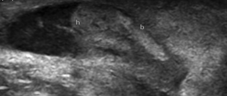
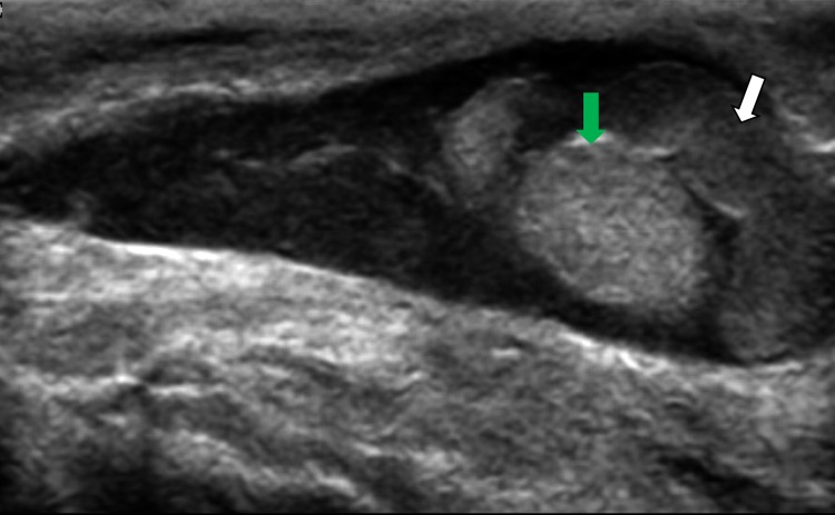
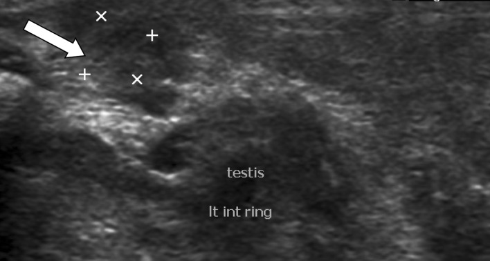
Sonography of the baby’s scrotum performed with a high-frequency linear transducer revealed a normal testis and epididymis with respect to its size and echotexture (Figs. 1, 2) in the right hemi-scrotum. There was fluid around the testis suggesting its presence in the tunica vaginalis. This fluid contained fine low-level echoes with few thin septae (Figs. 1, 3). Scanning toward the root of the scrotum revealed the extension of the scrotal fluid into the inguinal canal suggesting a patent processus vaginalis (Fig. 3). There was a non-luminal echogenic solid structure consistent with omentum floating freely within the fluid suggesting an associated congenital inguinal hernia. (Figs. 1, 3). The scrotal wall was grossly thickened and edematous (Fig. 1). Color Doppler examination of the right hemi-scrotum revealed normal vascularity of the testis and the epididymis, thus ruling out the possibility of either an acute inflammatory condition or torsion of the testis (Fig. 4). The left scrotal sac (Fig. 5) was empty and a normal-sized testis having normal echotexture was seen in the inguinal canal suggestive of an ectopic left testis.
Fig. 1.
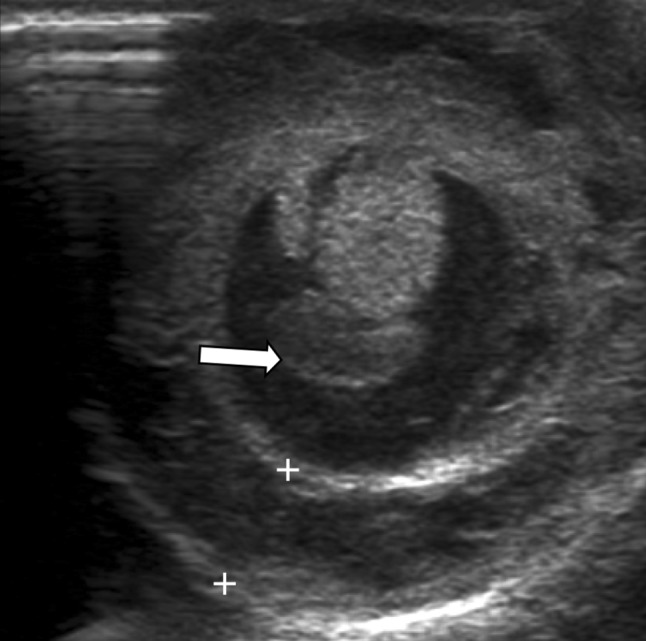
Transverse sonographic view of the right hemi-scrotum showing the normal testis and epididymal body surrounded by fluid. Adjacent omentum (arrow) is seen. Grossly edematous and thickened scrotal wall (within calipers) is also appreciated
Fig. 2.
Longitudinal sonographic view of the right hemi-scrotum showing normal right epididymis. h head of epididymis, b body of epididymis
Fig. 3.
Longitudinal sonographic view of the right hemi-scrotum showing fluid with fine low-level echoes and a few septae. The fluid is extending into the inguinal canal suggesting a patent processus vaginalis. An echogenic solid structure adjacent to the testis is the omentum (white arrow), which can be seen. The right testis (green arrow) appears normal
Fig. 4.
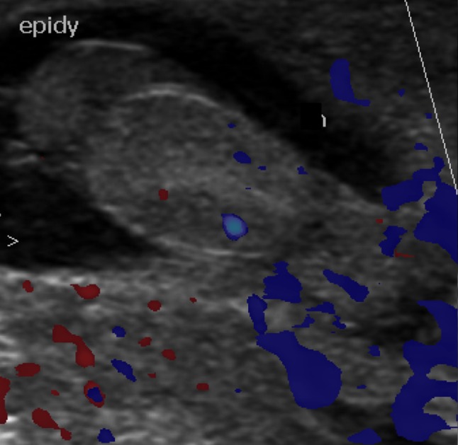
Longitudinal sonographic view of the right hemi-scrotum showing normal vascularity of the right testis on color Doppler. Epidy epididymal head
Fig. 5.
Left testis (white arrow) is present in the left inguinal canal and the left hemi-scrotum was empty, not seen in the image
The presence of free fluid in the peritoneal cavity indicated a concurrent abdominal pathology and thus an abdominal sonography was also conducted in consultation with the clinician. The ultrasound examination revealed normal solid intra-abdominal organs with normal bilateral kidneys. The right adrenal gland appeared bulky with loss of its normal central echogenic and outer hypoechoic appearance. A small hypoechoic lesion was present in one limb of the right adrenal gland (Fig. 6). The left adrenal gland appeared normal on sonography. Additionally, there was a mild to moderate amount of free fluid in the peritoneal cavity with low-level echoes and occasional septae. A sonographic diagnosis of adrenal hemorrhage with hemoperitoneum and right-sided congenital inguinal hernia (containing fluid and omentum) and the left undescended testis was made.
Fig. 6.
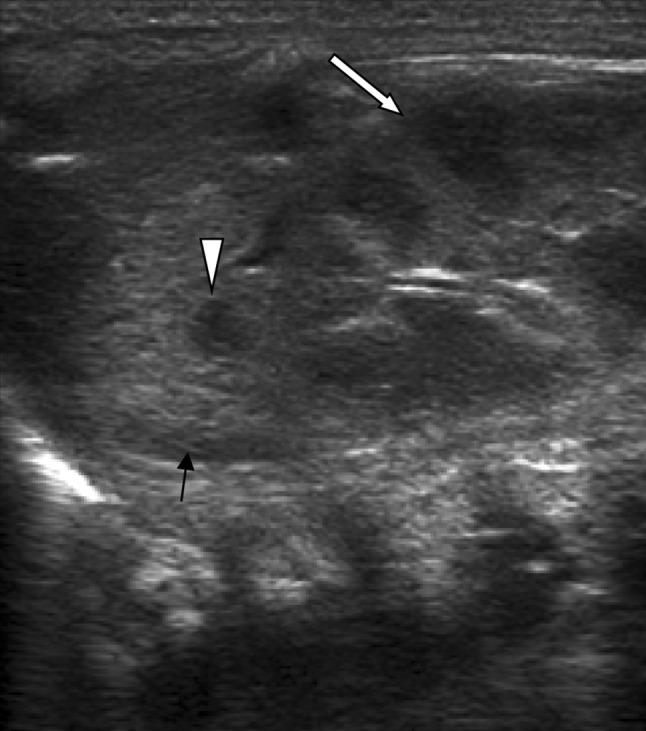
Abdominal sonography showing bulky right adrenal gland (black arrow) with loss of its normal appearance. There is a hypoechoic (arrow head) lesion seen in one limb. White arrow indicates the right kidney
Therefore, a concurrent sonographic examination of the scrotum and abdomen detected the extension of hemoperitoneum caused by right-sided adrenal hemorrhage into the right scrotal sac through the patent processus vaginalis.
The imaging diagnosis was immediately communicated to the pediatrician. Unfortunately, the baby developed a refractory shock not responding to medical treatment and died after 2 days.
Discussion
Adrenal hemorrhage usually occurs at birth or during the immediate postnatal period with the incidence ranging from approximately 1.7 to 2.1 per 1000 births [6]. It affects the right side more than the left and is bilateral in 10–15% of the cases [7]. The predilection for right adrenal hemorrhage is presumed to be secondary to the compression of gland between the liver and the spine which induces venous pressure changes in the right adrenal vein draining directly into the inferior vena cava [8]. In the newborn, adrenal gland is more vulnerable to vascular damage due to its large size and unique circulation. Any condition leading to hypoxia causes shunting of blood toward the vital organs including the adrenals. This phenomenon results in increased venous congestion in the gland and thus damage to the endothelial cell and resultant adrenal hemorrhage in the neonate [9].
The most common predisposing causes of neonatal adrenal hemorrhage (NAH) are birth trauma, prolonged labor, intrauterine infection, perinatal asphyxia or hypoxia, large birth weight, septicemia, hemorrhagic disorder, and hypothrombinemia [10–13]. In our patient, antepartum hemorrhage in the mother for which a cesarean section was done at 32 weeks could have caused perinatal asphyxia in the baby.
Clinical presentation is variable in NAH and depends on the volume of hemorrhage as well as the amount of adrenal cortex compromised. If bleeding is mild to moderate, it tends to remain within the adrenal capsule and may be clinically silent. Whereas massive bilateral hemorrhage may manifest as hemoperitoneum or hemoretroperitoneum and shock, in NAH anemia, abdominal mass, or persistent jaundice may be the presenting symptoms [5, 7, 14]. Scrotal hematoma [5], delayed persistent neonatal jaundice [15], or a multicystic adrenal mass [14] are also known but rare presentations of NAH. Addisonian crisis occurs only when 90% of the adrenal gland is destroyed by parenchymal hemorrhage [16]. In our patient also, adrenal hemorrhage leaked into the peritoneal cavity leading to shock in the neonate which was misinterpreted clinically as septic shock.
The prognosis depends on the extent of adrenal gland involvement, its volume, and the associated predisposing factor. Bilateral adrenal hemorrhage results in Addisonian crisis in 16 to 50% of the patients, which if not managed on emergent basis carries a grave prognosis [17]. Moderate hemorrhage also warrants early detection to improve the outcome by providing good supportive care to the baby.
Sonographic appearance of the normal adrenal gland is very typical in a neonate with the cortex appearing large and hypoechoic, whereas the medulla is relatively smaller and hyperechoic and remains so until 6 months beyond which sonographic differentiation between the cortex and medulla is lost. Loss of even the normal appearance should be viewed with suspicion of a definite adrenal pathology especially AH. Usually, the hemorrhage is seen as a homogeneous or an inhomogeneous echogenic mass within the adrenal gland which shows change in echogenicity as the blood degrades. With time, the central portion becomes hypoechoic and gradually replaces the solid mass in four to six weeks. There can be complete resolution or persistence of the hematoma as a pseudocyst, which appears as a mass with hypoechoic center and may or may not have calcification of its wall [15].
In our case also the finding of a small intra-adrenal hypoechoic lesion probably represented the residual resolving adrenal hematoma which may have occurred during the antenatal period (due to antepartum hemorrhage) and which was also responsible for the hemoperitoneum and the hematocele in the neonate. The bulky, echogenic right adrenal gland with altered echotexture likely represented the recent adrenal hemorrhage (due to prematurity and ischemia) and was responsible for the clinical shock-like state. As the baby was very sick, CT/MRI could not be done to confirm the findings. But in the present clinical scenario these sonographic findings can only represent two episodes of adrenal hemorrhage during the antepartum and the immediate postpartum period.
Initially, when seen as a solid echogenic lesion or even during the resolution phase when AH appears as a cystic or multicystic mass, it may be confused with a tumor of the adrenal gland. The closest differential is a neuroblastoma which warrants a CECT/MRI examination for further evaluation. Change in the sonographic appearance (both the size and the echotexture) of the lesion in a sick baby with an adrenal mass in a clinical setting of an adrenal hemorrhage suggests the diagnosis of the adrenal mass being a hematoma. Moreover, normal vanillylmandelic acid (VMA) levels also rule out a neuroblastoma which is detected in almost 90% cases [18].
Imaging, especially sonography, thus, plays a crucial role in detecting adrenal hemorrhage as it can also be carried out in an emergency setting besides being a quick, non-invasive, and a non-radiating modality. CECT or MRI is rarely utilized as they may not be feasible in a very sick patient. However, MRI is reserved for cases where diagnostic problems occur.
On NCCT, the presence of blood is indicated by an HU of 50–90 within the lesion in the acute phase which gradually reduces and the cystic component increases with degradation of the hematoma. Non-enhancement of the solid component rules out the possibility of an adrenal mass, and this finding corresponds to the absence of color signal on Doppler examination. However, CT being a radiating modality, its use is reserved.
On MRI, any hyperintensity on T1-weighted image suggests hemorrhage. A quickly altering signal intensity also suggests an evolving hemorrhage especially if the adreniform shape is preserved. Larger hematomas appear round or oval and have to be differentiated from a tumoral hemorrhage by lack of an associated enhancing mass. Bilateral affection and peri-adrenal infiltration also points toward hemorrhage rather than a tumor.
Diagnosis and follow-up of AH using USG is the most effective modality to gently evaluate our tender patients and avoid unnecessary laparotomy [9, 14].
With the occurrence of AH, the adrenal gland capsule may rupture and the blood enters the peritoneal cavity resulting in hemoperitoneum. The peritoneal blood may reach the scrotum via the patent processus vaginalis or by dissecting along the retroperitoneum, and presents as a swelling and bluish discoloration of the scrotum [9, 19]. The former phenomenon was also observed in our patient who presented with an acute, bluish scrotum and was clinically misdiagnosed as testicular torsion. Tracking of the hemoperitoneum into the scrotum through the patent processus vaginalis is the nature way of making the pathology apparent.
Scrotal swelling, although unusual in newborns, may result from different localized causes involving the testis (torsion, orchitis, tumor), epididymis (epididymo-orchitis), or the scrotal sac (cellulitis, abscess, hematoma, incarcerated or strangulated complete inguinal hernia) [1, 2]. Another important differential requiring an emergency surgical intervention is acute torsion of testis [20]. Non-scrotal abdominal causes especially NAH should be considered and ruled out in an acutely developing scrotal swelling to avoid making a misdiagnosis [21–24] and, thus, delay the treatment, as happened in our case. Radiologists should have the knowledge of this unusual presentation of AH and always evaluate the adrenal gland on sonography, especially if the baby is sick. Even a subtle abnormality in the adrenal gland or loss of the normal appearance warrants ruling out the possibility of adrenal hemorrhage. In fact, the presence of peritoneal fluid or retroperitoneal collection along with the adrenal abnormality is highly suspicion of adrenal hemorrhage.
These acute scrotal causes are commonly found in adolescents and adults unlike in neonates and small children, whereas the non-scrotal causes of acute scrotal swelling are uncommon at any age, although pain in scrotum due to pathology elsewhere is sometimes encountered in adults. The most frequent non-scrotal causes of acute scrotum (pain with or without associated swelling) are ureteric colic, inguinal hernia, acute pancreatitis, as well as non-traumatic adrenal hemorrhage. Ruptured abdominal aortic aneurysm should also be considered in elderly patients presenting with acute testicular pain [25].
Careful and detailed clinical history and examination may give a clue to the etiology. Imaging plays an essential part in the workup of acute scrotum at any age. Usually, ultrasound with or without a Doppler examination is adequate to evaluate the scrotum and its contents and characterize the varied scrotal causes, while ultrasound and CECT and/or MRI are required to assess the abdomen to detect the non-scrotal causes.
Presence of a normal-sized testis and epididymis of normal echotexture with normal vascularity on Doppler rules out any inflammatory pathology or possibility of torsion. Ultrasound can also show the presence and the extent of inguinal hernia, its contents, and the presence of any complications.
An acute scrotal hematoma is a genitourinary emergency requiring urgent attention. Trauma is the commonest cause for its occurrence and warrants drainage. In a neonate, NAH should always be a consideration in an appropriate clinical setting and ruled out by evaluating the adrenal glands.
In our case, a completely unsuspected clinical condition of NAH with hemoperitoneum extending into the scrotum was revealed. Sonographic characterization of the fluid also gave a clue to the diagnosis. The similar appearance of ascites and hydrocele (containing fine low-level echoes and fine septae) hinted at the same pathology with the extension of the peritoneal fluid (hemoperitoneum) into the right scrotal sac through the patent processus vaginalis. This finding in combination with an adrenal abnormality suggested NAH in the baby. In a neonate, acute scrotum due to meconium peritonitis can be ruled out by the absence of history of non-passage of meconium in the postnatal period and the absence of coarse echoes in the peritoneal fluid. Lack of fluid in the left scrotum was due to the associated undescended testis on the left side. The adrenal hemorrhage explained the shock in the baby which had been misinterpreted as septic shock and this caused delay in treatment.
Thus, sonography besides successfully ruling out the clinical possibility of either an acute epididymoorchitis or testicular torsion revealed an unsuspected NAH to be the cause of the acute scrotum.
Conclusion
Ultrasound is the modality of choice to diagnose and characterize the various scrotal causes and also to suspect the non-scrotal causes of acute scrotum, which may be delineated on simultaneous abdominal sonography. It is important to correctly identify the surgical causes to salvage the underlying testicular tissue. The non-scrotal causes of acute abdomen should be known to the clinician as well as the radiologists across all age groups and possibility of adrenal hemorrhage considered when concurrent imaging abnormality is detected in the adrenal gland/s. This is essential, especially in neonates with predisposing factors, and warrants quick management to save the baby.
Compliance with ethical standards
Conflict of interest
There is no conflict of interest (financial of non-financial) declared by any author/s.
Ethical approval
This case report complies with the ethical standards laid down in the Helsinki Declaration of 1975 and its late amendments. The confidentiality of the patient has been maintained. The manuscript has been read and approved by all the authors and all have significantly contributed towards the article.
Informed consent
The written informed consent had been obtained from the patient’s parents for publication of the case details and the images as well.
References
- 1.Kreeftenberg HG, Jr, Zeebregts CJ, Tamminga RY, de Langen ZJ, Zijlstra RJ. Scrotal hematoma, anemia and jaundice as manifestations of adrenal neuroblastoma in a newborn. J Pediatr Surg. 1999;34:1856–1857. doi: 10.1016/S0022-3468(99)90331-7. [DOI] [PubMed] [Google Scholar]
- 2.Sencan A, Karaca I, Bostanci Sencan A, Mir E. Inguinoscrotal hematocele of the newborn. Turk J Pediatr. 2000;42:84–86. [PubMed] [Google Scholar]
- 3.Adorisio O, Mattei R, Ciardini E, Centonze N, Noccioli B. Neonatal adrenal hemorrhage mimicking an acute scrotum. J Perinatol. 2007;27:130–132. doi: 10.1038/sj.jp.7211638. [DOI] [PubMed] [Google Scholar]
- 4.Noviello C, Cobellis G, Muzzi G, Pieroni G, Amici G, Martino A. A Neonatal adrenal hemorrhage presenting as contralateral scrotal hematoma. Minerva Pediatr. 2007;59:157–159. [PubMed] [Google Scholar]
- 5.Velaphi SC, Perlman JM. Neonatal adrenal hemorrhage: clinical and abdominal sonographic findings. Clin Pediatr (Phila) 2001;40:545–548. doi: 10.1177/000992280104001002. [DOI] [PubMed] [Google Scholar]
- 6.Mangurten HH. Birth injuries. In: Martin RJ, Fanaroff AA, Walsh MC, editors. Fanaroff and Martin’s neonatal perinatal medicine-diseases of the fetus and newborn. 8. Philadelphia: Mosby Elsevier; 2006. pp. 529–559. [Google Scholar]
- 7.Avolio L, Fusillo M, Ferrari G, Chiara A, Bragheri R. Neonatal adrenal hemorrhage manifesting as acute scrotum: timely diagnosis prevents unnecessary surgery. Urology. 2002;59:601viii–601x. doi: 10.1016/S0090-4295(01)01610-7. [DOI] [PubMed] [Google Scholar]
- 8.Tulassay T, Seri I, Evans J. Renal vascular disease in the newborn. In: Taeugah HW, Ballard RA, Avery ME, editors. Schaffer’s and Avery’s Diseases of Newborn. 7. Philadelphia: WB Saunders Company; 1998. pp. 1177–1187. [Google Scholar]
- 9.Miele V, Galluzzo M, Patti G, Mazzoni G, Calisti A, Valenti M. Scrotal hematoma due to neonatal adrenal hemorrhage: the value of ultrasonography in avoiding unnecessary surgery. Pediatr Radiol. 1997;27:672–674. doi: 10.1007/s002470050209. [DOI] [PubMed] [Google Scholar]
- 10.Duman N, Oren H, Gülcan H, Kumral A, Olguner M, Ozkan H. Scrotal hematoma due to neonatal adrenal hemorrhage. Pediatr Int. 2004;46:360–362. doi: 10.1111/j.1442-200x.2004.01898.x. [DOI] [PubMed] [Google Scholar]
- 11.Rumińska M, Welc-Dobies J, Lange M, Maciejewska J, Pyrzak B, Brzewski M. Adrenal hemorrhage in neonates: risk factors and diagnostic and clinical procedure. Med Wieku Rozwoj. 2008;12:457–462. [PubMed] [Google Scholar]
- 12.Chang TA, Chen CH, Liao MF, Chen CH. Asymptomatic neonatal adrenal hemorrhage. Clin Neonatal. 1998;5:23–26. [Google Scholar]
- 13.Black J, Williams DI. Natural history of adrenal hemorrhage in the newborn. Arch Dis Child. 1973;48:183–190. doi: 10.1136/adc.48.3.183. [DOI] [PMC free article] [PubMed] [Google Scholar]
- 14.Wang CH, Chen SJ, Yang LY, Tang RB. Neonatal adrenal hemorrhage presenting as a multiloculated cystic mass. J Chin Med Assoc. 2008;71:481–484. doi: 10.1016/S1726-4901(08)70153-9. [DOI] [PubMed] [Google Scholar]
- 15.Qureshi UA, Ahmad N, Rasool A, Choh S. Neonatal adrenal hemorrhage presenting as late onset neonatal jaundice. J Indian Assoc Pediatric Surg. 2009;14(4):221–223. doi: 10.4103/0971-9261.59607. [DOI] [PMC free article] [PubMed] [Google Scholar]
- 16.Simon DR, Palese MA. Clinical update on the management of adrenal hemorrhage. Curr Urol Rep. 2009;10:78–83. doi: 10.1007/s11934-009-0014-y. [DOI] [PubMed] [Google Scholar]
- 17.Jordan E, Poder L, Courtier J, Sai V, Jung A, Coakley FV. Imaging of nontraumatic adrenal hemorrhage. AJR Am J Roentgenol. 2012;199(1):91–98. doi: 10.2214/AJR.11.7973. [DOI] [PubMed] [Google Scholar]
- 18.Tuchman M, Ramnaraine ML, Woods WG, Krivit W. Three years of experience with random urinary homovanillic and vanillylmandelic acid levels in the diagnosis of neuroblastoma. Pediatrics. 1987;79:203–205. [PubMed] [Google Scholar]
- 19.Huang CY, Lee YJ, Lee HC, Huang FY. Picture of the month. Neonatal adrenal hemorrhage. Arch Pediatr Adolesc Med. 2000;154:417–418. doi: 10.1001/archpedi.154.4.417. [DOI] [PubMed] [Google Scholar]
- 20.Francis XS, Mark FB (2007) Abnormalities of the testes and scrotum and their surgical management. In: Wein AJ, Kavoussi LR, Novick AC, Peters CA (eds) Campbell-Walsh urology 9, vol. 4. Elsevier, Philadelphia, pp 3789–3793
- 21.O’Neill JMD, Hendry GMA, MacKinlay GA. An unusual presentation of neonatal adrenal hemorrhage. Eur J Ultrasound. 2002;16:261–264. doi: 10.1016/S0929-8266(02)00081-2. [DOI] [PubMed] [Google Scholar]
- 22.Bergami G, Malena S, Di Mario M, Fariello G. Sonographic follow-up of neonatal adrenal hemorrhage. Fourteen case reports. Radiol Med. 1990;79:474–478. [PubMed] [Google Scholar]
- 23.Putnam MH. Neonatal adrenal hemorrhage presenting as a right scrotal mass. JAMA. 1989;261:2958. doi: 10.1001/jama.1989.03420200048034. [DOI] [PubMed] [Google Scholar]
- 24.Karpe B, Nybonde T. Adrenal hemorrhage versus testicular torsion-a diagnostic dilemma in the neonate. Pediatr Surg Int. 1989;4:337–340. doi: 10.1007/BF00183402. [DOI] [Google Scholar]
- 25.Sufi PA. A rare case of leaking abdominal aneurysm presenting as isolated right testicular pain. Can J Emerg Med. 2007;9:124–126. doi: 10.1017/s1481803500014925. [DOI] [PubMed] [Google Scholar]


