Abstract
Dynactin is a dynein-regulating protein that increases the processivity of dynein movement on microtubules. Recent studies have shown that a tripartite complex of dynein–dynactin–Bicaudal D2 is essential for highly processive movement. To elucidate the regulation of dynein motility by dynactin, we focused on two isoforms (A and B) of dynactin 1 (DCTN1), the largest subunit of dynactin that contains both microtubule- and dynein-binding domains. The only difference between the primary structures of the two isoforms is that DCTN1B lacks the K-rich domain, a cluster of basic residues. We measured dynein motility by single molecule observation of recombinant dynein and dynactin. Whereas the tripartite complex containing DCTN1A exhibited highly processive movement, the complex containing DCTN1B dissociated from microtubules with no apparent processive movement. This inhibitory effect of DCTN1B was caused by reductions of the microtubule-binding affinities of both dynein and dynactin, which was attributed to the coiled-coil 1 domain of DCTN1. In DCTN1A, the K-rich domain antagonized these inhibitory effects. Therefore, dynactin has two antagonistic domains and promotes or suppresses dynein motility to accomplish correct localization and functions of dynein within a cell.
Introduction
Dynactin is a very large multi-subunit complex that links dynein with its cargo and is known as a cytoplasmic dynein modulator by binding dynein to specific vesicles or organelles [1–3]. Cytoplasmic dynein is a minus end-directed multi-subunit microtubule motor protein [4]. Dynactin is involved in many cellular functions, including vesicle transport [2,5], organelle positioning [6,7], spindle assembly [8] and microtubule plus end localization [9–11] with dynein. Dynactin abnormalities cause several diseases, including Perry syndrome [12,13] and amyotrophic lateral sclerosis [14,15]. Dynein–dynactin (DD) complexes are distributed in a broad range in cells and their spatial distribution changes throughout the cell cycle [16]. Correct localization of the DD complex is important for maintenance of cellular functions. However, the regulatory mechanism of the DD complex in determining its cellular distribution has been poorly understood.
Previous studies have indicated that dynactin enhances microtubule binding of dynein and induces processive movement of dynein using beads coated with multiple dynein molecules [17,18]. More recently, it has been reported that the DD complex itself does not exhibit unidirectional and highly processive movements. However, BICD2 forms the dynein–dynactin–BICD2 (DDB) complex and then induces unidirectional and highly processive movements in single molecule observations [19,20].
Dynactin itself is a very large multi-subunit complex including dynactin 1 (DCTN1) [21], p50, actin-related protein 1 (Arp1) [22], and other eight kinds of subunits [1]. DCTN1, also known as p150Glued, forms a dimer and extrudes the Arp rod as the arm and shoulder, containing a microtubule-binding region (N-terminal region) [23], dynein-binding region [coiled-coil 1 (CC1) domain] [24], and Arp1-binding region (C-terminal region) [3]. The N-terminal region consists of a Cap-Gly domain [25,26] and K-rich domain [27]. The Cap-Gly domain exhibits equimolar binding to microtubules [28], and the K-rich domain is involved in supporting this interaction [28], which is similar to the K-loop of kinesin [29,30]. The N-terminal region of DCTN1 is different between spliced isoforms, DCTN1A (1A isoform) and DCTN1B (1B isoform). The 1A isoform contains both Cap-Gly and K-rich domains, whereas the 1B isoform lacks the K-rich domain [27]. These isoforms are differentially expressed in tissues [27,31]. However, the roles of these isoforms have not been elucidated in DD and DDB complexes.
In this study, we used recombinant DCTN1 to isolate the dynein complex with one DCTN1 isoform and conducted single molecule observations to quantify the microtubule-binding ratio of dynein and dynactin. Consequently, we demonstrate that the two DCTN1 isoforms (1A and 1B) exert different effects on dynein motility. The 1A isoform induces unidirectional movement, whereas the 1B isoform reduces the microtubule-binding affinity to inhibit unidirectional movement. By comparing the properties of the two isoforms and several mutants, we show that both Cap-Gly and K-rich domains are essential for microtubule binding of dynein to promote unidirectional movement. Furthermore, we found that the CC1 domain is responsible for reductions of the microtubule-binding affinities of both dynein and dynactin and that the K-rich domain antagonizes these effects. We conclude that DCTN1 has two antagonistic regulatory domains that interact intramolecularly with each other, and then exerts opposing effects on dynein motility.
Materials and methods
Construction of expression vectors
cDNAs encoding DCTN1 isoforms and BICD2 were amplified from HEK293 cells by RT-PCR. The PCR primers used for cloning DCTN1 isoforms were as follows: 5´-ATGGCACAGAGCAAGAGGCAC-3´ and 5´-TTAGGAGATGAGGCGACTGTG-3´ for DCTN1 (NM_004082), and 5´-ATGTCGGCGCCGTCGGAGGAG-3´ and 5´-CTACAGGCTCGGTGTGGCTGGCTTGG-3´ for BICD2 (NM_015250). Note that our cloned DCTN1 sample from HEK293 cells was the 1B isoform that lacked 21 amino acids of the K-rich domain (Δexon 5–7) as reported by Dixit et al [32]. cDNA for the 1A isoform was obtained by inverse PCR using the following PCR primers: 5´-GCCCGAAAGACCACAACTCGGCGACCCAAGCCCACGCGCCCAGCCAGTACT-3´ and 5´-TGTCGGTGCCTTCTTAGGCTTCAGTCCCCGCAGTTTGCTAGTCTTTGCAGT-3´. For 1BΔCC1 mutant construction, the CC1 domain-coding region was deleted using the following PCR primers: 5´-CCACCTCCAGAGACCTTTGAC-3´ and 5´-TGGGGAAGGAAGCGGGGGGAC-3´.
The PCR products were cloned into pcDNA5/FRT/TO (LifeTechnologies, Carlsbad, CA), a tetracycline-inducible mammalian expression vector. For protein purification, the streptavidin-binding peptide (SBP)-tag-inserted multifunctional green fluorescent protein (GFP)-tag [33] was fused to the N-terminus of DCTN1. The insertions of these tags or deletions of specific domains were achieved by inverse PCR.
Generation of stable inducible HEK293 cell lines
Flp-In T-REx HEK293 cells (Life Technologies, Carlsbad, CA) were maintained in Dulbecco’s modified Eagle’s medium (Nacalai, Tokyo, Japan) supplemented with 10% fetal bovine serum and 2 mM L-glutamine. Stable inducible cell lines were generated by co-transfection of the pcDNA5/FRT/TO vector containing recombinant DCTN1 isoforms or their mutants with the pOG44 vector encoding Flp recombinase according to the manufacturer’s instructions. The transfectants were screened by selection with 100 μg/ml hygromycin, followed by harvesting hygromycin-resistant colonies.
Protein purification
HEK293 cells expressing DCTN1 isoforms, dynein heavy chain, or BICD2 were cultured in 20 150-mm culture dishes, and then collected and rinsed twice with phosphate-buffered saline. Cell lysates were prepared by homogenizing the cells in buffer B (10 mM Pipes-NaOH, pH 7.0, 200 mM NaCl, 10% sucrose, and 1 mM dithiothreitol) containing 0.05% Triton X-100 and complete mini protease inhibitor cocktail (Roche, Basel, Swiss). After centrifugation and filtration, the lysates were applied to a StrepTrap HP column (GE healthcare, Amersham, UK) that had been equilibrated with buffer B. After washing with buffer A (10 mM Pipes-NaOH, pH 7.0, 150 mM NaCl, and 1 mM dithiothreitol), bound proteins were eluted with buffer B containing 2.5 mM desthiobiotin. Purified DCTN1 isoforms and mutants were confirmed by sodium dodecyl sulfate-polyacrylamide gel electrophoresis (SDS-PAGE) (S1 Fig).
The CC1 fragment consisted of 214–547 a.a. of p150Glued. The 1BN-GCN4 fragment consisted of 1–193 a.a. of the 1B isoform and the GCN4 sequence [34] to dimerize this fragment. These fragments were fused with GFP and a His-tag at the C-terminus. CC1 and 1BN-GCN4 fragments were expressed in pET and pCold expression systems, respectively. These fragments were then purified using profinity IMAC Ni-charged Resin (Bio-Rad, Hercules, CA).
Tubulin was purified from porcine brain as described by Weingarten et al [35]. The tubulin was incubated for 30 min at 37°C with 1 mM GTP and 5 mM MgSO4 to polymerize. After the incubation, taxol was added to a final concentration of 40 μM.
Single molecule observation
Flow chambers were constructed with silanized glass, which were coated with a 5% anti-β tubulin antibody solution and then blocked with 10% Pluronic F-127 and casein. Taxol-stabilized microtubules were introduced into the flow chamber to obtain a constant density of microtubules on the glass surface in each experiment, followed by Alexa 647-labeled dynein and GFP-labeled dynactin in SM buffer (12 mM Pipes-KOH, pH 6.8, 25 mM KAce, 1 mM ATP, 2 mM DTT, 10% saturated casein, and an oxygen-scavenging system). Single molecule observations were performed at 25°C using a total internal reflection fluorescence (TIRF) microscope (IX71, Olympus, Tokyo, Japan) equipped with a 100×/1.45 PlanApo objective lens (Olympus). Images were acquired with a back illumination EMCCD camera (iXon, DV887DCS-BV, Andor, UK) at an exposure time of 100 ms.
Dynein and dynactin behaviors were analyzed by recording fluorescent spots that were visible along microtubules for more than 1 s. The total residence time of molecules on microtubules was measured in a window of 10 s and 10 μm of the microtubule length under the condition of 1 nM dynein or dynactin. Spots of dynein, which moved bidirectionally for more than 400 nm (five pixels), were classified as “diffusive”, those that moved bidirectionally for less than 400 nm were classified as “stationary”, and those that moved to the minus end by more than 400 nm were classified as “unidirectional” according to Schlager et al [20].
Protein binding assay
Dynein and dynactin were mixed in assay buffer (10 mM Pipes-NaOH, pH 7.0, 75 mM NaCl, 45 mM imidazole, and 0.01% Tween 20), and then the mixture was incubated for 10 min at 25°C. TALON Dynabeads (Life Technologies) were added to the mixture, followed by incubation for 10 min at 25°C. The Dynabeads were collected by a magnet. Unbound protein was recovered and washed twice with assay buffer. The bound protein was eluted by elution buffer (10 mM Pipes-NaOH, pH 7.0, 150 mM NaCl, and 300 mM imidazole). Unbound and bound fractions were analyzed by SDS-PAGE.
Results
Motility of the DDB complex containing 1A or 1B isoforms
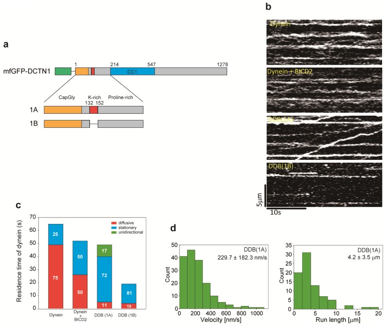
Previous studies have indicated that the DDB complex exhibits highly processive and unidirectional movements [19,20]. However, the difference in DCTN1 isoforms of dynactin remains unknown in the DDB complex. To investigate whether DCTN1 isoforms influence the highly processive movement of DDB complexes, we observed the behavior of DDB complexes with different DCTN1 isoforms (1A and 1B) by TIRF microscopy (Fig 1). To this end, each DCTN1 isoform was fused with a multifunctional GFP-tag at the N-terminus and expressed in HEK293 cells (Fig 1a). These DCTN1 isoforms were incorporated into the endogenous dynactin complex and successfully purified by affinity chromatography (S1 Fig).
Fig 1. Behaviors of DDB complexes with different DCTN1 isoforms.
(a) N-terminal regions of DCTN1 isoforms. Amino acid numbering is based on p150glued. (b) Kymographs of dynein movement on microtubules in the presence of 1 nM Alexa 647-labeled dynein, 2 nM 1A or 1B isoforms, and 20 nM BICD2. (c) Quantification of the behavior of dynein molecules on microtubules. The quantification data are presented as segmented vertical bars: the fraction of diffusive (red), stationary (blue), or unidirectional (green) dynein molecules on microtubules. Quantification of dynein molecules on microtubules in the presence of each dynactin isoform or mutant. The total residence time of dynein on microtubules under the condition of 1 nM Alexa 647-labeled dynein and 2 nM 1A or 1B isoforms. Mean ± S.D., n = 9 windows. (d) Histograms of the velocity and run length of unidirectional movement of the DDB complex (1A). Mean ± S.D., n = 168 particles (velocity) and n = 80 particles (run length).
We observed the behavior of Alexa 647-labeled dynein on microtubules (Fig 1b) and classified it into three types, i.e., diffusive, stationary and unidirectional movement (Fig 1c). GFP-DCTN1 was added at twice the concentration of dynein to easily observe the change in behavior of dynein on microtubules. Therefore, a large amount of GFP-DCTN1 bound to the microtubules and discrimination of individual DCTN1 molecules was difficult. Thus, we quantified and analyzed the total residence time of dynein on microtubules in these experiments. The total residence time of dynein exhibiting each type of movement was calculated based on our counting criteria (see Materials and Methods). In the absence of dynactin and BICD2, the total residence time of dynein on microtubules was 64.9 ± 15.6 s (Fig 1c). Seventy-five percent of the observed dynein exhibited diffusive motion and 25% of bound dynein molecules were stationary (Fig 1c and S1 Table). The total residence time of dynein on microtubules in the presence of BICD2 (dynein + BICD2) was 51.8 ± 7.4 s. Dynein was either stationary (50%) or diffusive (50%) and no processive movement was observed (Fig 1b and 1c and S1 Table). BICD2 may have an effect that increases stationary dynein. Addition of the 1A isoform (DDB 1A) did not change the total residence time of dynein on microtubules (49.1 ± 9.5 s). However, a significant fraction of dynein (17%) moved unidirectionally with marked reduction in the diffusive fraction (11%) and an increase in the stationary fraction (72%) compared with the absence of dynactin. The unidirectional movement occurred with a mean velocity of 229.7 ± 182.3 nm/s and a mean run length of 4.2 ± 3.5 μm (Fig 1d). The highly processive movement of the DDB complex with the 1A isoform is consistent with previous reports [19,20]. The fraction of dynein moving unidirectionally in our experiment with full-length BICD2 was lower than that in previous studies employing C-terminal truncated BICD2 (BICD2N) [19,20]. Similarly, the mean run length was slightly shorter than in previous studies, which might be caused by the use of full-length BICD2. Therefore, the effect of BICD2 on dynein motility would be slightly different from that of BICD2N.
In contrast, addition of the 1B isoform (DDB 1B) resulted in a significantly shorter residence time of dynein on microtubules (19.2 ± 6.4 s) than without dynactin or with the 1A isoform. Most dynein was stationary (81%) and the remaining fraction was diffusive (19%). No unidirectional movement was observed (Fig 1b and 1c). These results suggest that the 1A isoform is essential for unidirectional processive movement of the DDB complex. Furthermore, the fact that addition of the 1B isoform reduced the total residence time of dynein on microtubules suggests that the 1B isoform may inhibit microtubule binding of dynein in the DDB complex.
Role of the DCTN1 isoform in the behavior of DD complexes on microtubules
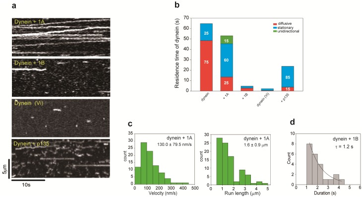
To investigate the effect of DCTN1 isoforms on dynein motility in a more simple system, we observed the behavior of DD complexes on microtubules in the absence of BICD2 (Fig 2a). The total residence time of dynein on microtubules was slightly decreased in the presence of the 1A isoform (53.2 ± 9.5 s) compared with that of dynein alone (64.9 s, Fig 1c). The fraction of stationary dynein molecules was increased to 60%, and 25% of dynein molecules exhibited diffusive motion. Surprisingly, 15% of dynein molecules exhibited unidirectional processive movement (S1 Movie). This ratio was larger than in the previous study (only 1%) [20], because dynactin in our experiment was not a mixture of the 1A and 1B isoforms. Therefore, the promotive effect of the 1A isoform was observed efficiently. The mean velocity and run length of the unidirectional movement were 130.0 ± 79.5 nm/s and 1.6 ± 0.9 μm, respectively (Fig 2c), which were lower than those of the DDB complex (see Fig 1d). Conversely, in the presence of the 1B isoform, the total residence time of dynein on microtubules was drastically decreased to 4.7 ± 3.9 s (Fig 2a and 2b). This total residence time was similar to that in the presence of vanadate (2.2 ± 2.4 s, Fig 2b). Whereas 79% of dynein in the presence of the 1A isoform did not dissociate from microtubules during the observation (30 s) (Fig 2a), the duration of dynein molecules on microtubules was greatly reduced in the presence of the 1B isoform (τ = 1.2 s) compared with the 1A isoform (Fig 2d). These findings indicate that the 1A isoform is necessary and sufficient for the unidirectional movement, and that the 1B isoform inhibits the microtubule binding ability of dynein.
Fig 2. Behaviors of DD complexes with different DCTN1 isoforms.
(a) Kymographs of dynein movement on microtubules in the presence of each dynactin isoform. (b) The total residence time of dynein on microtubules was the same as that in Fig 1c under the condition of 1 nM Alexa 647-labeled dynein and 2 nM of each dynactin isoform. Mean ± S.D., n = 18 windows (dynein alone), n = 15 windows in the presence of 1A or 1B isoforms, n = 30 windows in the presence of vanadate, n = 9 windows in the presence of the p135 isoform. The total residence time of dynein under each condition was altered significantly compared with dynein alone. Quantification of the behavior of dynein molecules on MTs is presented as segmented vertical bars: the fraction of diffusive (red), stationary (blue), or unidirectional (green) dynein molecules on microtubules. The total residence time of dynein on microtubules was the same as that in Fig 1b. Mean ± S.D. (c) Histograms of the velocity and run length of unidirectional movement of dynein molecules in the presence of the 1A isoform. n = 92 particles. (d) Duration of dynein interacting with MTs in the presence of 1B. n = 23 particles (1B isoform).
The above results clearly demonstrated that the 1B isoform inhibits the microtubule-binding ability of dynein in both the DDB complex (Fig 1) and DD complex (Fig 2). We hypothesized that the binding of dynactin with the 1B isoform to dynein affects its microtubule-binding ability.
The CC1 domain inhibits dynein motility
It is known that the CC1 domain of DCTN1 binds to the dynein intermediate chain [24], and the Arp1 rod interacts with the dynein tail via BICD2 [36]. Therefore, we investigated the effect of the CC1 domain on the microtubule-binding ability of dynein.
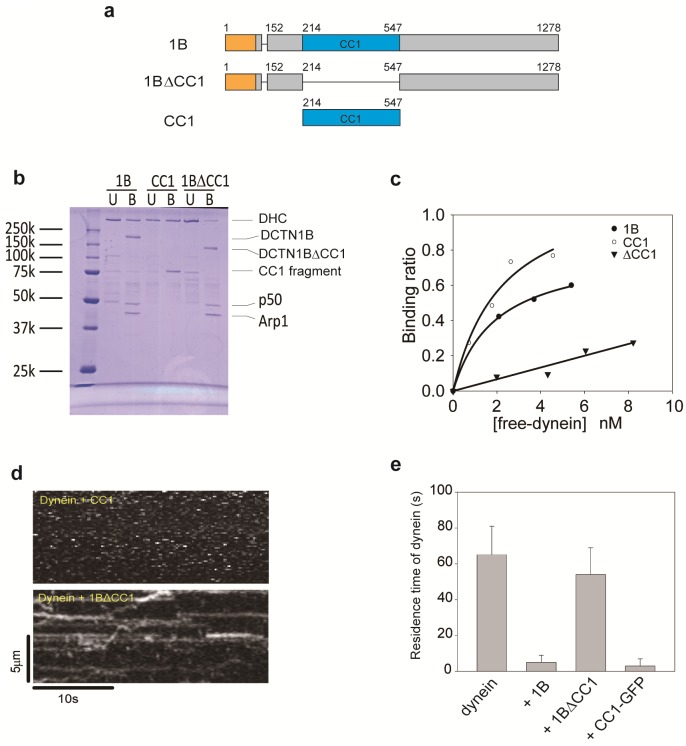
We generated the CC1 fragment and the 1B isoform lacking the CC1 domain (1BΔCC1) (Fig 3a) and initially examined the binding ability of the CC1 domain to dynein by pull-down assays with the purified proteins (Fig 3b and 3c). The 1B isoform bound to dynein in a simple 1:1 binding ratio with a dissociation constant (kd) of 2.3 nM. The engineered CC1 fragment also exhibited a similar binding ability with a dissociation constant (kd) of 2.0 nM. Conversely, the 1BΔCC1 mutant did not specifically bind to dynein, although the 1BΔCC1 mutant had the Arp1 rod (S1 Fig). Therefore, the CC1 domain is the primary dynein-binding site of dynactin in the absence of BICD2, which mediates binding between the dynein tail and Arp1 rod.
Fig 3. The CC1 fragment binds to dynein and inhibits microtubule binding of dynein.
(a) Mutant of the 1B isoform and the CC1 fragment. Amino acid numbering is based on p150glued. (b) Interactions between dynein and dynactin determined by TALON Dynabeads pull-down assays of purified proteins. The protein bands of dynein heavy chain (DHC) in SDS-PAGE gels were quantified by densitometry. (c) Binding ratio of dynein with the 1B isoform (○), CC1 fragment (●), and 1BΔCC1 mutant (▼). (d) Kymographs of dynein motility on microtubules in the presence of the 1BΔCC1 mutant or CC1 fragment. (e) Quantification of dynein molecules on microtubules in the presence of the 1BΔCC1 mutant or CC1 fragment. The total residence time of dynein on microtubules was the same as that in Fig 1b under the condition of 1 nM Alexa 647-labeled dynein and 2 nM 1BΔCC1 mutant or CC1 fragment. Mean ± S.D., n = 15 windows.
We next examined the effect of the CC1 fragment and 1BΔCC1 mutant on the microtubule-binding ability of dynein (Fig 3d). The CC1 fragment greatly reduced the total residence time of dynein on microtubules (3.0 ± 4.0 s, Fig 3e) to a level similar to that with the 1B isoform (4.7 ± 3.9 s, Fig 2b). In contrast, the total residence time of dynein in the presence of the 1BΔCC1 mutant did not significantly differ (54.0 ± 14.6 s, Fig 3e) compared with dynein alone (64.9 ± 15.6 s, Fig 2b). Seventy-one percent of the observed dynein exhibited diffusive motion in the presence of 1BΔCC1 and this ratio was almost the same in comparison with dynein alone (75%) (S1 Table). These results demonstrate that the CC1 domain binds to dynein and inhibits the microtubule-binding ability of dynein.
Microtubule-binding affinities of DCTN1 isoforms
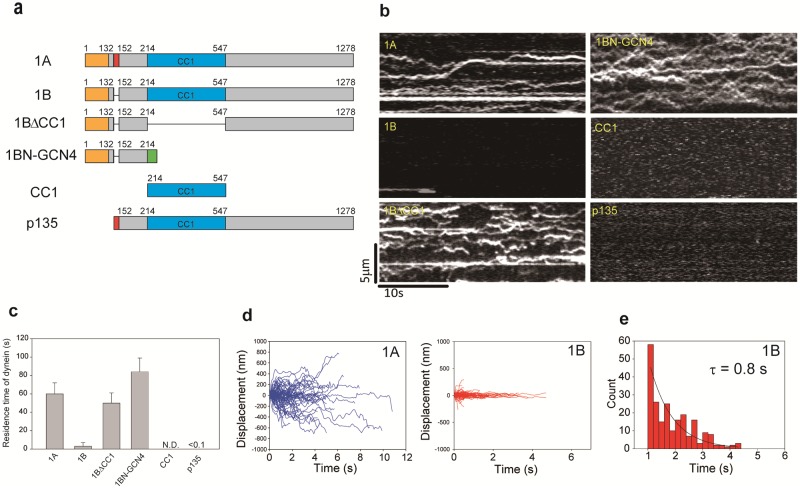
The fact that the CC1 domain binds to dynein and inhibits its microtubule binding in the 1B isoform raises an important issue: why the 1A isoform does not inhibit the microtubule binding of dynein and enables unidirectional processive movement. Dynactin itself interacts with microtubules via the N-terminal region of DCTN1 which contains a Cap-Gly domain [23,28]. Because the K-rich domain is reported to increase the microtubule-binding ability of the Cap-Gly domain, we hypothesized that the microtubule-binding ability of each DCTN1 isoform might be different. To estimate the microtubule-binding abilities of DCTN1 isoforms and mutants (Fig 4a), we observed the behavior of dynactin on microtubules by TIRF microscopy. The 1A isoform was frequently observed on microtubules (Fig 4b) and the total residence time of dynactin on microtubules was 60.1 ± 11.8 s (Fig 4c). In contrast, the 1B isoform exhibited a 20-fold reduction in the total residence time of dynactin on microtubules (3.1 ± 4.4 s). A previous study hardly observed the microtubule binding of dynactin [19], probably because their preparation of dynactin might be almost all the 1B isoform.
Fig 4. Single molecule behavior of dynactin on microtubules.
(a) Mutant of the 1B isoform and the fragments of DCTN1. Amino acid numbering is based on p150glued. (b) Kymographs of dynactin including each isoform and mutant behavior on microtubules. (c) Quantification of each isoform and mutant on microtubules. The total residence time of dynactin on microtubules was the same as that in Fig 1b. Mean ± S.D., n = 9 windows (1A isoform, 1BΔCC1 mutant, 1BN-GCN4 fragment, CC1 fragment and p135 isoform) and n = 12 windows (1B isoform). (d) Time course of displacement of 1A (left panel) and 1B (right panel) isoforms on microtubules. The diffusion coefficients of 1A and 1B isoforms were 55.4 × 10² and 1.9 × 10² nm2/s, respectively. (e) Duration of 1B isoforms on microtubules. n = 454 particles.
The 1A isoform exhibited highly diffusive movements with a diffusion coefficient of 46.9 × 10² nm2/s (Fig 4d and S2 Fig) and 72% of the 1A isoform did not dissociate from microtubules during the observation (30 s). Conversely, the 1B isoform moved much less diffusively with a diffusion coefficient of 2.0 × 10² nm2/s (Fig 4d and S2 Fig) and the dwell time on microtubules was 0.8 s (Fig 4e).
These results suggest that the 1B isoform has a much lower microtubule-binding ability than the 1A isoform. Thus, the K-rich domain represses the inhibitory effect of the CC1 domain on the microtubule-binding ability of the 1A isoform.
To examine the effect of the CC1 domain on the microtubule-binding ability of dynactin, we deleted the CC1 domain from the 1B isoform (1BΔCC1) (Fig 4a). Interestingly, the 1BΔCC1 mutant interacted with microtubules (50.0 ± 11.3 s), which was comparable to the 1A isoform (60.1 ± 11.8 s). To test whether the CC1 domain inhibited the N-terminal region, we generated the N-terminal fragment of the 1B isoform. Because the N-terminal region does not have dimerization sites, we generated the 1BN-GCN4 fragment which was the dimerized N-terminal fragment of the 1B isoform by the GCN4 sequence as a substitute for the CC1 domain. The 1BN-GCN4 fragment interacted with microtubules at a similar level (83.5 ± 14.8 s, Fig 4c). The CC1 fragment itself did not interact with microtubules (Fig 4c). These results suggest that the N-terminal region is a unique site in the dynactin complex to interact with microtubules. Similarly, the p135 isoform which does not have Cap-Gly domain hardly bound to microtubules (Fig 4c). Thus, the Cap-Gly domain is essential to bind microtubules. Nevertheless, the ratio of the stationary fraction on microtubules was increased in the presence of the p135 isoform (85%, Fig 2b and S1 Table) compared with dynein alone (25%, Fig 2b and S1 Table). However, the total residence time of dynein in the presence of the p135 isoform (23.8 ± 3.7 s, Fig 2b) was shorter than that of dynein alone (64.9 s, Fig 2b). Because the p135 isoform has the partial K-rich domain unlike the 1B isoform, the partial K-rich domain may repress the inhibitory effect of the CC1 domain, but the repression may not be sufficient.
Collectively, these results indicate that the microtubule-binding ability of the CapGly domain is reduced by the CC1 domain and the K-rich domain antagonizes the inhibitory effect of the CC1 domain.
Discussion
In this study, we found that dynactin has two agonistic regulatory domains (CC1 and K-rich domains) and exerts opposing effects on dynein motility depending on the DCTN1 isoform. The function of dynactin to increase dynein processivity, which has already been shown in previous reports [19,20], is considered to be attributed to the 1A isoform. Other than neurons, most tissues express more of the 1B isoform than the 1A isoform [27,31]. Both the 1A and the 1B isoforms of DCTN1 may have coexisted in the previous reports [19,20], and the observed highly processive movement was achieved by the fraction with the 1A isoform. While the effect of the 1A isoform has been observed in previous studies, the properties of the 1B isoform were not revealed. In this study, we used recombinant DCTN1 isoforms and each isoform was investigated separately. Recombinant DCTN1 was incorporated into the dynactin complex and affinity purified using the SBP-tag in the fused multifunctional GFP [33] (S1 Fig). This purification method made it possible to isolate the dynactin complex with one DCTN1 isoform or mutant to compare the functions of the isoforms in dynein motility. Moreover, we characterized the motion of dynein on microtubules and quantified the populations of each behavior of dynein molecules including the microtubule-unbinding state. Namely, we would focus on the quantification of the population of each behavior of dynein. As a result, we were able to determine the difference in binding to microtubules of both DD complexes with 1A or 1B isoform. Our strategies revealed new functions of several domains within DCTN1.
In the presence of the 1A isoform, we detected a decrease in the fraction of diffusive molecules of the DD complex, but an increase in that of stationary molecules compared with dynein alone (Fig 2b). The increase in stationary dynein might be due to the increase in microtubule-binding affinity. Dynein might be loaded by thedynactin-mediated tethering to microtubule, and then the cross-bridge cycle-related binding of dynein might be induced, leading to the increase in stationary fractions. As another possibility, both the cross-bridge cycle-independent microtubule binding of dynein and the diffusive microtubule-binding of dynactin are involved in the microtubule-binding of the DD complex and then the number of microtubule-binding sites in the DD complex increases compared with dynein alone. Therefore, the interference between two diffusive motions may decrease the diffusion coefficient of the DD complex, and then the motion of the DD complex may change to stationary. The DD complex with the 1A isoform underwent unidirectional movement in a manner similar to that of multimolecular dyneins [37]. Because the DDB complex showed a higher velocity and processivity (Fig 1d) than the DD complex (Fig 2c), BICD2 appears to regulate the complex to achieve a stable state that is favorable for the unidirectional movement and then increases processivity.
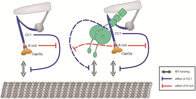
Conversely, the 1B isoform reduced the microtubule-binding affinity of dynein in both DDB and DD complexes (Figs 1c and 2b). Thus, the 1B isoform has an inhibitory effect on dynein motility. Consequently, the inhibition of dynein motility could be executed by non-N-terminal regions. Therefore, we focused on the CC1 domain that contains the dynein-binding site. The CC1 fragment, CC1-GFP, reduced the microtubule-binding affinity of dynein (Fig 3e), suggesting that the CC1 domain is a regulatory unit that inhibits dynein motility (Fig 5, right, blue dashed line). Based on previous studies, the CC1 fragment is known to be a dynein inhibitor because the addition of excess CC1 fragments disrupts the DD complex by competitive inhibition with endogenous dynactin in cells [38,39]. Our finding of the inhibitory effect of CC1 on dynein motility in vitro suggests that the inhibition of dynein motility in a cell is not only caused by disruption of the DD complex but also by the direct inhibitory effect of CC1.
Fig 5. Relationships between regulatory domains.
Left: The CC1 domain inhibits the microtubule-binding ability of the Cap-Gly domain within the same molecule, and this inhibition is repressed by the K-rich domain. Right: The effect on dynactin itself still remains. The CC1 domain, which is bound to dynein, inhibits the microtubule-binding ability of the DD complex, and this inhibition is repressed by the K-rich domain.
Because the only difference between the two isoforms is that the 1B isoform lacks the K-rich domain, this domain is important to support the microtubule-binding ability of the Cap-Gly domain. Furthermore, the lack of the K-rich domain induced the dominance of the CC1 domain and then caused dissociation of the DDB complex (DDB 1B) (Fig 1c) and DD complex (Fig 2b). Therefore, the K-rich domain represses the dissociation of DDB and DD complexes by the CC1 domain (Fig 5, right, red dashed line). Repression of the inhibitory effect by the CC1 domain is necessary to maintain the microtubule-binding ability of dynein, and the K-rich domain is involved in this repression. Although the p135 isoform does not have the Cap-Gly domain, the stationary dynein molecules were increased in the presence of the p135 isoform (Fig 2b). Thus, the partial K-rich domain tends to repress the inhibitory effect of the CC1 domain. However, unidirectional movements were not exhibited by the DD complex with the p135 isoform, and the total residence time of dynein on microtubules in the presence of the p135 isoform was shorter than that of dynein alone (Fig 2b), which is different from previous studies [40][41]. Because yeast dynein does not exhibit bidirectional diffusive movement, which is exhibited in the behavior of mammalian dynein on microtubules [40], the regulatory mechanism of dynein may differ between yeast and mammalian dyneins. Another previous study showed unidirectional movement using both dynein- and p135-coated beads [41]. However, the configuration of dynein and the p135 fragment on the beads was uncertain. Thus, the effect of the CC1 domain may be unclear in that study. We suggest that the p135 isoform has a role that increases to stationary dynein on microtubules similarly to the 1A isoform, but it can not induce unidirectional movement of dynein because it does not have the Cap-Gly domain.
The inhibitory mechanism of the microtubule-binding ability of dynein by the CC1 domain is unknown. The CC1 domain is reported to contain the primary dynein-binding site that binds to the dynein intermediate chain [24,42,43]. Because the dynein heavy chain is involved in microtubule binding [44], it is plausible that the CC1 domain also interacts with the heavy chain. Similar to the effect of vanadate, the CC1 domain would change the conformation of dynein, which may influence the microtubule-binding site of dynein, such as the stalk [45] or strut [46].
We further found that the CC1 domain affected the microtubule-binding ability of dynactin itself. The total residence time of the 1BΔCC1 mutant on microtubules (50.0 s) was higher than that of the 1B isoform (3.0 s) (Fig 4c), and the microtubule-binding affinity of the 1BΔCC1 mutant was similar to that of the 1BN-GCN4 fragment as the dimerized Cap-Gly fragment (Fig 4c). Thus, the CC1 domain may inhibit the microtubule-binding ability of the Cap-Gly domain (Fig 5, left, blue solid line). In contrast, the total residence time of the 1A isoform on microtubules was 60.1 s, which was much higher than that of the 1B isoform (3.0 s) (Fig 4c). Because the 1A isoform has the K-rich domain, the K-rich domain represses the inhibitory effect of the CC1 domain (Fig 5, left, red solid line). Thus, DCTN1 has two regulatory domains, K-rich and CC1, which interact intramolecularly with each other and change the microtubule-binding affinity of dynactin.
Although the 1A isoform induced unidirectional movement of the DD complex, the total residence time of dynein on microtubules with the 1A isoform was slightly lower than that of dynein alone (Fig 2b). Moreover, we detected an increase in the fraction of stationary molecules of the DDB complex with the 1B isoform (Fig 1c). These results suggest that the properties of the two isoforms are not exclusive, and that dynactin possesses opposing properties and the distinctive function of each isoform is constituted by a predominate one of opposing properties of the regulatory domains in DCTN1 (Fig 5, left).
In a cell, the DD complex localizes at microtubule plus ends [10,16,47] or vesicles [16]. A previous in vitro reconstitution study only found the dynein–dynactin–EB1 complex at the microtubule plus end [48], and it is known that EB1 binds to the Cap-Gly domain [49,50]. Our findings suggest that the CC1 domain of the 1B isoform prevents the dynein–dynactin–EB1 complex from minus end-directed movement by dynein, and the complex was found only at the plus end by the aid of +TIPs. In vesicle transport, vesicles with both dynein and kinesin can exhibit plus end directional movement along microtubules [16], because the 1B isoform suppresses dynein motility. The inhibitory effect of the CC1 domain of the 1B isoform on the microtubule-binding ability of the DD complex is involved in the passive transportation toward the plus end direction. Our preliminary experiment showed even when dynein-binding vesicles moved toward the minus end direction, dynactin still bound to the vesicles in cells that only expressed the 1B isoform (S2 Movie). To explain the minus end directional movement of the vesicles, there seems to be the following possibilities. One is that the 1B isoform and dynein are not associated, and they might independently bind to the same vesicle. Another is that the inhibitory effect of the 1B isoform might be repressed by another regulator. The promotion and suppression of dynein motility may be crucial for correct localization and functions of dynein within a cell.
Supporting information
Lane 1: marker; lane 2: 1A isoform; lane 3: 1B isoform; lane 3: 1BΔCC1 mutant, lane 4: CC1 fragment, lane 5: SNAP-BICD2, lane 6: dynein, lane 7: 1BN-GCN4.
(TIF)
MSD plots of 1A (●) and 1B (○) isoforms. Mean ± S.D. The diffusion coefficient (D) was calculated by the following formula: MSD = 2Dt.
(TIF)
(PDF)
Left panel: Alexa 647-labeled dynein; middle panel: GFP fused 1A isoform; right panel: merged movie. The playback speed is 10-fold faster than the acquisition speed. Scale bar is 1 μm.
(MOV)
HeLa cells that expressed both mcherry-fused dynein intermediate chain and GFP-fused dynactin p50 were observed by laser confocal microscopy [16]. Left panel: dynein intermediate chain; middle panel: dynactin p50; right panel: merged movie. The playback speed is 3-fold faster than the acquisition speed. Scale bar is 10 μm.
(MOV)
Acknowledgments
We thank K. Matsuda for technical assistance and K. Saito for helpful discussions.
Data Availability
All relevant data are within the paper and its Supporting Information files.
Funding Statement
This work was supported by JSPS KAKENHI Grant Numbers 15H01310 and 16H04772 to YYT. The funder had no role in study design, data collection and analysis, decision to publish, or preparation of the manuscript.
References
- 1.Schroer TA. Dynactin. Annu Rev Cell Dev Biol [Internet]. 2004;20:759–79. Available from: http://www.ncbi.nlm.nih.gov/entrez/query.fcgi?cmd=Retrieve&db=PubMed&dopt=Citation&list_uids=15473859 [DOI] [PubMed] [Google Scholar]
- 2.Kwinter DM, Lo K, Mafi P, Silverman M a. Dynactin regulates bidirectional transport of dense-core vesicles in the axon and dendrites of cultured hippocampal neurons. Neuroscience [Internet]. 2009;162(4):1001–10. Available from: 10.1016/j.neuroscience.2009.05.038 [DOI] [PubMed] [Google Scholar]
- 3.Kumar S, Zhou Y, Plamann M. Dynactin-membrane interaction is regulated by the C-terminal domains of p150(Glued). EMBO Rep [Internet]. 2001;2(10):939–44. Available from: http://www.ncbi.nlm.nih.gov/pubmed/11571270 [DOI] [PMC free article] [PubMed] [Google Scholar]
- 4.Kardon JR, Vale RD. Regulators of the cytoplasmic dynein motor. Nat Rev Mol Cell Biol [Internet]. 2009;10(12):854–65. Available from: 10.1038/nrm2804 [DOI] [PMC free article] [PubMed] [Google Scholar]
- 5.Muresan V, Stankewich MC, Steffen W, Morrow JS, Holzbaur EL, Schnapp BJ. Dynactin-dependent, dynein-driven vesicle transport in the absence of membrane proteins: a role for spectrin and acidic phospholipids. Mol Cell [Internet]. 2001;7(1):173–83. Available from: http://www.ncbi.nlm.nih.gov/pubmed/11172722 [DOI] [PubMed] [Google Scholar]
- 6.Deacon SW, Serpinskaya AS, Vaughan PS, Lopez Fanarraga M, Vernos I, Vaughan KT, et al. Dynactin is required for bidirectional organelle transport. J Cell Biol [Internet]. 2003;160(3):297–301. Available from: http://www.pubmedcentral.nih.gov/articlerender.fcgi?artid=2172679&tool=pmcentrez&rendertype=abstract [DOI] [PMC free article] [PubMed] [Google Scholar]
- 7.Allan V, A V. Organelle movement, Dynactin: portrait of a dynein regulator. Curr Biol [Internet]. 1994;4(11):1000–2. Available from: http://www.sciencedirect.com/science?_ob=ArticleURL&_udi=B6VRT-4DGVHYY-5C&_user=655616&_coverDate=11/30/1994&_rdoc=1&_fmt=high&_orig=search&_sort=d&_docanchor=&view=c&_acct=C000035458&_version=1&_urlVersion=0&_userid=655616&md5=e019aaad15101d9c8b7ee431370 [DOI] [PubMed] [Google Scholar]
- 8.Siller KH, Doe CQ. Lis1/dynactin regulates metaphase spindle orientation in Drosophila neuroblasts. Dev Biol. 2008;319(1):1–9. 10.1016/j.ydbio.2008.03.018 [DOI] [PMC free article] [PubMed] [Google Scholar]
- 9.Ligon LA, Shelly SS, Tokito M, Holzbaur EL. The microtubule plus-end proteins EB1 and dynactin have differential effects on microtubule polymerization. Mol Biol Cell [Internet]. 2003;14(4):1405–17. Available from: http://www.ncbi.nlm.nih.gov/entrez/query.fcgi?cmd=Retrieve&db=PubMed&dopt=Citation&list_uids=12686597 [DOI] [PMC free article] [PubMed] [Google Scholar]
- 10.Zhang J, Li S, Fischer R, Xiang X. Accumulation of cytoplasmic dynein and dynactin at microtubule plus ends in Aspergillus nidulans is kinesin dependent. Mol Biol Cell [Internet]. 2003;14(4):1479–88. Available from: http://www.pubmedcentral.nih.gov/articlerender.fcgi?artid=153116&tool=pmcentrez&rendertype=abstract [DOI] [PMC free article] [PubMed] [Google Scholar]
- 11.Watson P, Stephens DJ. Microtubule plus-end loading of p150(Glued) is mediated by EB1 and CLIP-170 but is not required for intracellular membrane traffic in mammalian cells. J Cell Sci [Internet]. 2006;119(Pt 13):2758–67. Available from: http://www.pubmedcentral.nih.gov/articlerender.fcgi?artid=1630633&tool=pmcentrez&rendertype=abstract [DOI] [PMC free article] [PubMed] [Google Scholar]
- 12.Farrer MJ, Hulihan MM, Kachergus JM, Dächsel JC, Stoessl AJ, Grantier LL, et al. DCTN1 mutations in Perry syndrome. Nat Genet [Internet]. 2009;41(2):163–5. Available from: http://www.pubmedcentral.nih.gov/articlerender.fcgi?artid=2813485&tool=pmcentrez&rendertype=abstract [DOI] [PMC free article] [PubMed] [Google Scholar]
- 13.Newsway V, Fish M, Rohrer JD, Majounie E, Williams N, Hack M, et al. Perry syndrome due to the DCTN1 G71R mutation: a distinctive levodopa responsive disorder with behavioral syndrome, vertical gaze palsy, and respiratory failure. Mov Disord [Internet]. 2010;25(6):767–70. Available from: http://www.ncbi.nlm.nih.gov/pubmed/20437543 [DOI] [PMC free article] [PubMed] [Google Scholar]
- 14.Münch C, Rosenbohm A, Sperfeld AD, Uttner I, Reske S, Krause BJ, et al. Heterozygous R1101K mutation of the DCTN1 gene in a family with ALS and FTD. Ann Neurol. 2005;58(5):777–80. 10.1002/ana.20631 [DOI] [PubMed] [Google Scholar]
- 15.Holzbaur ELF. Axonal Transport and ALS. Encycl Neurosci. 2010;1181–7. [Google Scholar]
- 16.Kobayashi T, Murayama T. Cell cycle-dependent microtubule-based dynamic transport of cytoplasmic dynein in mammalian cells. PLoS One [Internet]. 2009;4(11):e7827 Available from: papers://4c48fe13-7c69-4b65-a0fa-467dccdedc3f/Paper/p13984 10.1371/journal.pone.0007827 [DOI] [PMC free article] [PubMed] [Google Scholar]
- 17.King SJ, Schroer T a. Dynactin increases the processivity of the cytoplasmic dynein motor. Nat Cell Biol. 2000;2(1):20–4. 10.1038/71338 [DOI] [PubMed] [Google Scholar]
- 18.Culver-Hanlon TL, Lex S a, Stephens AD, Quintyne NJ, King SJ. A microtubule-binding domain in dynactin increases dynein processivity by skating along microtubules. Nat Cell Biol [Internet]. 2006;8(3):264–70. Available from: http://www.ncbi.nlm.nih.gov/pubmed/16474384 [DOI] [PubMed] [Google Scholar]
- 19.McKenney RJ, Huynh W, Tanenbaum ME, Bhabha G, Vale RD. Activation of cytoplasmic dynein motility by dynactin-cargo adapter complexes. Science (80-) [Internet]. 2014;345(6194):337–41. Available from: http://www.sciencemag.org/content/345/6194/337.full [DOI] [PMC free article] [PubMed] [Google Scholar]
- 20.Schlager M a, Hoang HT, Urnavicius L, Bullock SL, Carter AP. In vitro reconstitution of a highly processive recombinant human dynein complex. EMBO J [Internet]. 2014;33(17):1–14. Available from: http://www.ncbi.nlm.nih.gov/pubmed/24986880 [DOI] [PMC free article] [PubMed] [Google Scholar]
- 21.Tokito MK, Holzbaur EL. The genomic structure of DCTN1, a candidate gene for limb-girdle muscular dystrophy (LGMD2B). Biochim Biophys Acta [Internet]. 1998;1442(2–3):432–6. Available from: http://www.ncbi.nlm.nih.gov/pubmed/9805007 [DOI] [PubMed] [Google Scholar]
- 22.Holleran E a., Tokito MK, Karki S, Holzbaur ELF. Centractin (ARP1) associates with spectrin revealing a potential mechanism to link dynactin to intracellular organelles. J Cell Biol. 1996;135(6 II):1815–29. [DOI] [PMC free article] [PubMed] [Google Scholar]
- 23.Waterman-Storer CM, Karki S, Holzbaur EL. The p150Glued component of the dynactin complex binds to both microtubules and the actin-related protein centractin (Arp-1). Proc Natl Acad Sci U S A. 1995;92(5):1634–8. [DOI] [PMC free article] [PubMed] [Google Scholar]
- 24.Siglin AE, Sun S, Moore JK, Tan S, Poenie M, Lear JD, et al. Dynein and dynactin leverage their bivalent character to form a high-affinity interaction. PLoS One [Internet]. 2013;8(4):e59453 Available from: http://www.pubmedcentral.nih.gov/articlerender.fcgi?artid=3618186&tool=pmcentrez&rendertype=abstract [DOI] [PMC free article] [PubMed] [Google Scholar]
- 25.Weisbrich A, Honnappa S, Jaussi R, Okhrimenko O, Frey D, Jelesarov I, et al. Structure-function relationship of CAP-Gly domains. Nat Struct Mol Biol [Internet]. 2007;14(10):959–67. Available from: http://www.ncbi.nlm.nih.gov/pubmed/17828277 [DOI] [PubMed] [Google Scholar]
- 26.Li S, Finley J, Liu ZJ, Qiu SH, Chen H, Luan CH, et al. Crystal structure of the cytoskeleton-associated protein glycine-rich (CAP-Gly) domain. J Biol Chem. 2002;277:48596–601. 10.1074/jbc.M208512200 [DOI] [PubMed] [Google Scholar]
- 27.Zhapparova ON, Bryantseva S a, Dergunova L V, Raevskaya NM, Burakov A V, Bantysh OB, et al. Dynactin subunit p150Glued isoforms notable for differential interaction with microtubules. Traffic [Internet]. 2009;10(11):1635–46. Available from: http://www.ncbi.nlm.nih.gov/pubmed/19778315 [DOI] [PubMed] [Google Scholar]
- 28.Kobayashi T, Shiroguchi K, Edamatsu M, Toyoshima YY. Microtubule-binding properties of dynactin p150 expedient for dynein motility. Biochem Biophys Res Commun [Internet]. 2006;340(1):23–8. Available from: http://www.ncbi.nlm.nih.gov/pubmed/16343429 [DOI] [PubMed] [Google Scholar]
- 29.Okada Y, Hirokawa N. Mechanism of the single-headed processivity: diffusional anchoring between the K-loop of kinesin and the C terminus of tubulin. Proc Natl Acad Sci U S A [Internet]. 2000;97(2):640–5. Available from: http://www.pubmedcentral.nih.gov/articlerender.fcgi?artid=15383&tool=pmcentrez&rendertype=abstract [DOI] [PMC free article] [PubMed] [Google Scholar]
- 30.Rogers KR, Weiss S, Crevel I, Brophy PJ, Geeves M, Cross R. KIF1D is a fast non-processive kinesin that demonstrates novel K-loop-dependent mechanochemistry. EMBO J [Internet]. 2001;20(18):5101–13. Available from: http://emboj.embopress.org/content/20/18/5101.abstract [DOI] [PMC free article] [PubMed] [Google Scholar]
- 31.Lazarus JE, Moughamian AJ, Tokito MK, Holzbaur ELF. Dynactin Subunit p150(Glued) Is a Neuron-Specific Anti-Catastrophe Factor. PLoS Biol [Internet]. 2013;11(7):e1001611 Available from: http://www.pubmedcentral.nih.gov/articlerender.fcgi?artid=3712912&tool=pmcentrez&rendertype=abstract [DOI] [PMC free article] [PubMed] [Google Scholar]
- 32.Dixit R, Levy JR, Tokito M, Ligon LA, Holzbaur EL. Regulation of dynactin through the differential expression of p150Glued isoforms. J Biol Chem [Internet]. 2008;283(48):33611–9. Available from: http://www.ncbi.nlm.nih.gov/entrez/query.fcgi?cmd=Retrieve&db=PubMed&dopt=Citation&list_uids=18812314 [DOI] [PMC free article] [PubMed] [Google Scholar]
- 33.Kobayashi T, Morone N, Kashiyama T, Oyamada H, Kurebayashi N, Murayama T. Engineering a novel multifunctional green fluorescent protein tag for a wide variety of protein research. PLoS One [Internet]. 2008;3(12):e3822 Available from: http://journals.plos.org/plosone/article?id=10.1371/journal.pone.0003822 [DOI] [PMC free article] [PubMed] [Google Scholar]
- 34.Pensato S, Renda M, Leccia F, Saviano M, D’Andrea LD, Pedone C, et al. PNA zipper as a dimerization tool: Development of a bZip Mimic. Biopolymers. 2010;93(5):434–41. 10.1002/bip.21357 [DOI] [PubMed] [Google Scholar]
- 35.Weingarten MD, Lockwood AH, Hwo SY, Kirschner MW. A protein factor essential for microtubule assembly. Proc Natl Acad Sci U S A [Internet]. 1975;72(5):1858–62. Available from: http://www.pnas.org/content/72/5/1858\nhttp://www.pubmedcentral.nih.gov/articlerender.fcgi?artid=432646&tool=pmcentrez&rendertype=abstract [DOI] [PMC free article] [PubMed] [Google Scholar]
- 36.Urnavicius L, Zhang K, Diamant AG, Motz C, Schlager MA, Yu M, et al. The structure of the dynactin complex and its interaction with dynein. Science (80-) [Internet]. 2015;347(6229):1441–6. Available from: http://www.sciencemag.org/cgi/doi/10.1126/science.aaa4080 [DOI] [PMC free article] [PubMed] [Google Scholar]
- 37.Torisawa T, Ichikawa M, Furuta A, Saito K, Oiwa K, Kojima H, et al. Autoinhibition and cooperative activation mechanisms of cytoplasmic dynein. Nat Cell Biol [Internet]. 2014;16(11):1118–24. Available from: http://www.nature.com/doifinder/10.1038/ncb3048 [DOI] [PubMed] [Google Scholar]
- 38.Shrum CK, Defrancisco D, Meffert MK. Stimulated nuclear translocation of NF-kappaB and shuttling differentially depend on dynein and the dynactin complex. Proc Natl Acad Sci U S A [Internet]. 2009;106(8):2647–52. Available from: http://www.pubmedcentral.nih.gov/articlerender.fcgi?artid=2650318&tool=pmcentrez&rendertype=abstract [DOI] [PMC free article] [PubMed] [Google Scholar]
- 39.Brocard CB, Boucher KK, Jedeszko C, Kim PK, Walton P a. Requirement for microtubules and dynein motors in the earliest stages of peroxisome biogenesis. Traffic [Internet]. 2005;6(5):386–95. Available from: http://www.ncbi.nlm.nih.gov/pubmed/15813749 [DOI] [PubMed] [Google Scholar]
- 40.Kardon JR, Reck-Peterson SL, Vale RD. Regulation of the processivity and intracellular localization of Saccharomyces cerevisiae dynein by dynactin. Proc Natl Acad Sci U S A [Internet]. 2009;106(14):5669–74. Available from: http://www.pubmedcentral.nih.gov/articlerender.fcgi?artid=2657088&tool=pmcentrez&rendertype=abstract [DOI] [PMC free article] [PubMed] [Google Scholar]
- 41.Tripathy SK, Weil SJ, Chen C, Anand P, Vallee RB, Gross SP. Autoregulatory mechanism for dynactin control of processive and diffusive dynein transport. Nat Cell Biol [Internet]. 2014;16(12):1192–201. Available from: http://www.nature.com/doifinder/10.1038/ncb3063 [DOI] [PMC free article] [PubMed] [Google Scholar]
- 42.Karki S, Holzbaur EL. Affinity chromatography demonstrates a direct binding between cytoplasmic dynein and the dynactin complex. J Biol Chem [Internet]. 1995;270(48):28806–11. Available from: http://www.ncbi.nlm.nih.gov/pubmed/7499404 [DOI] [PubMed] [Google Scholar]
- 43.Vaughan KT, Vallee RB. Cytoplasmic dynein binds dynactin through a direct interaction between the intermediate chains and p150Glued. J Cell Biol [Internet]. 1995;131(6 Pt 1):1507–16. Available from: http://www.pubmedcentral.nih.gov/articlerender.fcgi?artid=2120689&tool=pmcentrez&rendertype=abstract [DOI] [PMC free article] [PubMed] [Google Scholar]
- 44.Gee M a, Heuser JE, Vallee RB. An extended microtubule-binding structure within the dynein motor domain. Nature. 1997;390(6660):636–9. 10.1038/37663 [DOI] [PubMed] [Google Scholar]
- 45.Gibbons IR, Garbarino JE, Tan CE, Reck-Peterson SL, Vale RD, Carter AP. The affinity of the dynein microtubule-binding domain is modulated by the conformation of its coiled-coil stalk. J Biol Chem [Internet]. 2005;280(25):23960–5. Available from: http://www.pubmedcentral.nih.gov/articlerender.fcgi?artid=1464088&tool=pmcentrez&rendertype=abstract [DOI] [PMC free article] [PubMed] [Google Scholar]
- 46.Kon T, Sutoh K, Kurisu G. X-ray structure of a functional full-length dynein motor domain. Nat Struct Mol Biol [Internet]. 2011;18(6):638–42. Available from: 10.1038/nsmb.2074 [DOI] [PubMed] [Google Scholar]
- 47.Hendricks AG, Lazarus JE, Perlson E, Gardner MK, Odde DJ, Goldman YE, et al. Dynein tethers and stabilizes dynamic microtubule plus ends. Curr Biol. 2012;22:632–7. 10.1016/j.cub.2012.02.023 [DOI] [PMC free article] [PubMed] [Google Scholar]
- 48.Duellberg C, Trokter M, Jha R, Sen I, Steinmetz MO, Surrey T. Reconstitution of a hierarchical +TIP interaction network controlling microtubule end tracking of dynein. Nat Cell Biol [Internet]. 2014;16(8):804–11. Available from: 10.1038/ncb2999 [DOI] [PubMed] [Google Scholar]
- 49.Yan S, Hou G, Schwieters CD, Ahmed S, Williams JC, Polenova T. Three-dimensional structure of CAP-gly domain of mammalian dynactin determined by magic angle spinning NMR spectroscopy: Conformational plasticity and interactions with end-binding protein EB1. J Mol Biol [Internet]. 2013;425(22):4249–66. Available from: 10.1016/j.jmb.2013.04.027 [DOI] [PMC free article] [PubMed] [Google Scholar]
- 50.Askham JM, Vaughan KT, Goodson H V, Morrison EE. Evidence that an interaction between EB1 and p150(Glued) is required for the formation and maintenance of a radial microtubule array anchored at the centrosome. Mol Biol Cell [Internet]. 2002;13(10):3627–45. Available from: http://www.molbiolcell.org/content/13/10/3627.short\nhttp://www.pubmedcentral.nih.gov/articlerender.fcgi?artid=129971&tool=pmcentrez&rendertype=abstract [DOI] [PMC free article] [PubMed] [Google Scholar]
Associated Data
This section collects any data citations, data availability statements, or supplementary materials included in this article.
Supplementary Materials
Lane 1: marker; lane 2: 1A isoform; lane 3: 1B isoform; lane 3: 1BΔCC1 mutant, lane 4: CC1 fragment, lane 5: SNAP-BICD2, lane 6: dynein, lane 7: 1BN-GCN4.
(TIF)
MSD plots of 1A (●) and 1B (○) isoforms. Mean ± S.D. The diffusion coefficient (D) was calculated by the following formula: MSD = 2Dt.
(TIF)
(PDF)
Left panel: Alexa 647-labeled dynein; middle panel: GFP fused 1A isoform; right panel: merged movie. The playback speed is 10-fold faster than the acquisition speed. Scale bar is 1 μm.
(MOV)
HeLa cells that expressed both mcherry-fused dynein intermediate chain and GFP-fused dynactin p50 were observed by laser confocal microscopy [16]. Left panel: dynein intermediate chain; middle panel: dynactin p50; right panel: merged movie. The playback speed is 3-fold faster than the acquisition speed. Scale bar is 10 μm.
(MOV)
Data Availability Statement
All relevant data are within the paper and its Supporting Information files.


