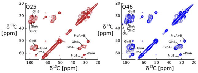
Figure 5.
The structure of the static amyloid core of HTTex1Q46 fibrils and HTTex1Q25 fibrils seeded with HTTex1Q46 is very similar. 2D 13C-13C DARR spectra recorded at 25 kHz MAS, 0°C, with a 50 ms mixing time. Amino acid type assignments are indicated. Both spectra give essentially the same cross peak pattern indicating that the structural motif of the amyloid core formed by the two proteins does not depend on the length of the polyQ region. The spectrum of HTTex1Q46 was previously published as Figure 2 by Isas et al.12

