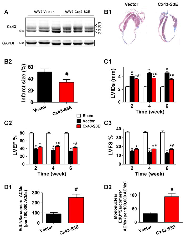
Figure 6. Cardiac AAV9-Cx43-S3E (ischemia resistant Cx43 mutant) therapy promotes ACM proliferation and redifferentiation and improves cardiac function in the post-MI heart.
AAV9-Cx43-S3E or AAV9-vector was administrated 3 days after MI. A: Altered Cx43 phosphorylation in post-MI mice with AAV9-Cx43-S3E treatment. Western blot analysis of whole-cell lysates prepared from left ventricles of mice 6 weeks post-MI. Cx43 lysates are shown in various major phosphorylated forms of Cx43 (P0, P1, P2, P3) with the indicated treatments, probed with polyclonal pan-Cx43 antibody. B: The representative images (B1) and quantification (B2) of heart infarct size 6 weeks post-MI. C: The cardiac structure and function were assessed by serially echocardiography. Left ventricle internal diameter in systole (LVIDs, C1), left ventricular ejection fraction (LVEF, C2) and fraction shortening (LVFS, C3) at 2–6 weeks post-MI were presented. D: ACM proliferation and redifferentiation detected by EdU incorporation analysis at 3 weeks post-MI. Total EdU+/sarcomere+/lacZ+ cells (D1) and mononuclear EdU+/sarcomere+/lacZ+ cells (D2) were quantified. N=10–14; * p<0.05 vs. Sham; # p<0.05 vs. AAV9-vector.

