Abstract
Imprinted genes show parent-specific activity (functional haploidy), which makes them particularly vulnerable to epigenetic dysregulation. Here we studied the methylation profiles of oppositely imprinted genes at single DNA molecule resolution by two independent parental allele-specific deep bisulfite sequencing (DBS) techniques. Using Roche (GSJunior) next generation sequencing technology, we analyzed the maternally imprinted MEST promoter and the paternally imprinted MEG3 intergenic (IG) differentially methylated region (DMR) in fetal cord blood, adult blood, and visceral adipose tissue. Epimutations were defined as paternal or maternal alleles with >50% aberrantly (de)methylated CpG sites, showing the wrong methylation imprint. The epimutation rates (range 2–66%) of the paternal MEST and the maternal MEG3 IG DMR allele, which should be completely unmethylated, were significantly higher than those (0–15%) of the maternal MEST and paternal MEG3 alleles, which are expected to be fully methylated. This hypermethylation of the non-imprinted allele (HNA) was independent of parental origin. Very low epimutation rates in sperm suggest that HNA occurred after fertilization. DBS with Illumina (MiSeq) technology confirmed HNA for the MEST promoter and the MEG3 IG DMR, and to a lesser extent, for the paternally imprinted secondary MEG3 promoter and the maternally imprinted PEG3 promoter. HNA leads to biallelic methylation of imprinted genes in a considerable proportion of normal body cells (somatic mosaicism) and is highly variable between individuals. We propose that during development and differentiation maintenance of differential methylation at most imprinting control regions may become to some extent redundant. The accumulation of stochastic and environmentally-induced methylation errors on the non-imprinted allele may increase epigenetic diversity between cells and individuals.
Introduction
Although it is generally assumed that the two alleles in a diploid genome are expressed to similar levels, 6–10% of all autosomal genes show monoallelic expression [1]. In most cases, silencing of one allele (allelic exclusion) occurs in a stochastic manner and different cells in a tissue randomly express either the maternal or the paternal copy. Only an estimated 100–200 imprinted genes (http://igc.otago.ac.nz/home.html) show parent-specific monoallelic expression, conferring an epigenetic asymmetry on the two parental genomes. In general, paternally expressed (maternally imprinted) genes such as the insulin-like growth factor 2 (Igf2) tend to enhance and maternally expressed (paternally imprinted) genes such as the Igf2 receptor (Igf2r) to restrict growth [2]. Apart from growth regulation, imprinted genes may be important for brain development [3] as well as behavioral and neurological functions after birth [4]. Most imprinted genes are located in clusters that contain differentially methylated regions (DMRs), which act as imprinting control regions (ICRs) [5]. Imprinted (i)DMRs are established in the male or female germline (primary DMRs), respectively, and/or during or shortly after fertilization (secondary DMRs). They escape genome-wide methylation reprogramming after fertilization and are stably inherited during somatic cell divisions [6,7]. In a narrow sense, imprinting disorders are a group of rare congenital syndromes which are caused by specific imprinting defects (failure of imprint establishment or maintenance), affecting growth, development, and metabolism [8]. Loss of imprinting (LOI) occurs in a large variety of human tumors and contributes to the multi-step process of tumorigenesis [9].
One important hallmark of DNA methylation patterns is their enormous plasticity. In addition to genome-wide epigenetic reprogramming in the germline and during preimplantation development [6,7], DNA methylation patterns are also influenced by environmental exposures [10,11]. According to the "developmental origins of health and disease (DOHAD)" or Barker hypothesis, adverse environmental exposures, in particular during periconceptional and/or intrauterine development confer life-long increased risks for metabolic and other complex diseases [12,13]. Persistent epigenetic changes are thought to be the mechanism by which environmental factors transmit risks to the exposed individual [14,15].
When studying the effects of environmental factors on the epigenome, usually the average methylation of a given locus is compared between exposed and non-exposed individuals. For example, previously we have shown that methylation of the mesoderm-specific transcript/paternally expressed gene 1 (MEST/PEG1) gene is slightly decreased in cord blood and placenta of children exposed to gestational diabetes mellitus [16]. However, since imprinted genes have one methylated and one unmethylated parental allele, the average methylation level (theoretically 50%) is a surrogate marker which is difficult to interpret. In addition, one cannot distinguish whether regional methylation changes are due to single CpG methylation errors in a large number of alleles (in a genomic DNA sample) or to a few allele methylation errors, where all or most CpGs in individual DNA molecules are aberrantly (de)methylated. Because it is usually the density of CpG methylation in a cis-regulatory region rather than individual CpGs that turns a gene "on" or "off” [17], single CpG methylation errors are most likely without functional consequences. In contrast, allele methylation errors must be considered as true epimutations, interfering with imprinted gene expression in the respective cell.
Materials and methods
Ethics statement
The Ethics Committee of the Medical Faculty at Würzburg University approved this study (votum no. 11/13 and 212/15). Written informed consent was obtained to use anonymized tissue samples for research purposes.
Studied samples
Fetal cord blood (FCB) samples were collected at the Department of Gynecology and Obstetrics, Municipal Clinics, Mönchengladbach, adult blood (AB) at the Institute of Human Genetics, University of Würzburg, visceral adipose tissue (VAT) at the IFB Adiposity Diseases, University of Leipzig, and sperm at the Fertility Center, Wiesbaden, Germany. Genomic DNAs were isolated with the DNeasy Blood and Tissue Kit (Qiagen, Hilden, Germany) and bisulfite converted with the Epitect Bisulfite Kit (Qiagen). DNA quality and concentration were determined with a NanoDrop 2000c spectrometer (Thermo Fisher Scientific, Massachusetts, USA).
Informative samples for deep bisulfite sequencing (DBS) were selected by genotyping of single nucleotide polymorphisms (SNPs) rs7159412 (T/C, MAF 0.13) in the MEG3 IG DMR, rs10134980 (A/C, MAF 0.17) in the MEG3 promoter, rs2301335 (G/A, MAF 0.49) in MEST, and rs2302376 (C/T, MAF 0.20) in PEG3. Primers (S1 Table) were designed with Primer3 version 4.0.0 [18]. The PCR reaction mixtures consisted of 2.5 μl 10x PCR buffer with 20 mM MgCl2, 0.5 μl 10 mM PCR Grade Nucleotide Mix, 0.2 μl (5 U/μl) FastStart Taq DNA Polymerase (Roche Diagnostics, Mannheim, Germany), 1.0 μl (10 pmol/μl) of forward and reverse primers (Metabion, Martinsried, Germany), 18.8 μl PCR-grade water, and 1 μl (approximately 100 ng) of genomic DNA. Amplifications were performed with an initial denaturation step at 95°C for 5 min, 38 cycles of 95°C for 30 s, primer-specific annealing temperature (59°C for MEG3 IG DMR, MEG3 promoter, and MEST; 62°C for PEG3) for 30 s, and 72°C for 45 s, and a final extension step at 72°C for 10 min. Pyrosequencing was performed on a PyroMark Q96 MD pyrosequencing system with the PyroMark Gold Q96 CDT reagent kit and PyroMD software (Qiagen).
DBS with Roche GSJunior
Primers (S2 Table) for sequencing bisulfite converted DNA were designed with the PyroMark Assay Design 2.0 software (Qiagen). Assays were established with the help of artificially methylated standard DNAs displaying 0%, 25%, 50%, 75%, and 100% methylation. For MEST, first-round PCR was performed in 25 μl reactions consisting of 12.5 μl HotStarTaq Master Mix (Qiagen), 0.5 μl (10 pmol/μl) of forward and reverse primers (Metabion), 10.5 μl PCR-grade water, and 1.0 μl (approximately 20 ng) bisulfite-converted template DNA. PCR was carried out with an initial denaturation step at 95°C for 15 min, 40 cycles of 95°C for 30 s, 54°C for 1 min, and 72°C for 1 min, and a final extension step at 72°C for 10 min. For the MEG3 IG DMR, the 25 μl reaction mixture consisted of 2.5 μl 10 x PCR buffer, 4 μl of extra MgCl2 (25 mM), 0.5 μl (10 mM) PCR Grade Nucleotide Mix, 0.2 μl (5 U/μl) FastStart Taq DNA Polymerase (Roche Diagnostics), 0.5 μl (10 pmol/μl) of forward and reverse primers, 15.8 μl of PCR-grade water, and 1 μl template DNA. PCR was performed with denaturation at 95°C for 5 min, 50 cycles of 95°C for 30 s, 60°C for 30 s, and 72°C for 45 s, and final elongation at 72°C for 10 min. In the second-round PCR, sample-specific multiplex identifiers (MIDs), 454 Titanium A and B sequences, and key (TCAG) sequences were added. The 50 μl reaction mixture consisted of 25 μl HotStarTaq Master Mix, 1 μl (10 pmol/μl) of forward and reverse primers, 20 μl PCR-grate water, and 3 μl first-round PCR product. Each sample was amplified with a unique MID primer pair. After initial denaturation at 95°C for 10 min, 40 cycles of 95°C for 20 s and 72°C for 45 s (annealing and elongation), and a final elongation step at 72°C for 7 min were performed. The second-round amplification products were purified with Agencourt AMPure XP Beads (Beckmann Coulter, Krefeld, Germany). DNA quality was checked on the 2100 Bioanalyzer using DNA 7500 LabChips (Agilent Technologies, Waldbronn, Germany) and quantification performed with the NanoDrop 2000c spectrophotometer (Thermo Fisher Scientific). All samples were pooled in equimolar amounts and then diluted to a final concentration of 1x 10⁶ molecules/μl.
After emulsion PCR (emPCR), amplification products were sequenced on a Roche/454 GSJunior system, following the Roche emPCR Amplification Method and Sequencing Method Manual. Sequences of poor quality and mixed sequences, resulting from more than one bead in one well or several DNA fragments attached to one bead [19], were removed by the Roche Genome Sequencer software. Reads were aligned to genomic reference sequences and sorted by the sample-specific MIDs. Standard flowgram files (SFF) were analyzed with the Amplikyzer software [20], providing individual CpG site methylation over entire reads and samples.
DBS with Illumina MiSeq
Bisulfite-converted DNA was amplified with amplicon-specific primers (S2 Table) in 50 μl PCR reactions, consisting of 5 μl 10 x PCR buffer with MgCl2 (20 mM), 1 μl PCR Grade Nucleotide Mix, 0.4 μl (5 U/μl) FastStart Taq DNA Polymerase (Roche Diagnostics), 2.0 μl (10 pmol/μl) of forward and reverse primers, 37.6 μl of PCR-grade water, and 2 μl template DNA. 1.6 μl of each primer and 38.4 μl PCR-grade water were used for the MEG3 promoter. After an initial denaturation at 95°C for 5 min, 43 (for MEG3 promoter and PEG3) and 45 cycles (MEG3 IG DMR and MEST), respectively, were performed using 95°C for 30 s, primer-specific annealing temperature (53°C for MEST, MEG3 IG DMR and promoter, and 57°C for PEG3) for 30 s, and 72°C for 60 s, and a final extension step at 72°C for 10 min. PCR products were purified with the QIAquick PCR Purification Kit (Qiagen) and quantified with the Qubit dsDNA BR Assay System (Life Technologies, Carlsbad, USA).
Samples were pooled in equimolar amounts with 24 index combinations and diluted to a final concentration of 0.2 ng/μl. Pools were purified with the Agencourt AMPure XP Beads (Beckmann Coulter). For adaptor ligation with NEBNext Multiplex Oligos for Illumina (Dual Index Primer Set 1; New England BioLabs, Frankfurt a. M., Germany), A-tailing was performed with Klenow Fragment (New England BioLabs) and ligation with T4 DNA Ligase (New England BioLabs), followed by another purification step. The optimal cycle number (ranging from 16 to 22) was determined with ligation efficiency PCR, using PfuTurbo Cx Hotstart DNA Polymerase (Agilent Technologies). During the final amplification step the pools were barcoded. After another purification, DNA concentration and fragment length were measured using the 2100 Bioanalyzer and the High Sensitivity DNA Kit (Agilent Technologies). For the final library, the 24 index pools were diluted to 4 nM and pooled. The resulting library was denatured, diluted to 12 pM, and mixed in a 5:1 ratio with 12 pM PhiX Control v3 (Illumina). Paired-end sequencing was performed on an Illumina MiSeq with a MiSeq Reagent Kit v3 (2 x 300 cycles) cartridge (Illumina, San Diego, USA).
After demultiplexing an initial quality assessment was performed with FastQC, v0.11.2 (http://www.bioinformatics.babraham.ac.uk/projects/fastqc/). Adapters and low quality reads were trimmed with TrimGalore, v0.4.0 (http://www.bioinformatics.babraham.ac.uk/projects/trim_galore/) powered by Cutadapt, v1.6 (https://cutadapt.readthedocs.io/en/stable/) [21]. TrimGalore parameters were set at:—paired,—trim1, -q 30,—length 50, -e 0. Trimmed paired reads were joined with the fastq-join option of ea-utils, v1.1.2–537 (https://expressionanalysis.github.io/ea-utils/) using the parameters–m 10 and–p 0. The reads were aligned to allele-specific human hg19 reference sequences with Bismark, v0.14.3 [22] and Bowtie2, v2.2.6 [23] with the parameters -n 1,—non_directional,—un—multicore 24,—score_min L,0,-0.1,—rdg 10,3,—rfg 10.3 in effect. Read alignments were processed with SAMtools v1.3 [24]. For methylation calling, the bismark_methylation_extractor was run with the parameters—comprehensive and—single-end. Alignments were visualized with the Integrative Genomics Viewer [25,26]. After sorting the samples by average allele-specific methylation, heatmaps were generated using the heatmap.2 function in the gplots R package with the cluster method “complete” and the distance method “euclidian”.
Reverse transcription quantitative real-time PCR (RT-qPCR)
Primers for RT-qPCR (S3 Table) were designed with Primer3Plus [18]. RNA was isolated from VAT using the RNeasy Lipid Tissue Mini Kit (Qiagen); cDNA was synthesized from 2 μg RNA with the SuperScript II Reverse Transcriptase (Thermo Fisher Scientific). The RT-qPCR reaction volume (10 μl) consisted of 6.75 μl PCR-grade water, 2 μl 5x HOT FIREPol EvaGreen qPCR Mix Plus (Solis BioDyne, Tartu, Estonia), and 1 μl forward and reverse primer mix (3.3 pmol/μl) and 0.25 μl cDNA as template. An initial denaturing step of 5 min at 95°C was followed by 40 cycles of 15 s at 95°C, 20 s at 60°C, and 20 s at 72°C (melt curve: 60°C to 95°C). RT-qPCR was performed on ABI Viia 7 System (Applied Biosystems). Evaluation of melt curve and amplification plots was done with the QuantStudio Real-Time PCR Software v1.2.4 (Applied Biosystems) using the ΔΔCt method. Each sample was analyzed in technical triplets. Negative controls without cDNA template were used in each run for each primer pair. GAPDH, HPRT1, IPO8, and RPLP0 [27] were used as reference genes for normalization.
Statistical analyses
Both descriptive and inferential statistical analyses were performed with IBM SPSS version 23 (http://www.spss.com). For group comparisons, depending on the data distribution either nonparametric Mann-Whitney U or parametric T tests were performed. A p-value <0.05 was considered as significant. Numerical values of all measurements (of DBS and expression analyes) and other relevant information of each studied sample are found in S4 Table.
Results
DBS was used to assess parental allele-specific methylation patterns of oppositely imprinted genes. Using Roche GSJunior technology, we studied the promoter of the maternally imprinted (maternally methylated) MEST promoter and the intergenic (IG) DMR of the paternally imprinted (paternally methylated) maternally expressed gene 3 (MEG3) in FCB, AB, and VAT. The more advanced MiSeq technology was then used to confirm the MEST promoter and MEG3 IG DMR results and to study two additional loci, namely the paternally imprinted MEG3 promoter and the maternally imprinted PEG3 promoter in FCB. To distinguish parental alleles, heterozygous samples were selected by genotyping informative SNPs in the regions of interest. Ideally, the SNP ratio that is the number of reads for one allele divided by the number of reads for the second allele should be one. SNP ratios deviating from one indicate preferential amplification of one parental allele.
DBS with the Roche GSJunior
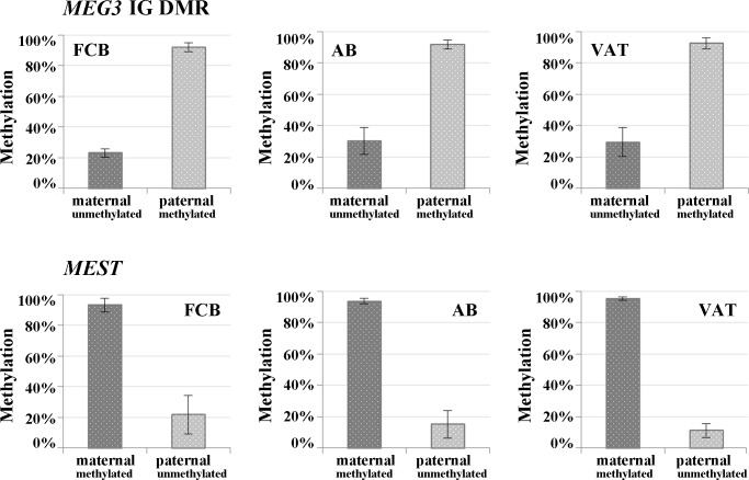
Altogether, 30 FCB, 23 AB, and 13 VAT samples were informative for the MEG3 IG DMR and 50 FCB, 36 AB, and 24 VAT samples for MEST. An average of 796±261 reads and a SNP ratio of 0.78±0.14 per sample were obtained for the MEG3 IG DMR and 1,036±355 reads and a SNP ratio of 0.72±0.2 for MEST, respectively. The methylated paternal allele of the MEG3 IG DMR showed >90% methylation (Fig 1, upper panel), relatively close to the theoretical 100%. The maternal allele, which was expected to be completely unmethylated, showed considerable hypermethylation (23–30%) in all three analyzed tissues. Similar results were observed for the oppositely imprinted MEST promoter (Fig 1, bottom panel), where the unmethylated paternal allele displayed an excess methylation (11–22%).
Fig 1. Parental allele-specific methylation of the MEG3 IG DMR and the MEST promoter.
Mean methylation levels and standard deviations of the paternal vs. maternal alleles were determined by DBS with the Roche GSJunior in FCB, AB, and VAT. For both genes, the non-imprinted allele, which is expected to be completely unmethylated, showed an aberrantly high methylation in all analyzed tissues, whereas the imprinted allele showed the expected (90–100%) methylation.
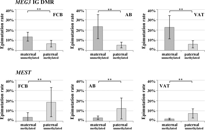
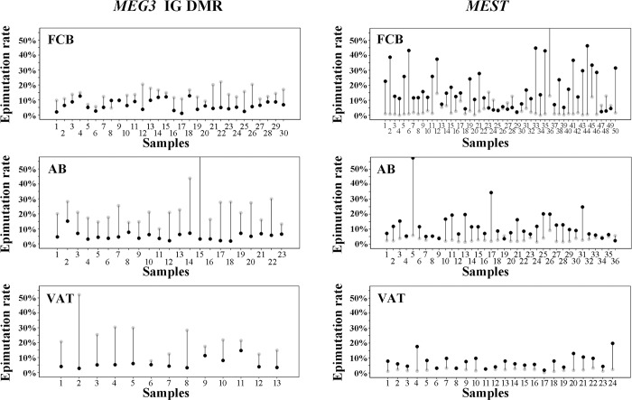
When looking at individual DNA molecules, the majority of the non-imprinted maternal MEG3 IG DMR and paternal MEST alleles were unmethylated (0–20% methylation), however the observed allele methylation values covered the whole range from 0% to 100% with a high number of alleles displaying >50% methylation (S1A Fig). In contrast, the vast majority of the imprinted paternal MEG3 and maternal MEST alleles were fully methylated (81–100% methylation) with a low number of alleles in the 0–80% methylation range. Alleles with >50% aberrantly (de)methylated CpGs were classified as epimutations. Epimutations were 3-4-fold more frequent on the normally unmethylated maternal MEG3 (13–23%) and paternal MEST alleles (8–19%), compared to the normally methylated paternal MEG3 (6–7%) and maternal MEST alleles (2–4%) (Fig 2; S5 Table). Irrespective of parental origin, the epimutation rate (ER) on the normally unmethylated non-imprinted allele was significantly (Mann-Whitney U test; p<0.001) higher than that on the normally methylated imprinted allele. Fig 3 displays the MEG3 IG DMR and MEST ERs of the paternal versus the maternal allele for each individual FCB, AB, and VAT sample. Despite considerable variation in allele-specific methylation between samples, the ER on the non-imprinted (unmethylated) maternal MEG3 IG DMR and paternal MEST allele was consistently and with very few execeptions considerable higher than that on the imprinted (methylated) allele. In individual samples, the ERs of the unmethylated allele ranged from 2% to 66% and of the methylated allele from 0% to 15% (S5 Table).
Fig 2. Parental allele-specific epimutation rates of the MEG3 IG DMR and the MEST promoter.
The percentage of alleles with >50% aberrantly (de)methylated CpGs was demined by DBS with the Roche GSJunior in FCB, AB, and VAT samples. The unmethylated alleles of the paternally imprinted MEG3 and the maternally imprinted MEST genes displayed significantly higher epimutation rates than the methylated alleles.
Fig 3. Epimutation rates of the MEG3 IG DMR and the MEST promoter in individual samples.
The percentage of paternal (black dots) versus maternal alleles (gray dots) with >50% aberrantly (de)methylated CpGs was determined by DBS with the Roche GSJunior in individual FCB, AB, and VAT samples. With very few exceptions, the unmethylated alleles of the paternally imprinted MEG3 and the maternally imprinted MEST genes displayed much higher epimutation rates than the methylated alleles.
The paternal and maternal alleles of iDMRs are expected to maintain their germline methylation patterns in somatic cells. As a reference for the ERs on the paternal allele we analyzed sperm samples. Unfortunately, oocytes as reference for the maternal allele were not available. It is striking that mean methylation of the methylated paternal MEG3 allele varied only 2–3% between sperm and different somatic tissues, whereas methylation variation of the unmethylated paternal MEST allele was between 10% and 20% (S2 Fig). The sperm ER was 0.9±1.6% for the MEG3 IG DMR and 0.5±1.4% for MEST. The mean ERs of the paternal allele in FCB, AB, and VAT were significantly higher (5–7% for MEG3 IG DMR and 8–19% for MEST). Thus, compared to somatic cells, the methylation imprints of paternally and maternally imprinted genes are strictly controlled in sperm and, to the extent of present knowledge, also in oocytes. Published single molecule analyses [28,29] suggest an ER of approximately 3% for the unmethylated MEG3 IG DMR in mature human oocytes (from assisted reproduction), compared to a mean ER of 13–23% of the maternal allele in the somatic tissues analyzed here.
DBS with the Illumina MiSeq
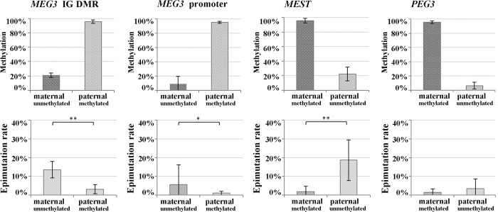
To increase sequencing power, DBS protocols were adopted to the Illumina MiSeq. Forty-five informative FCB samples were analyzed for the MEG3 IG DMR, 31 for the MEG3 promoter DMR, 58 for MEST, and 21 for PEG3. The mean read number per sample was 46,836±28,743 for the MEG3 IG DMR, 13,116±6,593 for the MEG3 promoter, 19,928±19,771 for MEST, and 42,429±17,053 for PEG3. The corresponding SNP ratios were 0.71±0.12, 0.58±0.21, 0.71±0.15, and 0.60±0.13. Both the primary MEG3 IG DMR and the secondary MEG3 promoter DMR are paternally imprinted. Consistent with the Roche GSJunior data set (Fig 1), the paternal MEG3 IG DMR showed 95.5±2.2% and the non-imprinted maternal allele 21.1±3.1% methylation (Fig 4, upper panel). For the secondary MEG3 promoter DMR, paternal versus maternal allele methylation was 94.8±1.4% and 9.0± 10.6%, indicating that the demethylated state of the maternal allele is more strictly maintained than for the primary IG DMR. The maternally imprinted genes MEST and PEG3 displayed 95.7±3.1% and 95.6±1.8% methylation on the maternal allele and 22.3±9.4% and 6.3±4.7% on the non-imprinted paternal allele.
Fig 4. Mean FCB methylation and epimutation rates of maternal versus paternal alleles of the MEG3 IG DMR, MEG3 promoter, MEST, and PEG3.
DBS was performed with the Illumina MiSeq. The non-imprinted allele of MEG3 (both primary and secondary DMR) and MEST exhibited significantly higher epimutation rates than the imprinted allele.
For the MEG3 IG DMR and MEST the distribution of single molecule methylation levels generated with Roche GSJunior and the MiSeq technology were virtually identical (S1 Fig), revealing a broad range (20–100%) of hypermethylated non-imprinted alleles, whereas the fully (81–100%) methylated state of the imprinted allele was more strictly maintained. For the secondary MEG3 promoter and PEG3 the number of hypermethylated non-imprinted alleles was much lower than for the MEG3 IG DMR and the MEST promoter. For the MEG3 IG DMR, the MEG3 promoter, and MEST, the ER on the non-imprinted (unmethylated) allele was significantly (p<0.001) higher than on the imprinted (methylated) allele (Fig 4, lower panel). The secondary MEG3 promoter DMR exhibited significantly (p<0.001) lower ERs on both the non-imprinted and imprinted allele than the primary MEG3 IG DMR.
Expression analysis
To test possible regulatory effects of hypermethylation of the non-imprinted allele (HNA), MEST and MEG3 expression were quantified by RT-qPCR in 20 and 7 VAT samples, respectively. Unfortunately, these were the only samples available for RNA analyses. The difference between the ERs on the non-imprinted and the imprinted parental allele (ERNI-ERI) corresponds to the percentage of cells with biallelic methylation. The expression of MEST assays 1 and 2 (both targeting isoform 2) correlated with each other and were highly variable among the studied samples (S3 Fig). Unexpectedly, there was a positive correlation (Spearman rho = 0.5; p = 0.03 and 0.02 for assay 1 and 2, respectively) between methylation and expression. In addition, expression was positively correlated with BMI (rho = 0.4; p = 0.11 and 0.05, respectively), ranging from 21 to 54 kg/m2 in the analyzed individuals. For the MEG3 IG DMR, expression and methylation appeared to be inversely correlated, but due to the low sample size the results were not significant.
Discussion
DBS for the study of imprinted genes
DBS including a user-friendly analysis software was first established on the Roche GSJunior [20,30], yielding approximately 1000 reads per gene and sample. DBS with the Illumina MiSeq increased the average read number to 20,000–40,000. The developed protocols for library preparation and data analysis can be relatively easily adopted to high-throughput Illumina sequencers, which will further increase power (number of genes analyzed simultaneously, number of reads). Single molecule methylation analysis by DBS provides novel insights into imprinting mechanisms. Combined with SNP typing, it allows one to determine the methylation profiles of several thousand individual paternal versus maternal alleles in larger numbers of samples.
For most (but not all) iDMRs, the allele-specific methylation patterns were not strictly maintained in somatic tissues and the non-imprinted alleles were more susceptible to epimutations. A considerable number of blood and fat samples from normal individuals displayed ERs of the non-imprinted MEG3 IG DMR and MEST alleles between 30% and 60%. Although minor differences in cell composition may contribute to methylation variation between tissue samples (blood and VAT, respectively), they cannot account for the observed huge range (from 2% to 66%) in HNA between individuals. The results were highly consistent, using different library preparation and NGS techniques (Roche and Illumina). This largely excludes experimental or bioinformatical artefacts. Direct measurement of ERs of the non-imprinted versus the imprinted allele is technically challenging. The sequence divergence between methylated and unmethylated molecules (of the same allele) after bisulfite conversion may lead to an amplification bias towards one, i.e. the unmethylated T-rich product [31]. In addition, DNA molecules with incomplete bisulfite conversion may mimick hypermethylated alleles. Reduced sequence complexity, asymmetric C to T alignments, and increased searching space, compared to the original reference sequence, can result in false-positive matches [32]. To overcome these problems, we excluded all reads with low sequencing quality or sequencing errors. However, improved protocols with single molecule tagging before PCR amplification may be necessary to exclude any amplification bias with 100% confidence.
Hypermethylation of the non-imprinted allele (HNA)
In this study we assessed the allele-specific methylation profile of four iDMRs at single molecule resolution. Only the PEG3 promoter displayed the expected black and white pattern of allele-specific methylation, with comparably low (1.5% and 3.4%) ERs on both parental alleles. The maternally imprinted MEST promoter and the paternally imprinted MEG3 IG DMR showed significantly higher ERs on the unmethylated allele than on the methylated allele. The secondary MEG3 promoter DMR showed an intermediate pattern. Although the ER was significantly higher on the non-imprinted maternal allele, its unmethylated state was much better maintained than for the MEG3 IG DMR.
Classical plasmid cloning studies [28] revealed that the methylation imprint of the MEG3 IG DMR is strictly maintained in oocytes (fully demethylated) and sperm (fully methylated), whereas a considerable proportion of alleles in preimplantation embryos and somatic tissues (amniocytes, fetal and adult blood) showed intermediate methylation patterns. Although parental alleles were not distinguished, it is plausible to assume that most intermediate patterns represented hypermethylated maternal alleles. Collectively, these results suggest that relaxation of the primary MEG3 IG DMR occurs after imprinting of the secondary MEG3 promoter DMR has been established in the early embryo. Then, the IG DMR, which functions as an upstream regulator, may at least partially become redundant.
Previous studies have shown loss/relaxation of imprinting (LOI/ROI) at the transcriptional level. Biallelic expression of IGF2 was observed in 10–20% of blood samples from normal healthy individuals [33,34]. Reports that biallelic IGF2 expression in normal colonic mucosa and blood was severalfold more frequent in colorectal cancer patients than in healthy individuals have promoted the idea that LOI predisposes to cancer [35,36]. Interestingly, cord bloods from healthy newborns did not show an association of LOI/ROI with increased IGF2 mRNA levels or abnormal methylation levels [34]. Our results revealed an enormous variation of allele-specific methylation of at least some imprinted genes in normal tissues/individuals. In essentially all studied samples, the MEG3 IG DMR and MEST ERs were severalfold higher on the unmethylated than on the methylated allele, implying biallelic methylation in a considerable number (up to 50%) of cells. Comparable HNA was observed in different somatic tissues (blood and fat) but not in germ cells and there was no obvious increase in the percentage of abnormally methylated alleles with age (between fetal cord blood and adult blood). Thus, HNA most likely originated during early somatic development (before separation of the different embryonal layers). Some iDMRs were more susceptible to HNA than others, irrespective of parental origin.
Intuitively, HNA of the studied iDMRs should be associated with transcriptional gene silencing. However, MEST showed a positive correlation between methylation and expression, indicating that its regulation in VAT is more complex. The human MEST gene is endowed with two promoters generating transcripts with alternative first exons. The promoter studied here is thought to control monoallelic expression of isoform 1, whereas the isoform 2 under control of the second promoter is biallelically expressed [37]. In addition to the imprinted isoform 1, the MEST iDMR controls the promoter of an antisense transcript. In a variety of tumors hypermethylation of this DMR was associated with upregulation of the sense gene, most likely by downregulation of the antisense transcript [38]. A similar mechanism may be responsible for MEST upregulation in fat tissue. It has been reported that the imprinted isoform 1 is upregulated in fat tissue of obese individuals in both humans and mice [39,40]. In the VAT samples studied here expression of isoform 2 also increased with BMI. In rodent models, Mest expression levels correlated with the development of diet-induced adiposity and fat mass expansion [41,42]. Mest upregulation and MEST hypomethylation have been linked to an early overnutritional environment in mice [43] and humans [16], respectively. Thus, MEST is a primary candidate for developmental programming of a metabolic phenotype(s).
The maternally expressed gene 3 (MEG3) is controlled by a primary germline DMR and a secondary DMR, which is established after fertilization. The primary MEG3 IG DMR lies in the intergenic region 13 kb upstream and the secondary DMR in the MEG3 promoter 1.5 kb upstream of the transcription start [44]. The maternal MEG3 locus expresses a dozen or more alternatively spliced long non-coding RNAs as well as microRNAs and small nucleolar RNAs [45,46]. In VAT methylation of the MEG3 IG DMR appeared to be associated with decreased expression, although the results were not significant.
Imprinted genes, metastable epialleles, and developmental programming
During early development the epigenome is highly susceptible to environmental factors, i.e. assisted reproductive technologies [47,48] or maternal nutrition [14,15]. Imprinted genes may be particularly susceptible to adverse environmental exposures because they escape genome-wide epigenetic reprogramming after fertilization and are functionally haploid. To maintain functionally important parent-specific methylation patterns during many cell divisions in the developing embryo, the unmethylated parental allele must be protected very efficiently against de novo methylation. At the H19-IGF2 locus, differential methylation is maintained by binding of CTCF zinc-finger proteins to the unmethylated allele [49,50]. In addition, histone proteins may be involved. Enrichment of H3K4me2 and H3K4me3 on the unmethylated parental chromosome may prevent binding of DNA methyltransferases and de novo methylation. Aberrantly acquired methylation marks may be enzymatically removed by ten-eleven translocation proteins [51]. Thus, maintenance of an unmethylated state at one parental allele requires an interplay of different mechanisms. Stochastic and/or environmentally induced errors in this complex process may lead to HNA during somatic development.
At first glance, it may be surprising that the iDMRs of MEST and MEG3, genes controlling somatic growth, display such enormous epigenetic variation. Previously, it has been shown that IGF2/Igf2, another imprinted region involved in growth regulation, shows considerable variation of allelic expression between normal individuals [33,34] and tissue-specific variation in allelic methylation during development [52]. Interestingly, growth-regulating genes, including MEST and MEG3, in an imprinted gene network become transcriptionally downregulated in multiple organs during postnatal growth deceleration/cessation [53]. Thus, HNA may be part of a physiological process for regulation of an imprinted gene network to limit body size. This process may be susceptible to developmental programming through environmental factors to enhance variation at the population level.
In many aspects HNA resembles metastable epialleles (ME), which can display variable methylation and expression in the absence of genetic variation and thus lead to phenotypic variation in genetically identical individuals [54]. ME candidates have also been identified in humans [55]. Epigenetic programming of MEs by environmental exposures such as maternal nutrition [56] and assisted reproduction [57] provides a link to common adult disease (DOHAD).
Conclusions
Some growth-regulating imprinted genes such as MEST and MEG3, are susceptible to HNA during development and differentiation, whereas the iDMRs of others (i.e. PEG3) are strictly maintained. HNA leads to a variable number of cells with biallelic ICR methylation (somatic mosaicism). Similar to MEs, the extent of HNA is highly variable between individuals, most likely due to stochastic and environmental programming during early development, and may contribute an additional layer of complexity to phenotypic diversity in mammals.
Supporting information
(PDF)
(PDF)
(PDF)
(PDF)
(PDF)
(PDF)
(PDF)
(PDF)
Acknowledgments
The authors thank Dr. Daniel Liedtke and Sabine Knaup, Würzburg Human Genetics Institute for help with the RT-qPCR analyses.
Data Availability
All relevant data are within the paper and its Supporting Information.
Funding Statement
There was no grant support. The study was funded by revenues of the Wuerzburg Human Genetics Institute. The funders had no role in study design, data collection and analysis, decision to publish, or preparation of the manuscript.
References
- 1.Massah S, Beischlag TV, Prefontaine GG. Epigenetic events regulating monoallelic gene expression. Crit Rev Biochem Mol Biol. 2015;50:337–358. doi: 10.3109/10409238.2015.1064350 [DOI] [PubMed] [Google Scholar]
- 2.Reik W, Constância M, Fowden A, Anderson N, Dean W, Ferguson-Smith A, et al. Regulation of supply and demand for maternal nutrients in mammals by imprinted genes. J Physiol. 2003;547:35–44. doi: 10.1113/jphysiol.2002.033274 [DOI] [PMC free article] [PubMed] [Google Scholar]
- 3.Gregg C, Zhang J, Weissbourd B, Luo S, Schroth GP, Haig D, et al. High-resolution analysis of parent-of-origin allelic expression in the mouse brain. Science. 2010;329:643–648. doi: 10.1126/science.1190830 [DOI] [PMC free article] [PubMed] [Google Scholar]
- 4.Plasschaert RN, Bartolomei MS. Genomic imprinting in development, growth, behavior and stem cells. Development. 2014;141:1805–1813. doi: 10.1242/dev.101428 [DOI] [PMC free article] [PubMed] [Google Scholar]
- 5.Edwards CA, Ferguson-Smith AC. Mechanisms regulating imprinted genes in clusters. Curr Opin Cell Biol. 2007;19:281–289. doi: 10.1016/j.ceb.2007.04.013 [DOI] [PubMed] [Google Scholar]
- 6.Reik W, Dean W, Walter J. Epigenetic reprogramming in mammalian development. Science. 2001;293:1089–1093. doi: 10.1126/science.1063443 [DOI] [PubMed] [Google Scholar]
- 7.Messerschmidt DM, Knowles BB, Solter D. DNA methylation dynamics during epigenetic reprogramming in the germline and preimplantation embryos. Genes Dev. 2014;28:812–828. doi: 10.1101/gad.234294.113 [DOI] [PMC free article] [PubMed] [Google Scholar]
- 8.Eggermann T, Perez de Nanclares G, Maher ER, Temple IK, Tümer Z, Monk D, et al. Imprinting disorders: a group of congenital disorders with overlapping patterns of molecular changes affecting imprinted loci. Clin Epigenetics. 2015;7:123 doi: 10.1186/s13148-015-0143-8 [DOI] [PMC free article] [PubMed] [Google Scholar]
- 9.Jelinic P, Shaw P. Loss of imprinting and cancer. J Pathol. 2007;211:261–268. doi: 10.1002/path.2116 [DOI] [PubMed] [Google Scholar]
- 10.Cortessis VK, Thomas DC, Levine AJ, Breton CV, Mack TM, Siegmund KD, et al. Environmental epigenetics: prospects for studying epigenetic mediation of exposure-response relationships. Hum Genet. 2012;131:1565–1589. doi: 10.1007/s00439-012-1189-8 [DOI] [PMC free article] [PubMed] [Google Scholar]
- 11.Feil R, Fraga MF. Epigenetics and the environment: emerging patterns and implications. Nat Rev Genet. 2012;13:97–109. doi: 10.1038/nrg3142 [DOI] [PubMed] [Google Scholar]
- 12.Gillman MW. Developmental origins of health and disease. N Engl J Med. 2005;353:1848–1850. doi: 10.1056/NEJMe058187 [DOI] [PMC free article] [PubMed] [Google Scholar]
- 13.Barker DJ. The origins of the developmental origins theory. J Intern Med. 2007;261:412–417. doi: 10.1111/j.1365-2796.2007.01809.x [DOI] [PubMed] [Google Scholar]
- 14.Gluckman PD, Hanson MA, Buklijas T, Low FM, Beedle AS. Epigenetic mechanisms that underpin metabolic and cardiovascular diseases. Nat Rev Endocrinol. 2009;5:401–408. doi: 10.1038/nrendo.2009.102 [DOI] [PubMed] [Google Scholar]
- 15.El Hajj N, Schneider E, Lehnen H, Haaf T. Epigenetics and life-long consequences of an adverse nutritional and diabetic intrauterine environment. Reproduction. 2014;148:R111–R120. doi: 10.1530/REP-14-0334 [DOI] [PMC free article] [PubMed] [Google Scholar]
- 16.El Hajj N, Pliushch G, Schneider E, Dittrich M, Müller T, Korenkov M, et al. Metabolic programming of MEST DNA methylation by intrauterine exposure to gestational diabetes mellitus. Diabetes. 2013;62:1320–1328. doi: 10.2337/db12-0289 [DOI] [PMC free article] [PubMed] [Google Scholar]
- 17.Weber M, Hellmann I, Stadler MB, Ramos L, Pääbo S, Rebhan M, et al. Distribution, silencing potential and evolutionary impact of promoter DNA methylation in the human genome. Nat Genet. 2007;39:457–466. doi: 10.1038/ng1990 [DOI] [PubMed] [Google Scholar]
- 18.Untergasser A, Cutcutache I, Koressaar T, Ye J, Faircloth BC, Remm M, et al. Primer3-new capabilities and interfaces. Nucleic Acids Res. 2012;40:e115 doi: 10.1093/nar/gks596 [DOI] [PMC free article] [PubMed] [Google Scholar]
- 19.Mardis ER. Next-generation DNA sequencing methods. Annu Rev Genomics Hum Genet. 2008;9:387–402. doi: 10.1146/annurev.genom.9.081307.164359 [DOI] [PubMed] [Google Scholar]
- 20.Rahmann S, Beygo J, Kanber D, Martin M, Horsthemke B, Buiting K. Amplikyzer: automated methylation analysis of amplicons from bisulfite flowgram sequencing. PeerJ PrePrints. 2013;1:e122v2. [Google Scholar]
- 21.Martin M. Cutadapt removes adapter sequences from high-throughput sequencing reads. EMBnet.journal. 2011;17:10–12. [Google Scholar]
- 22.Krueger F, Andrews SR. Bismark: a flexible aligner and methylation caller for Bisulfite-Seq applications. Bioinformatics. 2011;27:1571–1572. doi: 10.1093/bioinformatics/btr167 [DOI] [PMC free article] [PubMed] [Google Scholar]
- 23.Langmead B, Salzberg SL. Fast gapped-read alignment with Bowtie 2. Nat Methods. 2012;9:357–359. doi: 10.1038/nmeth.1923 [DOI] [PMC free article] [PubMed] [Google Scholar]
- 24.Li H, Handsaker B, Wysoker A, Fennell T, Ruan J, Homer N, et al. The Sequence Alignment/Map format and SAMtools. Bioinformatics. 2009;25:2078–2079. doi: 10.1093/bioinformatics/btp352 [DOI] [PMC free article] [PubMed] [Google Scholar]
- 25.Robinson JT, Thorvaldsdóttir H, Winckler W, Guttman M, Lander ES, Getz G, et al. Integrative genomics viewer. Nat Biotechnol. 2011;29:24–26. doi: 10.1038/nbt.1754 [DOI] [PMC free article] [PubMed] [Google Scholar]
- 26.Thorvaldsdóttir H, Robinson JT, Mesirov JP. Integrative Genomics Viewer (IGV): high-performance genomics data visualization and exploration. Brief Bioinform. 2013;14:178–192. doi: 10.1093/bib/bbs017 [DOI] [PMC free article] [PubMed] [Google Scholar]
- 27.Hurtado del Pozo C, Vesperinas-García G, Gómez-Ambrosi J, Frühbeck G, Corripio-Sánchez R, Rubio MA, Obregon MJ. IPO8 and FBXL10: new reference genes for gene expression studies in human adipose tissue. Obesity (Silver Spring). 2010;18:897–903. [DOI] [PubMed] [Google Scholar]
- 28.Geuns E, de Temmerman N, Hilven P, van Steirteghem A, Liebaers I, de Rycke M. Methylation analysis of the intergenic differentially methylated region of DLK1-GTL2 in human. Eur J Hum Genet. 2007;15:352–361. doi: 10.1038/sj.ejhg.5201759 [DOI] [PubMed] [Google Scholar]
- 29.Kuhtz J, Romero S, de Vos M, Smitz J, Haaf T, Anckaert E. Human in vitro oocyte maturation is not associated with increased imprinting error rates at LIT1, SNRPN, PEG3 and GTL2. Hum Reprod. 2014;29:1995–2005. doi: 10.1093/humrep/deu155 [DOI] [PubMed] [Google Scholar]
- 30.Beygo J, Ammerpohl O, Gritzan D, Heitmann M, Rademacher K, Richter J, et al. Deep bisulfite sequencing of aberrantly methylated loci in a patient with multiple methylation defects. PLoS One. 2013;8:e76953 doi: 10.1371/journal.pone.0076953 [DOI] [PMC free article] [PubMed] [Google Scholar]
- 31.Warnecke PM, Stirzaker C, Melki JR, Millar DS, Paul CL, Clark SJ. Detection and measurement of PCR bias in quantitative methylation analysis of bisulphite-treated DNA. Nucleic Acids Res. 1997;25:4422–6442. [DOI] [PMC free article] [PubMed] [Google Scholar]
- 32.Xi Y., Li W. BSMAP: whole genome bisulfite sequence MAPping program. BMC Bioinformatics. 2009;10:232 doi: 10.1186/1471-2105-10-232 [DOI] [PMC free article] [PubMed] [Google Scholar]
- 33.Sakatani T, Wei M, Katoh M, Okita C, Wada D, Mitsuya K, et al. Epigenetic heterogeneity at imprinted loci in normal populations. Biochem Biophys Res Comm. 2001;283:1124–1130. doi: 10.1006/bbrc.2001.4916 [DOI] [PubMed] [Google Scholar]
- 34.Rancourt RC, Harris HR, Barault L, Michels KB. The prevalence of loss of imprinting of H19 and IGF2 at birth. FASEB J. 2013;27:3335–3343. doi: 10.1096/fj.12-225284 [DOI] [PMC free article] [PubMed] [Google Scholar]
- 35.Cui H, Horon IL, Ohlsson R, Hamilton SR, Feinberg AP. Loss of imprinting in normal tissue of colorectal cancer patients with microsatellite instability. Nat Med. 1998;4:1276–1280. doi: 10.1038/3260 [DOI] [PubMed] [Google Scholar]
- 36.Cui H, Cruz-Correa M, Giardiello FM, Hutcheon DF, Kafonek DR, Brandenburg S, et al. Loss of IGF2 imprinting: a potential marker of colorectal cancer risk. Science. 2003;299:1753–1755. doi: 10.1126/science.1080902 [DOI] [PubMed] [Google Scholar]
- 37.Riesewijk AM, Hu L, Schulz U, Tariverdian G, Höglund P, Kere J, et al. Monoallelic expression of human PEG1/MEST is paralleled by parent-specific methylation in fetuses. Genomics. 1997;42:236–244. doi: 10.1006/geno.1997.4731 [DOI] [PubMed] [Google Scholar]
- 38.Kim J, Bretz CL, Lee S. Epigenetic instability of imprinted genes in human cancers. Nucleic Acids Res. 2015;43:10689–10699. doi: 10.1093/nar/gkv867 [DOI] [PMC free article] [PubMed] [Google Scholar]
- 39.Kosaki K, Kosaki R, Craigen WJ, Matsuo N. Isoform-specific imprinting of the human PEG1/MEST gene. Am J Hum Genet. 2000;66:309–312. doi: 10.1086/302712 [DOI] [PMC free article] [PubMed] [Google Scholar]
- 40.Kamei Y, Suganami T, Kohda T, Ishino F, Yasuda K, Miura S, et al. Peg1/Mest in obese adipose tissue is expressed from the paternal allele in an isoform-specific manner. FEBS Lett. 2007;581:91–96. doi: 10.1016/j.febslet.2006.12.002 [DOI] [PubMed] [Google Scholar]
- 41.Koza RA, Nikonova L, Hogan J, Rim JS, Mendoza T, Faulk C, et al. Changes in gene expression foreshadow diet-induced obesity in genetically identical mice. PLoS Genet. 2006;2:e81 doi: 10.1371/journal.pgen.0020081 [DOI] [PMC free article] [PubMed] [Google Scholar]
- 42.Nikonova L, Koza RA, Mendoza T, Chao PM, Curley JP, Kozak LP. Mesoderm-specific transcript is associated with fat mass expansion in response to a positive energy balance. FASEB J. 2008;22:3925–3937. doi: 10.1096/fj.08-108266 [DOI] [PMC free article] [PubMed] [Google Scholar]
- 43.Kozak LP, Newman S, Chao PM, Mendoza T, Koza RA. The early nutritional environment of mice determines the capacity for adipose tissue expansion by modulating genes of caveolae structure. PLoS One. 2010;5:e11015 doi: 10.1371/journal.pone.0011015 [DOI] [PMC free article] [PubMed] [Google Scholar]
- 44.Murphy SK, Wylie AA, Coveler KJ, Cotter PD, Papenhausen PR, Sutton VR, et al. Epigenetic detection of human chromosome 14 uniparental disomy. Hum Mutat. 2003;22:92–97. doi: 10.1002/humu.10237 [DOI] [PubMed] [Google Scholar]
- 45.Zhang X, Rice K, Wang Y, Chen W, Zhong Y. Nakayama Y, et al. Maternally expressed gene 3 (MEG3) noncoding ribonucleic acid: isoform structure, expression, and functions. Endocrinology. 2010;151:939–947. doi: 10.1210/en.2009-0657 [DOI] [PMC free article] [PubMed] [Google Scholar]
- 46.Kameswaran V, Bramswig NC, McKenna LB, Penn M, Schug J, Hand NJ, et al. Epigenetic regulation of the DLK1-MEG3 microRNA cluster in human type 2 diabetic islets. Cell Metab. 2014;19:135–145. doi: 10.1016/j.cmet.2013.11.016 [DOI] [PMC free article] [PubMed] [Google Scholar]
- 47.Van Montfoort AP, Hanssen LL, de Sutter P, Viville S, Geraedts JP, de Boer P. Assisted reproduction treatment and epigenetic inheritance. Hum Reprod Update. 2012;18:171–197. doi: 10.1093/humupd/dmr047 [DOI] [PMC free article] [PubMed] [Google Scholar]
- 48.El Hajj N, Haaf T. Epigenetic disturbances in in vitro cultured gametes and embryos: implications for human assisted reproduction. Fertil Steril. 2013;99:632–641. doi: 10.1016/j.fertnstert.2012.12.044 [DOI] [PubMed] [Google Scholar]
- 49.Schoenherr CJ, Levorse JM, Tilghman SM. CTCF maintains differential methylation at the Igf2/H19 locus. Nat Genet. 2003;33:66–69. doi: 10.1038/ng1057 [DOI] [PubMed] [Google Scholar]
- 50.Engel N, Thorvaldsen JL, Bartolomei MS. CTCF binding sites promote transcription initiation and prevent DNA methylation on the maternal allele at the imprinted H19/Igf2 locus. Hum Mol Genet. 2006;15:2945–2954. doi: 10.1093/hmg/ddl237 [DOI] [PubMed] [Google Scholar]
- 51.Kelsey G, Feil R. New insights into establishment and maintenance of DNA methylation imprints in mammals. Philos Trans R Soc Lond B Biol Sci. 2013;368:20110336 doi: 10.1098/rstb.2011.0336 [DOI] [PMC free article] [PubMed] [Google Scholar]
- 52.Weber M, Milligan L, Delalbre A, Antoine E, Brunel C, Cathala G, et al. Extensive tissue-specific variation of allelic methylation in the Igf2 gene during mouse fetal development: relation to expression and imprinting. Mech Dev. 2001;101:133–141. [DOI] [PubMed] [Google Scholar]
- 53.Lui JC, Finkielstain GP, Barnes KM, Baron J. An imprinted gene network that controls mammalian somatic growth is down-regulated during postnatal growth deceleration in multiple organs. Am J Physiol Regul Integr Comp Physiol. 2008;295:R189–196. doi: 10.1152/ajpregu.00182.2008 [DOI] [PMC free article] [PubMed] [Google Scholar]
- 54.Rakyan VK, Blewitt ME, Druker R, Preis JI, Whitelaw E. Metastable epialleles in mammals. Trends Genet. 2002;18:348–351. [DOI] [PubMed] [Google Scholar]
- 55.Harris RA, Nagy-Szakal D, Kellermayer R. Human metastable epiallele candidates link to common disorders. Epigenetics. 2013;8:157–163. doi: 10.4161/epi.23438 [DOI] [PMC free article] [PubMed] [Google Scholar]
- 56.Dominguez-Salas P, Moore SE, Baker MS, Bergen AW, Cox SE, Dyer RA, et al. Maternal nutrition at conception modulates DNA methylation of human metastable epialleles. Nat Commun. 2014;5:3746 doi: 10.1038/ncomms4746 [DOI] [PMC free article] [PubMed] [Google Scholar]
- 57.Estill MS, Bolnick JM, Waterland RA. Bolnick AD, Diamond MP, Krawetz SA. Assisted reproductive technology alters deoxyribonucleic acid methylation profiles in bloodspots of newborn infants. Fertil Steril. 2016;106:629–639. doi: 10.1016/j.fertnstert.2016.05.006 [DOI] [PubMed] [Google Scholar]
Associated Data
This section collects any data citations, data availability statements, or supplementary materials included in this article.
Supplementary Materials
(PDF)
(PDF)
(PDF)
(PDF)
(PDF)
(PDF)
(PDF)
(PDF)
Data Availability Statement
All relevant data are within the paper and its Supporting Information.


