Case Report
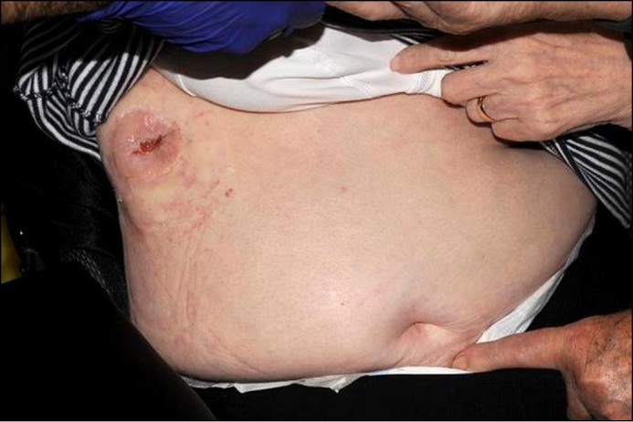
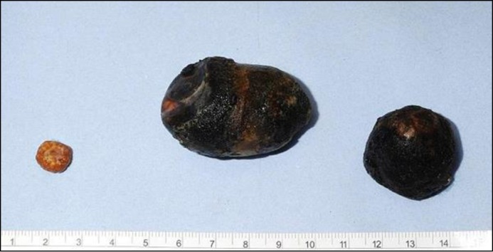
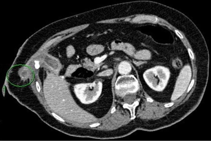
An 80-year-old woman presented with acute cholecystitis. On admission she was pyrexic and tachycardic with raised serum inflammatory markers. Computed tomography (CT) confirmed the diagnosis. She had significant morbidity from multiple sclerosis. She had left paresis and was wheelchair-bound. In view of her poor baseline function, the infection was managed with a percutaneous cholecystostomy drain. She recovered well with intravenous antibiotics and drainage of the infected gallbladder. After removal of the cholecystostomy, the drain track failed to heal and subsequently formed a cholecystocutaneous fistula (Figure 1). She managed well after the removal procedure, although the fistula was draining 200–300 mL turbid fluid daily. Two years later she passed 3 gallstones through the fistula, the largest being 5 cm in size (Figure 2). A follow-up CT scan showed an additional stone within the tract (Figure 3). The patient remains asymptomatic, so the plan is to wait for this to pass as well.
Figure 1.

Cholecystocutaneous fistula formation after the removal of the cholecystostomy.
Figure 2.

Three gallstones passed through the fistula, the largest 5 cm in size.
Figure 3.

Follow-up CT scan showing the presence of a stone within the tract.
Percutaneous cholecystostomy is a safe and effective way to manage acute cholecystitis in patients unsuitable for surgery.1 The incidence of cholecystocutaneous fistulas is low, and most form spontaneously due to untreated cholecystitis or are associated with malignancy of the gallbladder.2,3 An alternative management option for patients who are not suitable for surgery is to use endoscopic gallbladder drainage with a lumen-apposing metal stent (LAMS). This has been shown to be comparable to percutaneous drainage both in clinical outcomes and adverse events.4
There are few articles in the literature describing the passage of gallstones through cholecystocutaneous fistulas formed via previous percutaneous cholecystostomy.5,6 In these cases, the reported gallstones were smaller than the 5-cm stone seen in this patient. To our knowledge there are no reports of spontaneously passed gallstones 2 years after cholecystocutaneous fistula formation.
Disclosures
Author contributions: Both authors contributed equally to the manuscript. R. Date is the article guarantor.
Acknowledgements: We would like to thank Dr. Robert Stockwell for his assistance in producing the radiological images.
Financial disclosure: None to report.
Informed consent was obtained for this case report.
References
- 1.Tolan HK, Semiz Oysu A, Başak F, et al. Percutaneous cholecystostomy: A curative treatment modality forelderly and high ASA score acute cholecystitis patients. Ulus Travma Acil Cerrahi Derg. 2017; 23(1):34–8. [DOI] [PubMed] [Google Scholar]
- 2.Yüceyar S, Ertürk S, Karabiçak I, Onur E, Aydoğan F. Spontaneous cholecystocutaneous fistula presenting with an abscess containing multiple gallstones: A case report. Mt Sinai J Med. 2005; 72:402–4s. [PubMed] [Google Scholar]
- 3.Sodhi K, Athar M, Kumar V, Sharma ID, Husain N. Spontaneous cholecysto-cutaneous fistula complicating carcinoma of the gall bladder: A case report. Indian J Surg. 2012; 74(2):191–3. [DOI] [PMC free article] [PubMed] [Google Scholar]
- 4.Irani S, Ngamruengphong S, Teoh A, et al. Similar efficacies of endoscopic ultrasound gallbladder drainage with a lumen-apposing metal stent versus percutaneous transhepatic gallbladder drainage for acute cholecystitis. Clin Gastroenterol Hepatol. 2017; 15(5):738–45. [DOI] [PubMed] [Google Scholar]
- 5.Pripotnev S, Petrakos A. Cholecystocutaneous fistula after percutaneous gallbladder drainage. Case Rep Gastroenterol. 2014; 8(1):119–22. [DOI] [PMC free article] [PubMed] [Google Scholar]
- 6.Hariharan D, Lobo DN. Spontaneous extrusion of gallstones after percutaneous drainage. Ann R Coll Surg Engl. 2017; 99(3):e1–e2. [DOI] [PMC free article] [PubMed] [Google Scholar]


