Abstract
Purpose of Review
With the increase of publications available to the rehabilitation specialist, there is a need to identify a progression to safely progress the patient through their post-operative ACL reconstruction rehabilitation program. Rehabilitation after ACL reconstruction should follow an evidence-based functional progression with graded increase in difficulty in activities.
Recent Findings
Clinicians should be discouraged not to use strict time frames and protocols when treating patients following ACL reconstruction. Rather, guidelines should be followed that allow the rehabilitation specialists to progress the patient as improvements in strength, edema, proprioception, pain, and range of motion are demonstrated. Prior to returning to sport, specific objective quantitative and qualitative criteria should be met. The time from surgery should not be the only consideration.
Summary
The rehabilitation specialist needs to take into account tissue healing, any concomitant procedures, patellofemoral joint forces, and the goals of the patient in crafting a structured rehabilitation program. Achieving symmetrical full knee extension, decreasing knee joint effusion, and quadriceps activation early in the rehabilitation process set the stage for a safe progression. Weight bearing is begun immediately following surgery to promote knee extension and hinder quadriceps inhibition. As the patient progresses through their rehabilitative course, the rehabilitation specialist should continually challenge the patient as is appropriate based upon their goals, their levels of strength, amount of healing, and the performance of the given task.
Keywords: Knee, Cruciate, Rehabilitation, Progression, Guideline, Criteria
Introduction
Over 200,000 anterior cruciate ligament (ACL) injuries occur in the USA annually [1]. It is estimated that more than half of these injuries undergo surgical reconstruction [2]. Following ACL reconstruction (ACLR), under the direction of the orthopedic surgeon, the rehabilitation specialist is charged with the responsibility of returning the patient to their pre-injury level of function. Post-operative rehabilitation programs have changed dramatically over the past couple of decades. Strict protocols based on time elapsed from surgery have been replaced by criteria based guidelines (Table 1). These guidelines follow a progression where selective criteria are met prior to advancement in the program. This paper will discuss the progression of rehabilitation following ACL reconstruction.
Table 1.
Anterior cruciate ligament (BTB) rehabilitation guideline
| Post-operative phase I (weeks 0–2) |
| Goals: |
| ▪ Emphasis on full passive extension |
| ▪ Control post-operative pain/swelling |
| ▪ Range of motion 0° → 90° |
| ▪ Early progressive weight bearing |
| ▪ Prevent quadriceps inhibition |
| ▪ Independence in home therapeutic exercise program |
| Precautions: |
| ▪ Avoid active knee extension 40° → 0° |
| ▪ Avoid ambulation without brace locked at 0° |
| ▪ Avoid heat application |
| ▪ Avoid prolonged standing/walking |
| Treatment strategies: |
| ▪ Towel extensions, prone hangs, etc. |
| ▪ Quadriceps re-education (quad sets with EMS or EMG) |
| ▪ Progressive weight bearing |
| ▪ PWB → WBAT (patella tendon) with brace locked at 0° with crutches |
| ▪ Patella mobilization |
| ▪ Active flexion/active-assisted extension 90° → 0° exercise |
| ▪ SLR’s (all planes) |
| ▪ Brace locked at 0° for SLR (supine) |
| ▪ Short crank ergometry |
| ▪ Hip progressive resisted exercises |
| ▪ Proprioception board/balance system (bilateral weight bearing) |
| ▪ Leg press (bilateral/80° → 5° arc) (if ROM >90°) |
| ▪ Upper extremity cardiovascular exercises as tolerated |
| ▪ Cryotherapy |
| ▪ Home therapeutic exercise program: evaluation based |
| ▪ Emphasize patient compliance to home therapeutic exercise program and weight bearing precautions/progression |
| Criteria for advancement: |
| ▪ Ability to SLR without quadricep lag |
| ▪ ROM 0° → 90° |
| ▪ Demonstrate ability to unilateral (involved extremity) weight bear without pain |
| Post-operative phase 2 (weeks 2–6) |
| Goals: |
| ▪ ROM 0° → 130° |
| ▪ Good patella mobility |
| ▪ Minimal swelling |
| ▪ Restore normal gait (non-antalgic) |
| ▪ Ascend 8″ stairs with good control without pain |
| Precautions: |
| ▪ Avoid descending stairs reciprocally until adequate quadriceps control and lower extremity alignment |
| ▪ Avoid pain with therapeutic exercise and functional activities |
| Treatment strategies: |
| ▪ Progressive weight bearing/WBAT (patella tendon) with crutches brace opened 0° → 50°, if good quadriceps control (good quad set/ability to SLR without lag or pain) |
| ▪ D/C crutches when gait is non-antalgic |
| ▪ Brace changed to MD preference (OTS brace, patella sleeve, etc.) |
| ▪ Standard ergometry (if knee ROM >115°) |
| ▪ Leg press (90° → 0° arc) |
| ▪ AAROM exercises |
| ▪ Mini squats/weight shifts |
| ▪ Proprioception training: prop board/balance system/contralateral Theraband exercises |
| ▪ Initiate forward step-up program |
| ▪ Stairmaster |
| ▪ Aquaciser (gait training) if incision benign |
| ▪ SLR’s (progressive resistance) |
| ▪ Hamstring/calf flexibility exercises |
| ▪ Hip/hamstring PRE |
| ▪ Core stabilization exercises |
| ▪ Retrograde incline treadmill ambulation |
| ▪ Active knee extension to 40° |
| ▪ Home therapeutic exercise program: Individualized |
| Criteria for advancement: |
| ▪ ROM 0° → 125° |
| ▪ Normal gait pattern |
| ▪ Demonstrate ability to ascend 8″ step |
| ▪ Good patella mobility |
| Post-operative phase 3 (weeks 6–14) |
| Goals: |
| ▪ Restore Full ROM |
| ▪ Demonstrate ability to descend 8″ stairs with good leg control without pain |
| ▪ Improve ADL endurance |
| ▪ Improve lower extremity flexibility |
| ▪ Protect patellofemoral joint |
| Precautions: |
| ▪ Avoid pain with therapeutic exercise and functional activities |
| ▪ Avoid running and sport activity till adequate strength development and MD clearance |
| Treatment strategies: |
| ▪ Progress squat program |
| ▪ Initiate step down program |
| ▪ Leg press |
| ▪ Lunges |
| ▪ Isometric → isotonic knee extensions 90° → 40° |
| ▪ Advanced proprioception training (perturbations) |
| ▪ Agility exercises (sport cord) |
| ▪ Retrograde treadmill ambulation/running |
| ▪ Quadriceps stretching |
| ▪ KT 1000 knee ligament arthrometer exam at 3 months |
| ▪ Home therapeutic exercise program: evaluation based |
| Criteria for advancement: |
| ▪ ROM to WNL |
| ▪ Ability to descend 8″ stairs with good leg control/alignment without pain |
| ▪ Functional progression pending KT1000 and functional assessment |
| Post-operative phase 4 (weeks 14–22) |
| Goals: |
| ▪ Demonstrate ability to run pain free |
| ▪ Maximize strength and flexibility as to meet demands of activities of daily living |
| ▪ Isokinetic test ≥85% limb symmetry |
| Precautions: |
| ▪ Avoid pain with therapeutic exercise and functional activities |
| ▪ Avoid sport activity till adequate strength development and MD clearance |
| Treatment strategies: |
| ▪ Start forward running (treadmill) program when 8″ step down satisfactory |
| ▪ Continue LE strengthening and flexibility programs |
| ▪ Advance agility program/sport specific |
| ▪ Start plyometric program when strength base sufficient |
| ▪ Isotonic knee extension (full arc/pain and crepitus free) |
| ▪ Isokinetic training (fast → moderate velocities) |
| ▪ Home therapeutic exercise program: Individualized |
| Criteria for advancement: |
| ▪ Symptom-free running |
| ▪ Isokinetic test ≥85% limb symmetry |
| ▪ Lack of apprehension with plyometric and agility activities to date |
| Post-operative phase 5—return to sport (weeks 22–?) |
| Goals: |
| ▪ Lack of apprehension with sport specific movements |
| ▪ Maximize strength and flexibility as to meet demands of individual’s sport activity |
| ▪ Isokinetic test ≥90% limb symmetry |
| ▪ Hop test ≥90% limb symmetry |
| ▪ Acceptable quality movement assessment |
| Precautions: |
| ▪ Avoid pain with therapeutic exercise and functional activities |
| ▪ Avoid sport activity till adequate strength development and MD clearance |
| Treatment strategies: |
| ▪ Continue to advance LE strengthening, flexibility, and agility programs |
| ▪ Advance plyometric program |
| ▪ Brace for sport activity (MD preference) |
| ▪ Monitor patient’s activity level throughout course of rehabilitation |
| ▪ Reassess patient’s complaint’s (i.e., pain/swelling daily—adjust program accordingly) |
| ▪ Encourage compliance to home therapeutic exercise program |
| ▪ KT 1000 knee ligament arthrometer exam, isokinetic test, hop test(s), quality movement assessment at 6 months |
| ▪ Home therapeutic exercise program: Individualized |
| Criteria for discharge: |
| ▪ Isokinetic and functional hop test(s) ≥ 90% limb symmetry |
| ▪ Acceptable quality movement assessment |
| ▪ Lack of apprehension with sport specific movements |
| ▪ Flexibility to accepted levels of sport performance |
| ▪ Independence with gym program for maintenance and progression of therapeutic exercise program at discharge |
Adapted from “Anterior Cruciate Ligament Reconstruction” Cavanaugh JT, Postsurgical Rehabilitation Guidelines for the Orthopedic Clinician Cioppa-Mosca J, Cahill JB, Cavanaugh JT, Corradi-Scalise D, Rudnick H, Wollf AL, (eds) Elsevier Publishers pp.425–438, 2006
A functional progression was defined by Kegerreis [3] as an ordered sequence of activities enabling the acquisition or reacquisition of skills required for the safe and effective performance of athletic endeavors. In other words, the patient needs to master a simple activity before advancing to a more demanding activity. Programs are individualized, where some patients will be ready to advance sooner than others. Biological factors such as graft revascularization and maturation as well as fixation techniques are also considered to ensure a safe progression through the ACLR rehabilitation program.
Range of Motion
Following ACLR, achieving full knee extension range of motion (ROM) should be achieved as soon as possible. Extension loss results in abnormal joint arthrokinematics at both the tibiofemoral and patellofemoral joints. This in turn leads to abnormal articular cartilage contact pressures and quadriceps inhibition [4, 5•, 6].
Achieving full extension should ideally be achieved preoperatively. McHugh, et al. [7] found that patients with knee extension loss were 5× more likely to have extension loss issues after surgery.
Treatment strategies employed to achieve full extension include low load prolonged stretching (Fig. 1) and calf stretching. Patellofemoral joint mobilizations in a superior direction are utilized to encourage extension ROM [8] Sleeping in a post-operative brace locked at 0° extension is utilized to encourage extension and discourage the formation of a flexion contracture during the night hours. Full extension is one of several important criteria to meet to safely progress the patient off their crutches after surgery.
Fig. 1.
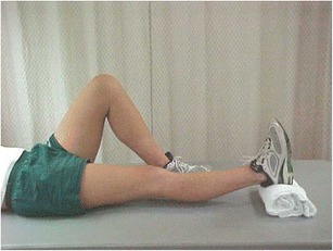
Passive low load prolonged stretching utilizing a rolled towel under the ankle
ROM exercises to facilitate flexion begin immediately after ACLR. ROM flexion goals of 120° should be met 4 weeks following surgery and full symmetrical flexion achieved by 12 weeks. Treatment strategies begin with active-assisted ROM exercises off the side of a plinth or bed (Fig. 2). Treatment strategies employed to further progress gains in flexion ROM include wall slides, active-assisted ROM sitting or on a step, and one half moon movement on a stationary bicycle. A short crank ergometer (Fig. 3) is utilized allowing patients to cycle earlier in the rehabilitative process and thus facilitate gains in flexion ROM [9]. Fleming and colleagues [10] demonstrated relatively low ACL peak strain values in vivo during stationary cycling.
Fig. 2.
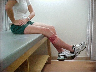
Active-assisted knee flexion/extension
Fig. 3.
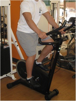
Short crank bicycle
Inferior-guided patellofemoral joint mobilizations are utilized to encourage gains in knee flexion [8]. When 120° of knee flexion is demonstrated, quadriceps stretching off the side of a plinth (Fig. 4) and eventually in a prone position is introduced to the patient’s program. Soft tissue massage can be of particular benefit throughout the progression to restore full symmetrical flexion ROM.
Fig. 4.
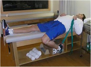
Quadricep stretching off the side of a plinth
Post-operative Weight Bearing
Weight bearing progression following ACLR is dictated by graft selection and surgeon preference. Advanced fixation techniques such as cancellous screw bone-to-bone fixation allow for immediate post-operative weight bearing. Following ACLR with an autologous bone-patellar tendon-bone (BTB) graft weight bearing is at first partial (50%) utilizing crutches and subsequently progressed to weight bearing as tolerated (WBAT) on successive days. This progression allows the knee joint to acclimate to increased loads. Ambulating in water, e.g., underwater treadmill (Fig. 5) can be utilized to gradually apply increased load through the knee joint and assist in the development of a normal gait pattern. Walking in chest-deep water results in a 60 to 75% reduction in weight bearing, while walking in waist-deep water results in a 40 to 50% reduction in weight bearing [11, 12].
Fig. 5.
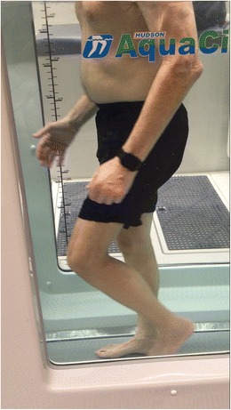
Underwater treadmill (Hudson Aquatic Systems LLC, Angola, IN)
A post-operative brace is initially locked at 0° for ambulation to protect the harvest site. The brace is opened when quadriceps control is demonstrated by the ability of the patient to straight leg raise (SLR) without a quadriceps lag or complaints of pain. Crutches are then discontinued upon meeting the criteria of demonstrating a normal non-antalgic gait.
A significant decrease in patellofemoral pain has been reported when an immediate progressive weight bearing guideline is utilized [13•]. ACLR’s utilizing hamstring or allografts are progressed at a slower rate secondary to the decreased strength of soft tissue fixation and biological considerations, respectively. Weight bearing may be delayed following ACLR’s with concomitant articular cartilage or meniscus repair procedures.
Strengthening
Re-establishing quadriceps control is an early goal of post-operative ACLR rehabilitation. Controlling post-operative effusion assists in discouraging quadriceps inhibition. Spencer et al. [14] identified that mechanoreceptors in the joint capsule respond to changes in tension and in turn inhibit motor nerves supplying the quadriceps muscles. Therapeutic interventions utilized include the use a commercial cold with compression device (Fig. 6), quadriceps setting with a towel under the knee, and weight bearing with the appropriate amount of load. Should a patient have difficulty eliciting a quadriceps contraction, a biofeedback unit or an electrical muscle stimulator can be used in conjunction with the quadriceps setting exercise to better facilitate a quadriceps contraction. Numerous studies [15–17] have demonstrated an earlier return of quadriceps strength after ACLR with the use of electrical stimulation. As quadriceps activation is demonstrated, a key observation in order to progress the patient is seeing the patient perform a straight leg raise without the assistance of the post-operative brace without any complaints of pain or quadriceps lag. When this criteria is met, the post-operative brace can be opened to allow normal knee range of motion during ambulation. Crutches are continued at this point until a non-antalgic gait is demonstrated. Closed kinetic chain exercises including leg press and squats inside a pain free arc of motion are introduced as these activities have been shown to minimize stress to the ACL [18–21, 22•, 23]. Limited evidence now demonstrates that open kinetic chain (OKC) exercises inside a 90°–0° arc of motion may not compromise graft laxity.[24–26]. Mikkelsen et al., randomized 44 ACLR (BTB) patients to either a closed chain rehabilitation only program and a closed chain program that added open chain exercises 6 weeks post-operatively. At 6-month follow up, KT-1000 knee ligament arthrometer values showed no significant difference in knee laxity between the groups. A significant increase in quadriceps strength in the open chain group was also identified. With the demonstration of 0°–130° ROM, OKC exercise progression begins with multiangle quadriceps isometrics inside a 90°–40° arc of motion (Fig. 7). Isometric exercises are progressed to isotonic exercises using progressive resistance (PRE). At 3 months, post-operatively isotonic exercise is allowed throughout a full arc of motion and progressed to isokinetic exercises utilizing moderate-fast speeds. Throughout this progression, the rehabilitation specialist should closely monitor the patellofemoral joint for crepitus and complaints of pain.
Fig. 6.
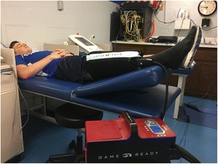
Gameready cold/compression device
Fig. 7.
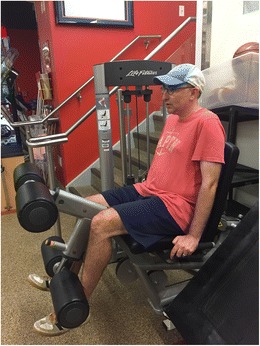
Isometric Knee Extension at 60° knee flexion
Neuromuscular Training
After ACLR, surgery afferent information is altered, which results in a disruption in the pathway between the patient’s center of gravity and base of support [27]. Improving neuromuscular reaction time to imposed loads enhances dynamic stabilization around the knee and thus protects the static reconstructed tissue from overstress or re-injury [28]. As soon as the patient achieves 50% weight bearing, neuromuscular/balance training is initiated on a dynamic balance system (Fig. 8) or proprioception device (foam cushion, rocker board, etc.). Balance activities are then increased progressively to include unilateral weight bearing, use of multiplanar support surfaces, and perturbation training [29]. Activities should attempt to eliminate or alter sensory information from the visual, vestibular, and somatosensory systems so as to challenge the other systems.
Fig. 8.
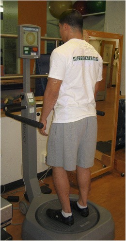
Dynamic balance system (Biodex Corporation, Shirley, NY)
Continued Progression
As range of motion and strength is demonstrated, the patient is instructed in a progressive step-up program, first mastering 6″ steps advancing to normal stair height 8″ steps. As further strength is demonstrated, a forward step down program is introduced (Fig. 9).
Fig. 9.
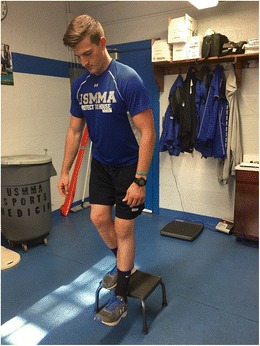
Forward step down exercise off an 8″ step
After 3 months post-op, if ROM is within normal limits and sufficient strength is demonstrated via a pain-free 8″ step down without deviation, a running program is started. Backward running is preceded by forward running, as retrograde running has been shown to generate lower patellofemoral joint compression forces than forward running [30]. An Alter G treadmill (Alter G, Inc., Fremont, CA) (Fig. 10) is utilized to incrementally add load during a forward running progression. A running program is progressed with an emphasis on speed over shorter distances vs. slower distance running.
Fig. 10.
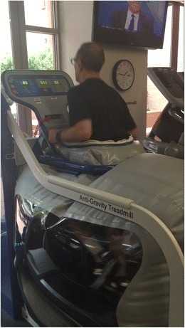
Unweighted treadmill (AlterG Inc., Fremont, CA)
Plyometric training is then incorporated only if full ROM, an adequate strength base and flexibility, is demonstrated. Plyometric training should follow a progression with its components of speed, intensity, load, volume, and frequency being monitored and progressed accordingly. Activities should begin with simple drills and advance to more complex exercises (e.g., double leg jumping vs. box drills). Agility and deceleratory training are important interventions to include in the later phases of rehabilitation in preparation for return to sport.
Return to Sport
When to return to sport following ACLR is a controversial issue. Pinczewski et al. [31] reported that one in four patients undergoing an ACLR will suffer a second tear within 10 years of their first. Paterno et al. [32•] reported an incidence rate of a second ACL injury within 2 years after returning to sports was nearly 6× greater then healthy controls.
More and more surgeons and rehabilitation specialists are utilizing numerous forms of assessment in determining an athlete’s readiness to return to play. Subjective rating scales, knee laxity testing, isokinetic testing, functional hop testing, balance testing, and qualitative movement assessment (Fig. 11) are utilized to provide evidence in the decision making process. Acceptable scores on these assessments are required to safely return the athlete to sport. Following discharge from a formal rehabilitation program, volume of athletic exposures needs to be modified.
Fig. 11.
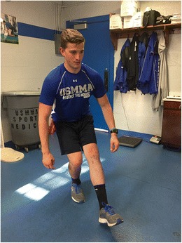
Quality movement assessment of a single leg squat exercise
Several studies have demonstrated deficits in muscular strength, kinesthetic sense, balance, and force attenuation 6 months to 2 years following reconstruction [32•, 33–35]. Return to sport 6 months following ACLR, therefore is no longer the expected norm.
Summary
Rehabilitation following ACL reconstruction has shifted from a paradigm based on protocols to a progression based program with gradient increases in difficulty. It is the rehabilitation specialist’s responsibility to consider the forces placed on the healing ACL graft and patellofemoral contact pressures generated during specific exercises and activities. Early in the rehabilitative process, the focus needs to be on gaining full knee extension, decreasing edema, and developing quadriceps strength. Therapeutic exercises should progress in difficulty often being performed in a variety of positions and settings. Neuromuscular training should be implemented into the rehabilitation program as early as deemed appropriate and progressed accordingly throughout the rehabilitative process. ACLR rehabilitation progression should be based on objective criteria and not just time frames. In order to achieve a successful outcome the rehabilitation specialist must continually assess the patient and select exercises that challenge the patient properly. When the patient is ready to return to sports, objective criteria must be met in order to reduce the risk of further injury. This transition is now considered to take place at greater than 6 months. Exposure/volume to athletic activity needs to be controlled in the post-rehabilitation period.
Compliance with Ethical Standards
Conflict of Interest
Both authors declare that they have no conflict of interest.
Human and Animal Rights and Informed Consent
This article does not contain any studies with human or animal subjects performed by any of the authors. Additional informed consent was obtained from all individual participants for whom identifying information is included in this article.
Footnotes
This article is part of the Topical Collection on ACL Rehab
References
Papers of particular interest, published recently, have been highlighted as: • Of importance •• Of major importance
- 1.Albrieght JC, Carpenter JE, Graf BK, Richmond JC, editors. Knee and leg: soft-tissue trauma. Rosemont: Orthopaedic Knowledge Update 6 American Academy of Orthopaedic Surgeons; 1999. [Google Scholar]
- 2.Centers for Disease Control & Prevention, National Center for Health Statistics . National hospital discharge survey: annual summary, 1996 [monograph on the Internet] Atlanta: Centers for Disease Control and Prevention; 1996. [Google Scholar]
- 3.Kegerreis S. The construction and implementation of a functional progression as a component of athletic rehabilitation. J Orthop Sports Phys Ther. 1983;5(1):14–19. doi: 10.2519/jospt.1983.5.1.14. [DOI] [PubMed] [Google Scholar]
- 4.Harner CD, Irrgang JJ, Paul J, Dearwater S, Fu FH. Loss of motion after anterior cruciate ligament reconstruction. Am J Sports Med. 1992;20(5):499–506. doi: 10.1177/036354659202000503. [DOI] [PubMed] [Google Scholar]
- 5.Shelbourne KD, Gray T. Minimum 10-year results after anterior cruciate ligament reconstruction: how the loss of normal knee motion compounds other factors related to the development of osteoarthritis after surgery. Am J Sports Med. 2009;37(3):471–480. doi: 10.1177/0363546508326709. [DOI] [PubMed] [Google Scholar]
- 6.Benum P. Operative mobilization of stiff knees after surgical treatment of knee injuries and post traumatic conditions. Acta Orthop Scand. 1982;53(4):625–631. doi: 10.3109/17453678208992269. [DOI] [PubMed] [Google Scholar]
- 7.McHugh MP, Tyler TF, Gleim GW, Nicholas SJ. Preoperative indicators of motion loss and weakness following anterior cruciate ligament reconstruction. J Orthop Sports Phys Ther. 1998;27:407–411. doi: 10.2519/jospt.1998.27.6.407. [DOI] [PubMed] [Google Scholar]
- 8.Fulkerson JP, Hungerford D. Disorders of the patellofemoral joint. 2. Williams & Wilkens: Baltimore; 1990. [Google Scholar]
- 9.Schwartz RE, Asnis PD, Cavanaugh JT, Asnis SE, Simmons JE, Lasinski PJ. Short crank cycle ergometry. J Orthop Sports Phys Ther. 1991;13(2):95–100. doi: 10.2519/jospt.1991.13.2.95. [DOI] [PubMed] [Google Scholar]
- 10.Fleming BC, Beynnon BD, Renstrom PA, et al. The strain behavior of the anterior cruciate ligament during bicycling. Am J Sports Med. 1998;26(1):109–118. doi: 10.1177/03635465980260010301. [DOI] [PubMed] [Google Scholar]
- 11.Bates A, Hanson N. The principles and properties of water. In: Aquatic exercise therapy. Philadelphia: WB Saunders, 1996:1–320.
- 12.Harrison RA, Hilman M, Bulstrode S. Loading of the lower limb when walking partially immersed: implications for clinical practice. Physiotherapy. 1992;78:164. doi: 10.1016/S0031-9406(10)61377-6. [DOI] [Google Scholar]
- 13.Tyler TF, MP MH, Gleim GW, Nicholas SJ. The effect of immediate weightbearing after anterior cruciate ligament reconstruction. Clin Orthop Relat Res. 1998;357:141–148. doi: 10.1097/00003086-199812000-00019. [DOI] [PubMed] [Google Scholar]
- 14.Spencer JD, Hayes KC, Alexander IJ. Knee joint effusion and quadriceps reflex inhibition in man. Arch Phys Med Rehabil. 1984;65(4):171–177. [PubMed] [Google Scholar]
- 15.Delitto A, Rose SJ, McKowen JM, Lehman RC, Thomas JA, Shively RA. Electrical stimulation versus voluntary exercise in strengthening thigh musculature after anterior cruciate ligament reconstructive surgery. Phys Ther. 1988;68:660–663. doi: 10.1093/ptj/68.5.660. [DOI] [PubMed] [Google Scholar]
- 16.Lossing I, Gremby G, Johnson T, Morelli B, Peterson L, Renstrom P. Effects of electrical muscle stimulation combined with voluntary contractions after knee ligament surgery. Med Sci Sports Exerc. 1988;20(1):93–98. doi: 10.1249/00005768-198802000-00014. [DOI] [PubMed] [Google Scholar]
- 17.Snyder-Mackler L, Ladin Z, Schepsis AA, Young JC. Electrical stimulation of the thigh muscles after reconstruction of the anterior cruciate ligament: effects of electrically elicited contraction of the quadriceps femoris and hamstring muscles on gait and on strength of the thigh muscles. J Bone Joint Surg Am. 1991;73(7):1025–1036. doi: 10.2106/00004623-199173070-00010. [DOI] [PubMed] [Google Scholar]
- 18.Henning CE, Lynch MA, Glick KR. An in vivo strain gauge study of elongation of the anterior cruciate ligament. Am J Sports Med. 1985;13(1):22–26. doi: 10.1177/036354658501300104. [DOI] [PubMed] [Google Scholar]
- 19.Lutz GE, Palmitier RA, An KN, et al. Comparison of tibiofemoral joint forces during open kinetic chain and closed kinetic chain exercises. J Bone Joint Surg Am. 1993;75(5):732–739. doi: 10.2106/00004623-199305000-00014. [DOI] [PubMed] [Google Scholar]
- 20.Yack HJ, Collins CE, Whieldon TJ. Comparison of closed and open kinetic chain exercise in the anterior cruciate ligament-deficient knee. Am J Sports Med. 1993;21(1):49–54. doi: 10.1177/036354659302100109. [DOI] [PubMed] [Google Scholar]
- 21.Ohkoshi Y, Yasuda K, Kaneda K, et al. Biomechanical analysis of rehabilitation in the standing position. Am J Sports Med. 1991;19(6):605–611. doi: 10.1177/036354659101900609. [DOI] [PubMed] [Google Scholar]
- 22.Wilk KE, Escamilla RF, Fleisig GS, et al. A comparison of tibiofemoral joint forces and electromyographic activity during open and closed kinetic chain exercises. Am J Sports Med. 1996;24(4):518–527. doi: 10.1177/036354659602400418. [DOI] [PubMed] [Google Scholar]
- 23.Bynum EB, Barrack RL, Alexander AH. Open versus closed chain kinetic exercises after anterior cruciate ligament reconstruction. A prospective randomized study. Am J Sports Med. 1995;23(4):401–406. doi: 10.1177/036354659502300405. [DOI] [PubMed] [Google Scholar]
- 24.Hooper D, Morrissey M, Drechsler W, Morrissey D, King J. Open and closed kinetic chain exercises in the early period after anterior cruciate ligament reconstruction. Am J Sports Med. 2001;297(2):167–174. doi: 10.1177/03635465010290020901. [DOI] [PubMed] [Google Scholar]
- 25.Mikkelsen C, Werner S, Eriksson E. Closed kinetic chain alone compared to combined open and closed kinetic chain exercises for quadriceps strengthening after anterior cruciate ligament reconstruction with respect to return to sport, a prospective matched follow-up study. Knee Surg Sports Traumatol Arthrosc. 2000;8:337–342. doi: 10.1007/s001670000143. [DOI] [PubMed] [Google Scholar]
- 26.Morrissey M, Drechsler W, Morrissey D, Knight P, Armstrong P, McAuliffe T. Effects of distally fixated versus nondistally fixated leg extensor resistance training on knee pain in the early period after anterior cruciate ligament reconstruction. Phys Ther. 2002;82:35–43. doi: 10.1093/ptj/82.1.35. [DOI] [PubMed] [Google Scholar]
- 27.Corrigan JP, Cashman WF, Brady MP. Proprioception in the cruciate deficient knee. J Bone Joint Surg Br. 1992;74(2):247–250. doi: 10.1302/0301-620X.74B2.1544962. [DOI] [PubMed] [Google Scholar]
- 28.Johansson H, Sjolander P, Soojka P. Activity in receptor afferents from the anterior cruciate ligament evokes reflex effects on fusimotor neurones. Neurosci Res. 1990;8(1):54–59. doi: 10.1016/0168-0102(90)90057-L. [DOI] [PubMed] [Google Scholar]
- 29.Cavanaugh JT, Moy RJ. Balance and postoperative lower extremity joint reconstruction. Orthop Phys Ther Clin NA. 2002; 11(1):75–99.
- 30.Flynn TW, Soutas-Little RW. Patellofemoral joint compressive forces in forward and backward running. J Orthop Sports Phys Ther. 1995;21(5):277–282. doi: 10.2519/jospt.1995.21.5.277. [DOI] [PubMed] [Google Scholar]
- 31.Pinczewski LA, Lyman J, Salmaon LJ, Russell VJ, Roe J, Linklater J. A 10-year study comparison of anterior cruciate ligament reconstruction with hamstring tendon and patellar tendon autograft: a controlled, prospective trial. Am J Sports Med. 2007;35:564–574. doi: 10.1177/0363546506296042. [DOI] [PubMed] [Google Scholar]
- 32.Paterno MV, Rauth MJ, Schmitt LC, Ford KR, Hewett TE. Incidence of second ACL injuries 2 years after primary ACL reconstruction and return to sport. Am J Sports Med. 2014;42(7):1567–1573. doi: 10.1177/0363546514530088. [DOI] [PMC free article] [PubMed] [Google Scholar]
- 33.Decker MJ, Torry MR, Noonan TH, Sterett WI, Steadman JR. Gait retraining after anterior cruciate ligament reconstruction. Arch Phys Med Rehabil. 2004;85(5):848–856. doi: 10.1016/j.apmr.2003.07.014. [DOI] [PubMed] [Google Scholar]
- 34.Ernst GP, Saliba E, Diduch DR, Hurwitz SR, Ball DW. Lower extremity compensations following anterior cruciate ligament reconstruction. Phys Ther. 2000;80(3):251–260. [PubMed] [Google Scholar]
- 35.Mattacola CG, Perrin DH, Gansneder BM, Gieck JH, Saliba EN, McCue FC., 3rd Strength, functional outcome, and postural stability after anterior cruciate ligament reconstruction. J Athl Train. 2002;37(3):262–268. [PMC free article] [PubMed] [Google Scholar]


