INTRODUCTION
The analogical description of a shape may be helpful in better understanding of anatomical structures during imaging. Seagull is a popular name for a seabird with a heavy body and two long wings. The “seagull sign “has been used in cardiology for evaluation of the mitral valve and in orthopedics for erosive osteoarthritis.[1,2,3] On a plain X-ray abdomen, a triradiate collection of a dark shadow of nitrogen gas within gallstone creates a “seagull sign”.[4]
“Seagull sign” has been commonly described for the left adrenal gland during endoscopic ultrasound (EUS).[5] The “seagull sign” in abdominal ultrasound identifies the celiac trunk and its division into hepatic and splenic arteries creates the wings of the seagull.[6] Wherever an artery divides into two smaller arteries or two veins unite to form a larger vein, they can give an appearance of a seagull. The union of the splenic vein, superior mesenteric vein and the formation of portal vein (PV) can also give an appearance of a seagull. Looking at the course of arteries and veins, application of color Doppler during EUS can provide a more extensive use of the “seagull sign”.[7,8,9] The size of wings may vary and the direction of wings may appear in different axis depending on the site of the division or union and the modality of evaluation. This article gives a detailed description of the “seagull sign” from different locations for unions and divisions. The understanding of these seagulls can help in quick and consistent identification of structures during EUS.
SEAGULLS OF STOMACH
It is usually possible to see the adrenal gland, celiac trunk, and PV forming three seagulls in the stomach.
Seagull of the left adrenal gland
The left adrenal gland is identified by anticlockwise rotation of about 45-90° after identifying the celiac artery [Figure 1]. The seagull-shaped left adrenal gland with a cranial and the caudal limb is easily identified at the upper border of the left kidney. The cranial limb lies posterior to the stomach and anterior to the left crus of diaphragm, whereas the caudal limb lies posterior to the pancreas and anterior to the left kidney. This seagull can be easily differentiated from other seagulls by absence of color flow on application of Doppler.
Figure 1.
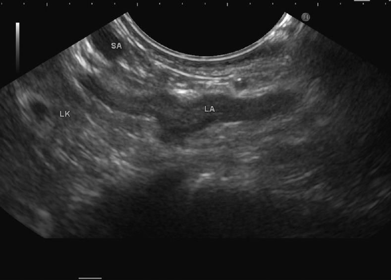
The seagull-shaped left adrenal gland with a cranial and caudal limb is identified at the upper border of the left kidney
The venous “seagull” of stomach
Union of the splenic vein, superior mesenteric vein and the formation of PV gives an appearance of a vascular seagull (venous) with the splenic vein forming the body of the seagull, the superior mesenteric vein forming one limb going parallel to the pancreas (7 to 10 o'clock) and the PV forming the limb going towards the liver (3 to 5 o'clock) [Figure 2].
Figure 2.
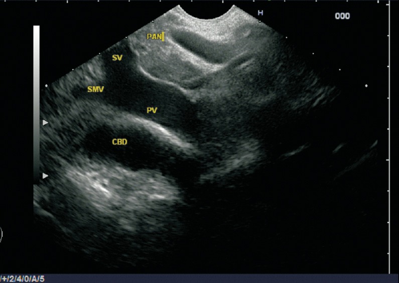
Union of the splenic vein (SV) with the superior mesenteric vein (SMV) and formation of portal vein (PV) gives an appearance of a venous vascular seagull. The common bile duct is identified beyond the confluence
The arterial “seagull” of stomach
The division of the celiac artery into the splenic artery and hepatic artery (HA) forms a seagull-shaped appearance with the celiac artery forming the body of the seagull, the splenic artery forming one limb going along the upper border of the pancreas (10 to 12 o'clock) and the common hepatic artery (CHA) forming the limb going towards the liver (5 to 7 o'clock) [Figures 3 and 4]. This has been also called as a “Whale's tail” sign.
Figure 3.
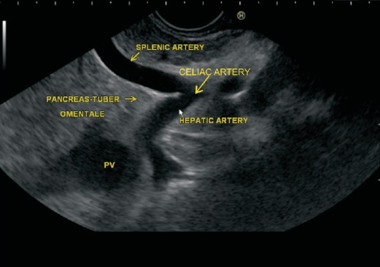
The division of the celiac artery (CA) into the splenic and hepatic artery forms a seagull-shaped appearance. The area between the bifurcations of these two arteries is occupied by the tuber omentale part of the pancreatic body
Figure 4.
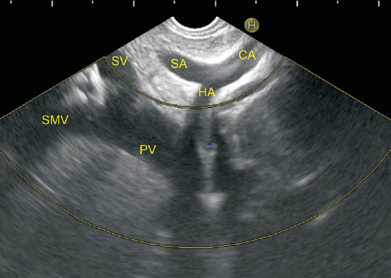
The CA is forming the body of the arterial seagull and the splenic artery (SA) is going towards the 12 o'clock position and the common hepatic artery (CHA) is going towards the liver at 7 o'clock position. The confluence of the SV and SMV to form PV is also seen
SEAGULLS OF DUODENUM
It is usually possible to see the CHA and the PV forming two vascular seagulls in the duodenal bulb.
The venous seagull of duodenum
The union of the superior mesenteric, splenic vein and the formation of PV gives an appearance of a vascular seagull (venous); with the PV forming the body of the seagull, the superior mesenteric vein forming the limb going the parallel to the pancreas (3 to 5 o'clock), and the splenic vein forming the limb going towards the left side of the screen (4 to 6 o'clock) [Figure 5].
Figure 5.
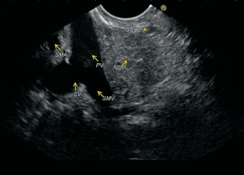
The union of the SMV and SV with formation of the PV gives an appearance of vascular seagull with the PV forming the body of the seagull. The superior mesenteric artery (SMA), gastroduodenal artery (GDA), and neck of pancreas (NOP) are also seen in the image
The arterial “seagull” of duodenum
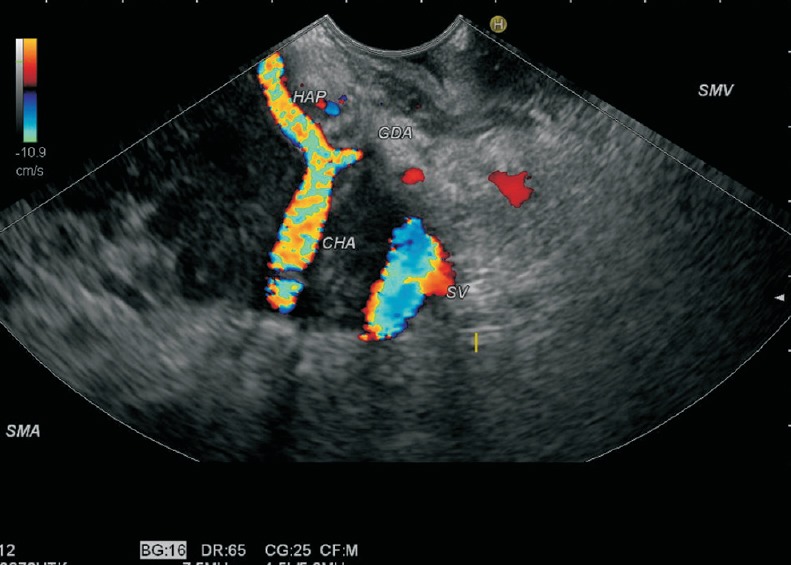
On EUS, the CHA is seen to indent the PV near the border away from the duodenal bulb (left border). The division of the CHA into the gastroduodenal artery (GDA) and the proper hepatic artery gives an appearance of vascular seagull (arterial) with the CHA forming the body of the seagull, the GDA forming the limb going towards the pancreas (3 o'clock) and the proper hepatic artery forming the limb going towards the liver (10 to 11 o'clock) [Figures 6 and 7].
Figure 6.
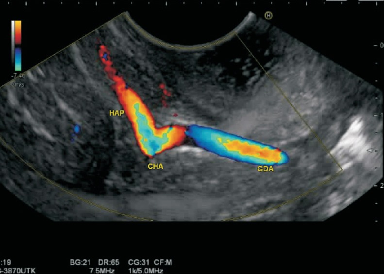
The division of the CHA into the GDA and proper hepatic artery (HAP) gives an appearance of arterial vascular seagull
Figure 7.
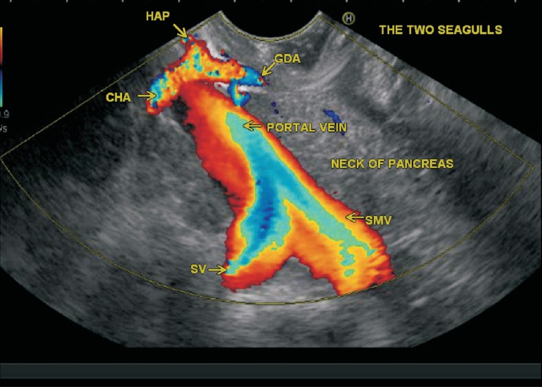
This image shows two vascular seagulls from duodenal bulb. The upper part of the image showing the division of the CHA into the GDA and HAP gives an appearance of smaller vascular seagull. A larger vascular seagull is also seen due to confluence of SV and SMV
DISCUSSION
“Seagull sign” has been popular among physicians to describe interesting anatomical structures. The “seagull sign” seems to be a fancy term, but on EUS it offers a wealth of information to the endosonographers. The advent of EUS with color Doppler imaging can help endosonographers in demonstration of vascular seagulls. “Seagull sign” serves as an easily identifiable anatomical landmark and helps in rapid and definite identification of many anatomical structures during EUS. This information is especially important in assessment of vascular invasion of tumors during EUS. This information is also pivotal in determination of staging and resectability of tumor [Figures 8 and 9].
Figure 8.
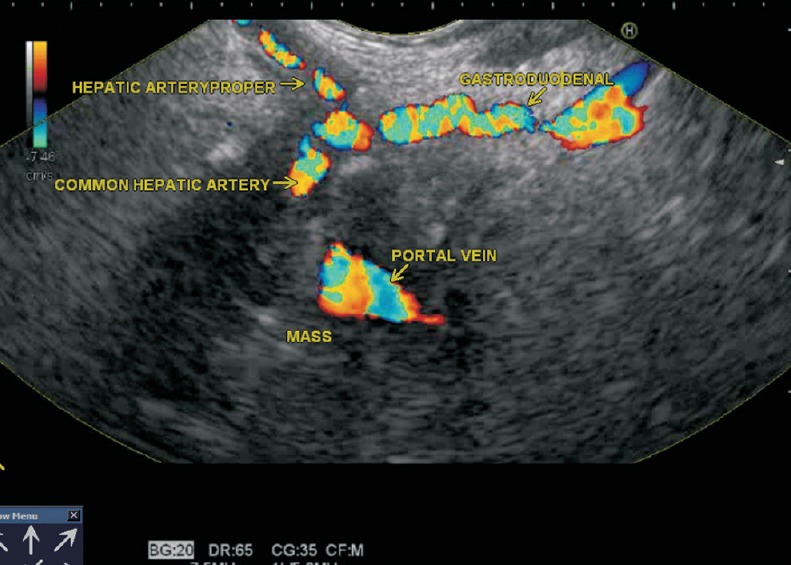
The arterial seagull is seen with the CHA forming the body of seagull. The CHA is traversing through an inflammatory collection of matted lymph nodes. The division into the GDA going towards pancreas (3 o'clock) and the HAP going towards liver (11 o'clock) is also seen
Figure 9.
The arterial seagull of the CHA dividing into the GDA going towards the pancreas (3 O'clock) and the HAP going towards the liver (11 O'clock) is seen. The CHA is traversing through a mass which is encasing the PV also
REFERENCES
- 1.Solorio S, Badui E, Yáñez M, et al. Double mitral valve orifice. Two-dimensional and Doppler echocardiographic diagnosis. Arch Med Res. 1996;27:491–4. [PubMed] [Google Scholar]
- 2.Steadman CD, Khoo J, McCann GP. The seagull sign. Postgrad Med J. 2010;86:253–4. doi: 10.1136/pgmj.2009.090332. [DOI] [PubMed] [Google Scholar]
- 3.Park MS, Yoon SJ, Park JH, et al. The management of the displaced medial wall in complex acetabular fractures using plates and additional cerclage. Hip Int. 2013;23:323–9. doi: 10.5301/hipint.5000027. [DOI] [PubMed] [Google Scholar]
- 4.Hay HR. Gas in gall stones: A rare radiological sign in the acute abdomen. Gut. 1966;7:387–91. doi: 10.1136/gut.7.4.387. [DOI] [PMC free article] [PubMed] [Google Scholar]
- 5.Gottschalk U, Jenssen U. Endosonographic evaluation of the adrenal glands: Part I. Video Journal and Encyclopedia GI Endoscopy [Google Scholar]
- 6.Cosby KS, Kendall JL. Cosby KS, Kendall JL, editors. Liver, gallbladder, and biliary tree. Practical Guide to Emergency Ultrasound. 2006:183–218. [Google Scholar]
- 7.Sharma M, Babu CS, Garg S, et al. Portal venous system and its tributaries: A radial endosonographic assessment. Endosc Ultrasound. 2012;1:96–107. doi: 10.7178/eus.02.008. [DOI] [PMC free article] [PubMed] [Google Scholar]
- 8.Rameshbabu CS, Wani ZA, Rai P, et al. Standard imaging techniques for assessment of portal venous system and its tributaries by linear endoscopic ultrasound a pictorial and video essay. Endosc Ultrasound. 2013;2:16–34. doi: 10.7178/eus.04.005. [DOI] [PMC free article] [PubMed] [Google Scholar]
- 9.Sharma M, Rai P, Mehta V, Rameshbabu CS. Techniques of imaging of aorta and its 1st order branches by Endoscopic Ultrasound. Endosc Ultrasound in Press. doi: 10.4103/2303-9027.156722. [DOI] [PMC free article] [PubMed] [Google Scholar]


