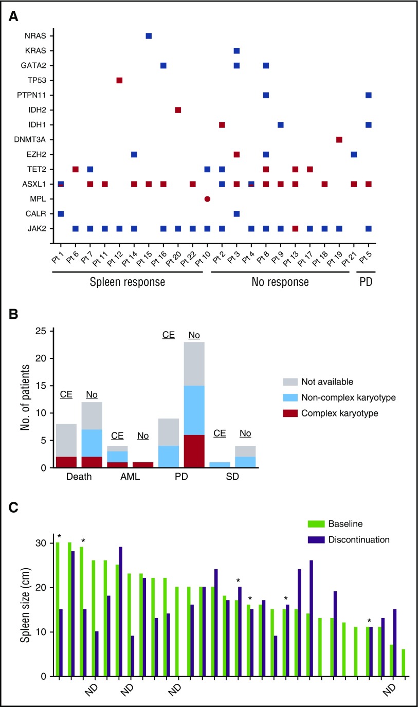
Figure 3.
Correlation between clonal evolution and disease status. (A) Mutations at baseline and follow-up in patients who acquired a mutation while receiving ruxolitinib. Red squares denote an acquired mutation. Red/blue squares denote the acquisition of a second, different mutation in the same gene. Note that at the time of follow-up, patient (Pt) 3 had lost the GATA2 and KRAS mutations present at baseline. (B) Bar graph depicting disease status at the time of ruxolitinib discontinuation in patients with (CE) and without (No) clonal evolution. Bars are colored according to karyotype: gray, sample was not available; light blue, noncomplex karyotype; red, complex karyotype. (C) Bar graph depicting spleen size at baseline (light green) and at the time of ruxolitinib discontinuation (purple). Asterisks indicate patients who acquired a new mutation while on therapy. ND, mutation status at discontinuation not determined. PD, progressive disease; SD, stable disease.

